




Abstract
Leptospirosis is a worldwide zoonosis caused by spirochetes from the genus Leptospira. In the present study, a visual and rapid method for detecting Leptospira interrogans was developed based on multiple cross-displacement amplification (MCDA) and nanoparticle-based lateral flow biosensor (LFB). A set of 10 primers was specifically designed to recognize 10 regions of the lipL 41 gene of L. interrogans. The MCDA reaction was optimized at 64°C for only 40 min, and the amplification products were directly applied to the biosensor. The entire process, including DNA extraction (25 min), MCDA reaction (40 min), and result interpretation (∼2 min), could be completed within 70 min. Amplification products were detectable from as few as 10 genomic equivalents per reaction of pure L. interrogans DNA. No cross-reaction with nonpathogenic Leptospira or other bacteria was observed. The MCDA-LFB method established in the current report is suitable for the rapid screening of L. interrogans in clinical, animal, and environmental samples.
Introduction
Leptospirosis is one of the most widespread zoonoses and is caused by infection with pathogenic spirochetes of the Leptospira genus (Levett 2001). Although leptospirosis is mainly prevalent in tropical and subtropical countries (Pappas et al. 2008), it is considered an emerging or reemerging zoonosis of global public health concern (Adler 2015). In China, leptospirosis is listed as a category B notifiable disease (Dai 1992).
Rodents are recognized as important mammalian reservoirs of Leptospira spp. (Guerra 2009, Meerburg et al. 2009). Most infected mammalian reservoir animals, such as rodents, present only mild chronic disease or are asymptomatic and shed infectious organisms in urine for their lifetime (Vinetz 2001), while humans can be infected through skin contact with contaminated water and develop acute leptospirosis, a mild to severe flu-like infection that can sometimes lead to a life-threatening disease with liver, lung, and kidney failure (Lacroix-Lamande et al. 2012). Pulmonary diffuse hemorrhaging, a serious clinical form of leptospirosis, is fatal in approximately 25% of patients (Viriyakosol et al. 2006). However, leptospirosis is frequently misdiagnosed due to its protean and nonspecific presentation resembling many other febrile diseases, notably viral hemorrhagic fevers such as dengue (Levett 2001). The leptospirosis problem remains underestimated due to a lack of awareness and underrecognition because of a lack of proper use of diagnostic tools (Ahmed et al. 2006). Early diagnosis is essential because antibiotic treatment is most effective during the initial course of the disease (Levett 2001). Therefore, availability of rapid and accurate point-of-care diagnostic tests is required to identify leptospirosis.
Traditionally, the microscopic agglutination test (MAT) is considered the “gold standard” for diagnosing leptospirosis, but it does not permit early diagnosis (Palaniappan et al. 2005), and some other diagnostic methods, such as dark-field microscopy, enzyme-linked immunosorbent assay, and Western blotting, have low sensitivity (McBride et al. 2005).
More rapid and sensitive methods, including the polymerase chain reaction (PCR), multiplex PCR, and real-time quantitative PCR methods, have also been reported to detect leptospirosis (Smythe et al. 2002, Levett et al. 2005, Kositanont et al. 2007, Ahmed et al. 2009, Bedir et al. 2010). However, the presence of false-positive PCR products complicates identification of true infection, and real-time PCR instruments are expensive and may not be readily available in many laboratories.
Instrument-independent loop-mediated isothermal amplification (LAMP) methods targeting lipL41 (Lin et al. 2009) and rss (Sonthayanon et al. 2011), a 16S rRNA gene of L. interrogans, have also been developed, but lipL41 LAMP has a detection limit of only l00 genomic equivalents (GEs) per reaction mixture, and the specificity of rrs LAMP is lower than that of lipL41 LAMP, thus hindering the clinical utility of rrs LAMP (Sonthayanon et al. 2011). A newly developed LAMP method to detect the rrs gene of pathogenic Leptospira spp. in urine was developed by Koizumi et al. (2012), with a detection sensitivity limit of 2 GEs per reaction mixture. Hsu et al. (2017) designed a lipL32 gene-based LAMP method that enables detection of 10 copies of L. interrogans.
Multiple cross-displacement amplification (MCDA), a novel nucleic acid amplification technique, has been applied to detect many bacterial agents (Wang et al. 2015, 2016, 2017a, 2017b, 2018a). MCDA assay was conducted under isothermal conditions (60–65°C); therefore, a simple heater or water bath that maintained a uniform temperature was sufficient. MCDA methods are simple, rapid, highly specific, and sensitive, and yield amplicons from as few as three bacterial cells. Detection of these amplicons can be achieved with disposable lateral flow biosensors (LFB) (Wang et al. 2016).
In the present report, an MCDA-LFB assay was established for the rapid detection of L. interrogans strains carrying the lipL41 gene (Lin et al. 2009). The analytical sensitivity and specificity were determined in pure cultures.
Materials and Methods
Reagents and instruments
The DNA Extraction Kit was provided by SBS Genetech Co., Ltd. (Beijing, China). Isothermal amplification kits and visual detection reagent (Malachite Green, MG) were purchased from BeiJing-HaiTaiZhengYuan Technology Co., Ltd. (Beijing, China). Biotin-BSA (biotinylated bovine serum albumin) and anti-FITC Ab (rabbit anti-fluorescein antibody) were purchased from the Abcam Co., Ltd. (Shanghai, China). Membrane backing materials, sample pads, conjugate pads, nitrocellulose (NC) membranes, and absorbent pads were purchased from the Jieyi Biotechnology Co., Ltd. (Shanghai, China). Streptavidin-immobilized 40-nm gold nanoparticles (SA-Gs) were obtained from BeiJing-HaiTaiZhengYuan Technology Co., Ltd.
Preparation of a gold nanoparticle-based dipstick biosensor
An LFB (4 × 60 mm) was developed as previously reported with some modifications (Wang et al. 2017a, 2018b). Briefly, the sample pad, conjugate pad, NC membrane, and absorbent pad were laminated onto a plastic adhesive backing card. The NC membrane was sprayed with the anti-FITC Ab (0.15 mg/mL) and biotin-BSA (2.5 mg/mL) conjugates to form the control line (CL) and test line (TL) and each line was separated by 5 mm. After the conjugate pad of the strip was deposited with SA-Gs in 0.01 M PBS (PH 7.4), the assembled cards were cut into 4-mm-wide strips (Deli No. 8012). The assembled biosensors were packaged in a plastic box with a desiccant gel and kept at room temperature.
Bacterial strains and genomic template preparation
The leptospiral strains used in this study were cultured in Ellinghausen–McCullough–Johnson–Harris medium, EMJH, and the leptospires were harvested as described previously (Table 1). The DNA of leptospires was extracted from cultures of isolated strains using the DNA Extraction Kit (SBS Genetech, Beijing, China) according to the manufacturer's directions. The extracted DNA was quantified with a NanoDrop ND-1000 instrument (Calibre, Beijing, China). Genomic DNA of L. interrogans 56601 was serially diluted (1 × 10⁵ GEs/μL; 1 × 10⁴ GEs/μL; 1 × 10³ GEs/μL; 1 × 10² GEs/μL; 1 × 101 GEs/μL; 1 × 100 GEs/μL; 1 × 10⁻¹ GEs/μL) for sensitivity analysis of L. interrogans-MCDA-LFB detection. Other bacterial strains (nonleptospiral strains) were stored in 10% (w/v) glycerol broth at −70°C and genomic templates of nonleptospiral strains were extracted from all culture strains using the QIAamp DNA Mini Kit (Qiagen, Germantown, MD).
Bacterial Strains Used in This Study
U, unidentified serotype; ATCC, American Type Culture Collection; GZCDC, Guizhou Provincial Center for Disease Control and Prevention.
Design of MCDA assay primers
A total of 10 MCDA primers, including F1, F2, CP1, CP2, C1, C2, D1, D2, R1, and R2, were designed using primer software PREMIER 5.0 and PrimerExplorer V4 (Eiken Chemical, Japan). Integrated DNA Technologies design tools were applied to analyze hairpin structures and hybrids of all the MCDA primers. The MCDA primers were further verified by performing Blast analysis, specific for L. interrogans. Biotin and fluorescein isothiocyanate (FITC) were labeled on the 5′ ends of the C1 and D1 primers, respectively. The primer information, including sequences, positions, and modifications of the primer pairs, is shown in Fig. 1 and Table 2. All of the HPLC purification grade oligomers were synthesized and purified by Tianyi-Huiyuan Biotech Co., Ltd. (Beijing, China).

Location and sequence of the pathogenic Leptospira-specific gene lipL41 used to design the MCDA primers. The nucleotide sequence of the sense strand of the lipL41 gene is shown. Right arrows and left arrows indicate sense and complementary sequences that were used, respectively. MCDA, multiple cross-displacement amplification. Color images are available online.
The Primers Used in This Study
C1*, 5′-labeled with biotin when used in the MCDA-LFB assay; D1*, 5′-labeled with FITC when used in the MCDA-LFB assay.
FITC, fluorescein isothiocyanate; mer, monomeric unit; nt, nucleotide; MCDA, multiple cross-displacement amplification; LFB, lateral flow biosensor.
The standard MCDA assay
MCDA reactions were performed in a 25-μL reaction system as described in previous studies (Wang et al. 2015, 2018c). Briefly, each reaction contained 0.4 μM each of F1 and F2 (displacement primers), 0.8 μM each of C1* and C2 (amplification primers), 1.2 μM each of R1, R2, D1,* and D2 (amplification primers), 2.4 μM each of CP1 and CP2 (cross primers), 1 μL of Bst DNA polymerase (8 U), 12.5 μL of 2 × reaction mix (isothermal amplification kit), and 1 μL of DNA template. Colorimetric indicator (MG), turbidimeter (LA-320C), and LFB detection were used to monitor and analyze the MCDA amplicons.
For the MG method, the amplified products caused a color change from colorless to light green, while the negative controls and blank control remained colorless. By using the LFB, two visible red lines (the TL and CL) could be observed in positive reactions, but only the CL was visual in negative and blank controls. The optimal reaction temperature was determined in the range of 60–67°C for 60 min. Mixtures with 1 μL of genomic templates of Shigella flexneri (S. flexneri) and Staphylococcus aureus strains were used as negative controls, and mixtures with 1 μL of distilled water (DW) were used as blank controls.
Sensitivity and specificity of the L. interrogans-MCDA-LFB assay
The DNA equivalent was defined by the genomic DNA amount of the templates and the limit of detection (LoD) was tested using serial dilutions (1 × 10⁴ GEs/μL; 1 × 10³ GEs/μL; 1 × 10² GEs/μL; 1 × 101 GEs/μL; 1 × 100 GEs/μL; 1 × 10⁻¹ GEs/μL). L. interrogans-MCDA-LFB detection result was compared with that of colorimetric indicator (MG) and turbidity analysis. Three replicates of each dilution were tested. The specificity of L. interrogans-MCDA-LFB was verified with DNA templates from 83 different bacterial strains (Table 1), which were repeated at least two times.
L. interrogans-MCDA-LFB detection in mouse kidney samples
L. interrogans-MCDA-LFB was used to detect L. interrogans DNA from the total DNA of field mouse kidneys, which has been detected by using leptospiral cultivation and isolation. Total DNA from uninfected mouse kidneys was used as a template in a negative control reaction. Simultaneously, G1/G2 primer (Gravekamp et al. 1993)-based PCR methods were applied for detection in the above mouse kidney samples. The specificity and sensitivity of L. interrogans-MCDA-LFB, G1/G2 primer-based PCR, and the conventional cultivation method detection of L. interrogans were compared.
Results
Confirmation and detection of L. interrogans MCDA products
To determine the availability of L. interrogans-MCDA primers (Table 2), L. interrogans-MCDA assays with DNA from pure cultures were carried out at 64°C for 1 h. Amplification occurred with DNA from L. interrogans 56601, but not with S. flexneri (GZCDC isolates), S. aureus (GZCDC isolates), and the DW control (Fig. 2). Therefore, the L. interrogans-MCDA primer set was a good candidate for the development of MCDA-LFB assay for L. interrogans detection.

Confirmation and detection of Leptospira interrogans-MCDA products.
Temperature optimization for the L. interrogans-MCDA-LFB assay
To verify the optimum amplification temperature, L. interrogans strain 56601 was used as positive control at a level of 10 pg per reaction and the reactions were monitored by the real-time turbidity method. All tested temperatures (60°C to 67°C with 1°C increments) produced typical kinetic graphs, with faster amplification achieved at assay temperatures of 64°C (Fig. 3). Thus, the amplification temperature of 64°C was used to perform the rest of the tests conducted in the current report.
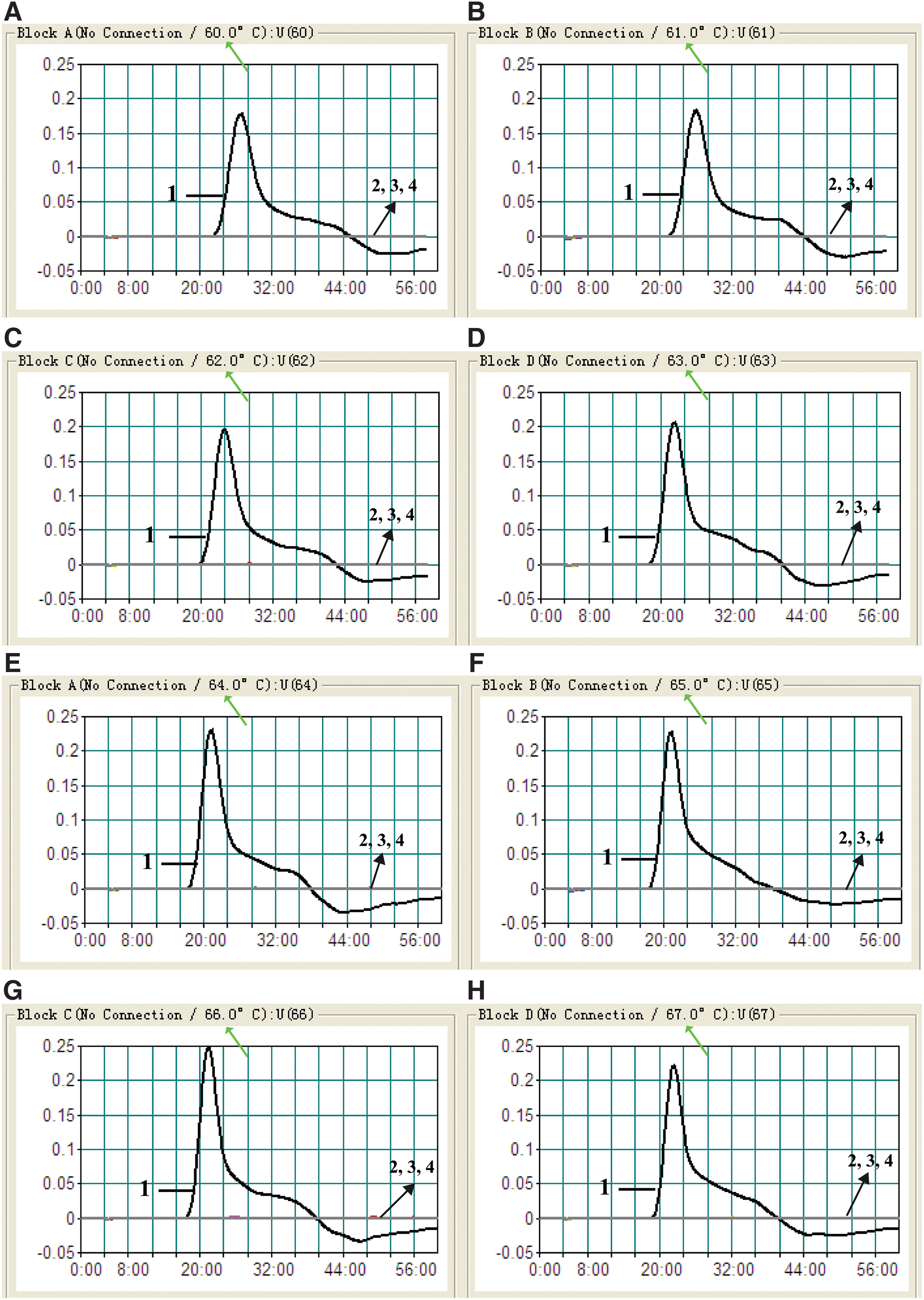
Optimal reaction temperature for L. interrogans-MCDA primer sets. The standard MCDA reactions for detection of L. interrogans were monitored by real-time measurement of turbidity, and the corresponding curves of DNA concentrations are marked in the figures. The threshold value was 0.1, and a turbidity >0.1 was considered positive. Eight kinetic graphs
Sensitivity of MCDA-LFB for L. interrogans detection
Serial dilutions of L. interrogans genomic DNA were used to examine the LoD of the MCDA-LFB assay. Our results demonstrated that the analytical sensitivity of the L. interrogans-MCDA-LFB assay was 10 GEs per reaction (Fig. 4A). Using MCDA by self-trial, the analytical sensitivity of the MCDA assay was further examined by turbidity detection and visual inspection of reaction products with MG reagents (Fig. 4B, C). By MG reagents and real-time turbidity, the LoD of the MCDA assay was also 10 GEs per reaction, which was in complete accordance with LFB detection.

Sensitivity analysis of the MCDA-LFB assay using serial dilutions of genomic DNA extracted from L. interrogans strain 56601. A total of three monitoring techniques, including the lateral flow biosensor
Specificity of MCDA-LFB for L. interrogans detection
When DNA from the bacteria listed in Table 1 was used in the MCDA-LFB assay, only the DNA from the L. interrogans strains provided positive results, with positive rate of 100% for L. interrogans detection. DNA from nonpathogenic Leptospira strains and from all non-Leptospira isolates did not lead to the production of detectable amplification products (Fig. 5). Two red lines, including the TL and CL, appeared on the strips for the positive tests, and only one red line (the CL) appeared on the biosensors, suggesting negative results for nonpathogenic Leptospira and other bacterial isolates and the blank control.

Specificity analysis of the L. interrogans-MCDA-LFB assay for different bacterial strains. MCDA reactions were carried out using different genomic DNA templates and were analyzed visually. Biosensors 1, 2, 3, 4, 5, 6, 7, 8, 9, 10, 11, 12, and 13 used L. interrogans strains 56601, 56602, 56603, 56605, 56606, 56607, 56608, 56609, 56610, 5662, S13, 56615, and 56635, respectively; biosensors 14, 15, and 16 used L. interrogans strains 56604 and 56655 and L. biflexa strain 57601 (nonpathogenic Leptospira strain), respectively; biosensors 17–38 used Listeria monocytogenes, Shigella flexneri, Salmonella, Staphylococcus aureus, enteropathogenic Escherichia coli, enterotoxigenic E. coli, invasive E. coli, enterohemorrhagic E. coli, enteroaggregative E.coli, Klebsiella pneumoniae, Streptococcus suis, Pseudomonas aeruginosa, Vibrio cholerae, Vibrio parahemolyticus, Streptococcus pneumoniae, Enterococcus faecalis, Enterococcus faecium, Bacillus cereus, proteus species, Neisseria meningitidis, and Enterobacter cloacae, in that order; and biosensor 39 was the blank control (DW). Color images are available online.
Application of MCDA-LFB for surveillance of leptospires from field mice
A total of 48 field mouse kidney samples were used for the application of L. interrogans-MCDA-LFB developed in the present study. Among the 48 field mouse kidney samples, 12 samples had been confirmed as infected by leptospires using the MAT method after leptospiral isolation and cultivation, and the other 36 samples were negative by using leptospiral isolation and cultivation. The results of MCDA-LFB detection showed that 14/48 field mouse kidney samples were positive after a 40-min amplification. The 14 MCDA-LFB-positive samples included 12 leptospiral isolation-positive and 2 leptospiral isolation-negative samples. However, only 8/48 field mouse kidney samples showed positive results when using conventional G1/G2 primer-based PCR, and all of the eight positive samples were from the leptospiral isolation-positive samples (Table 3).
Comparison of Conventional Polymerase Chain Reaction, Culture-Biotechnical, and Multiple Cross-Displacement Amplification for Detection of Leptospira interrogans in Kidney Samples
Discussion
The present study demonstrated that MCDA combined with a lateral flow biosensor (MCDA-LFB) utilizing the lipL41 gene as an amplification target can detect L. interrogans with excellent specificity and sensitivity. The assay's specificity was successfully examined using pure cultures. The test was positive for all L. interrogans isolates but negative for nonpathogenic Leptospira spp. and non-Leptospira isolates (Fig. 5). Therefore, the L. interrogans-MCDA-LFB method provided a high degree of selectivity for identifying L. interrogans strains.
In addition to its sufficient specificity, the newly established L. interrogans-MCDA-LFB method was able to detect as few as 10 GEs per reaction of L. interrogans DNA isolated from a pure culture (Fig. 4). The L. interrogans-MCDA-LFB assay was more sensitive than the L. interrogans-LAMP method (Lin et al. 2009), which only detects 100 GEs. Although the amplification products could be detected equally with other methods, the LFB is likely the preferred method as reading the results is less subjective and does not require instrumentation. The L. interrogans-MCDA-LFB assay only required simple incubation at 64°C for 40 min. Various portable user-friendly instruments adapted for MCDA reactions exist, such as the dry block heater (HDT-100C, HengAo, Tianjing, China). The portable (18 × 22 cm) battery-powered device supports 96 MCDA reactions per assay. MCDA can be conducted using commercial isothermal amplification kits (such as Isothermal Amplification Kits and NEB WarmStart Kits), and an MCDA reaction costs approximately 3.5 USD. The cost of the LFB is estimated to be 2 USD per test. In addition, labor costs are eliminated because trained personnel in a certified laboratory are not required. Therefore, our assay is more cost-effective.
The MCDA products were directly analyzed using the biosensor (Figs. 2–5). The entire procedure, including specimen processing (25 min), isothermal reaction (40 min), and detection (2 min), could be completed within 70 min. Detection of amplification products with a lateral flow device is not only fast but also simpler and less error prone than detection by the other methods.
The genus Leptospira is broadly divided into two species: Leptospira interrogans, comprising all pathogenic strains, and Leptospira biflexa, containing the nonpathogenic strains. The species L. interrogans comprises at least 250 antigenically distinct variants known as serovars belonging to 24 serogroups (Dutta and Christopher 2005). L. interrogans serovar Lai (56601) and serovar Pomona (56608) are the most common epidemic serovars in the South and North of China, respectively. In the current study, we used 15 reference strains representing 15 serovars of 15 serogroups of L. interrogans in China to develop MCDA-LFB detection methods. The results showed that the reference strains of 13 serovars representing 13 serogroups, including the two most common epidemic serogroup hemorrhage serovars Lai and serogroup Pomona serovar Pomona, were specifically covered in the scope of MCDA-LFB. To assess the ability of the L. interrogans MCDA-LFB method to detect leptospires in tissue specimens, we tested 48 mouse samples from environmental surveillance. The results of MCDA-LFB detection showed that 14/48 field mouse kidney samples were positive after a 40-min amplification (data not shown), while only 8/48 field mouse kidney samples showed positive results when using conventional G1/G2 primer-based PCR (Gravekamp et al. 1993), which is officially recommended for leptospiral detection in the Chinese System for Disease control and Prevention, and 12/48 samples were positive by using cultivation and isolation. These results demonstrated that the MCDA-LFB detection method is highly efficient and specific in tissue sample detection, reflecting its potential application in the surveillance of carrier status among host animals and early clinic diagnosis.
Conclusion
A reliable lipL41-MCDA-LFB assay was successfully developed for identification of L. interrogans, which could facilitate early diagnosis of leptospirosis patients and contribute to the control and prevention of leptospirosis in the localities. The MCDA-LFB approach devised here was sensitive, specific, and did not rely on complicated instruments and expensive reagents. The use of LFB could provide an objective, rapid, and easily interpretable readout of the method's results. Therefore, the lipL41-MCDA-LFB method could be regarded as a valuable tool for the rapid screening of L. interrogans isolates in clinical surveillance of carrier animal and environmental samples, especially in resource-limited areas of developing countries during epidemic periods.
Footnotes
Acknowledgments
We acknowledge the financial support of the special funds of the research team for experimental diagnostic techniques and molecular epidemiological studies of major infectious diseases in Guizhou Province (Program of Scientific and Technological Innovation Team of Guizhou Province. Grant no. Qian Ke He Platform talent [2018]5606), a grant from the National Natural Science Foundation of China (grant no. 81760366), Special Funds for High-Level Creative Talents Cultivation in Guizhou Province (Qian Ke He (2016)4021), and Special Funds for the Cultivation of Outstanding Youth Talents of Science and Technology in Guizhou Province (no. Qian Ke He Ren Word [2015] 09).
Authors' Contributions
S.L. and Jie Yan conceived and designed the experiments. Y.L. and M.W. performed the experiments. S.L., X.C., and Weilin Hu analyzed the data. S.L. contributed reagents/materials/analysis tools, performed the software, and wrote the article.
Author Disclosure Statement
Shijun Li and Ying Liu have filed for a patent from the State Intellectual Property Office of the People's Republic of China, which covers the novel method and sequences included in this article (application number: 201811040029.7). The other authors report no conflicts of interest in this work.