
Abstract
Leptospirosis and rabies are zoonotic diseases of public health importance and endemic diseases in tropical countries such as Costa Rica. Peridomestic wild animals such as raccoons (Procyon lotor) have been implicated as competent hosts of Leptospira spirochetes and rabies virus. This study focused on understanding the role of urban raccoons in the dynamics of leptospirosis and rabies in a tropical environment. A total of 97 specimens of the common raccoon were captured within the Greater Metropolitan Area of Costa Rica; 32.6% (31/95) of raccoons presented evidence of antibodies (> 1: 100) against Leptospira sp. Attempts to cultivate Leptospira failed, but 19 serovars were identified, which are also responsible for causing disease in humans in Costa Rica. Detected titers ranged from 1: 100 to 1: 6400. Lymphoid hyperplasia in kidneys and spirochetes were demonstrated in 3 of 20 necropsied cases (15%). Twenty brain samples were analyzed by hematoxylin and eosin stain for evidence of encephalitis and Negri body detection and simultaneously frozen brain material was employed to perform a rapid immunoassay test for rabies antigen. All tested samples were negative. This study is the first report of Leptospira seroprevalence in raccoons that cohabit urban areas in Costa Rica. We also highlight the importance of the raccoon as one of their natural competent host and sentinel animals within highly populated urban environments in tropical cities.
Introduction
L
Costa Rica is a tropical country in Central America, where Leptospira is considered an endemic pathogen. The infection route is direct contact with contaminated abiotic sources (such as water or mud) or by direct exposure to domestic and wild animals. Urine secretion from infected wild mammals such as raccoons is often the primary infection source (Richardson and Gauthier, 2003, Junge et al., 2007, Davis et al., 2008, Valverde et al., 2013). Reservoir animals either with or without clinical signs can be active or passive Leptospira carriers and shedders (Duncan et al., 2012, Adler, 2015).
On the other hand, rabies virus is a deadly pathogen affecting a wide variety of mammals, including humans, with some ∼60,000 global human cases per year (WHO, 2016).
The raccoon (Procyon lotor) is considered one of the main rabies reservoirs in North American temperate zones, for example, in the east coast of the United States. Raccoons carry the variant, RRV- ST1/GT1, which is one of the most important variants with regard to the raccoon vaccination program (Conover, 2002, Jackson, 2013). In Costa Rica, the main rabies reservoir is the common vampire bat (Desmodus rotundus). It causes death mainly in livestock, but cases have also been reported in humans in recent years. Although raccoons are important reservoirs in the northern hemisphere, so far there has been no evidence that the raccoon rabies strain is currently circulating in Costa Rica (Hutter et al., 2016).
The raccoon's close proximity to people has generated conflicts in the Great Metropolitan Area (GAM) of Costa Rica, a phenomenon that was recently reported (Baldi et al., 2016). Due to their interactions with humans in a common shared space, raccoons play an important role as effective pathogen spreaders for microorganisms such as Leptospira, rabies virus, and other zoonotic disease agents (Conover, 2002, Bradley and Altizer, 2007, Bondo et al., 2016). The presence of zoonotic pathogens in urban wildlife represents a potential source of infection for humans and is a serious concern that must be considered and addressed.
The aim of this study was to determine the seroprevalence of leptospirosis and to assess the presence of rabies virus in an urban raccoon population in the GAM of Costa Rica.
Materials and Methods
Study area
Animals were captured by placing Havahart live traps (Lititz, PA) in small wooded areas near rivers or in backyards and gardens within the GAM (Fig. 1). Traps were baited with bananas or canned cat food or the mix of both. The traps were set up at night and were visited the following morning. We used a total of 10 live traps at each sampling point for a period of three to seven consecutive nights every 2 months over a total 20-month period (from April 2013 to July 2016). In this period, we captured a total of 97 raccoons at 15 different sites. The first 20 captured individuals were euthanized for subsequent pathological examinations. The remaining 77 animals were anesthetized for sampling and ear-tagged and subsequently released.
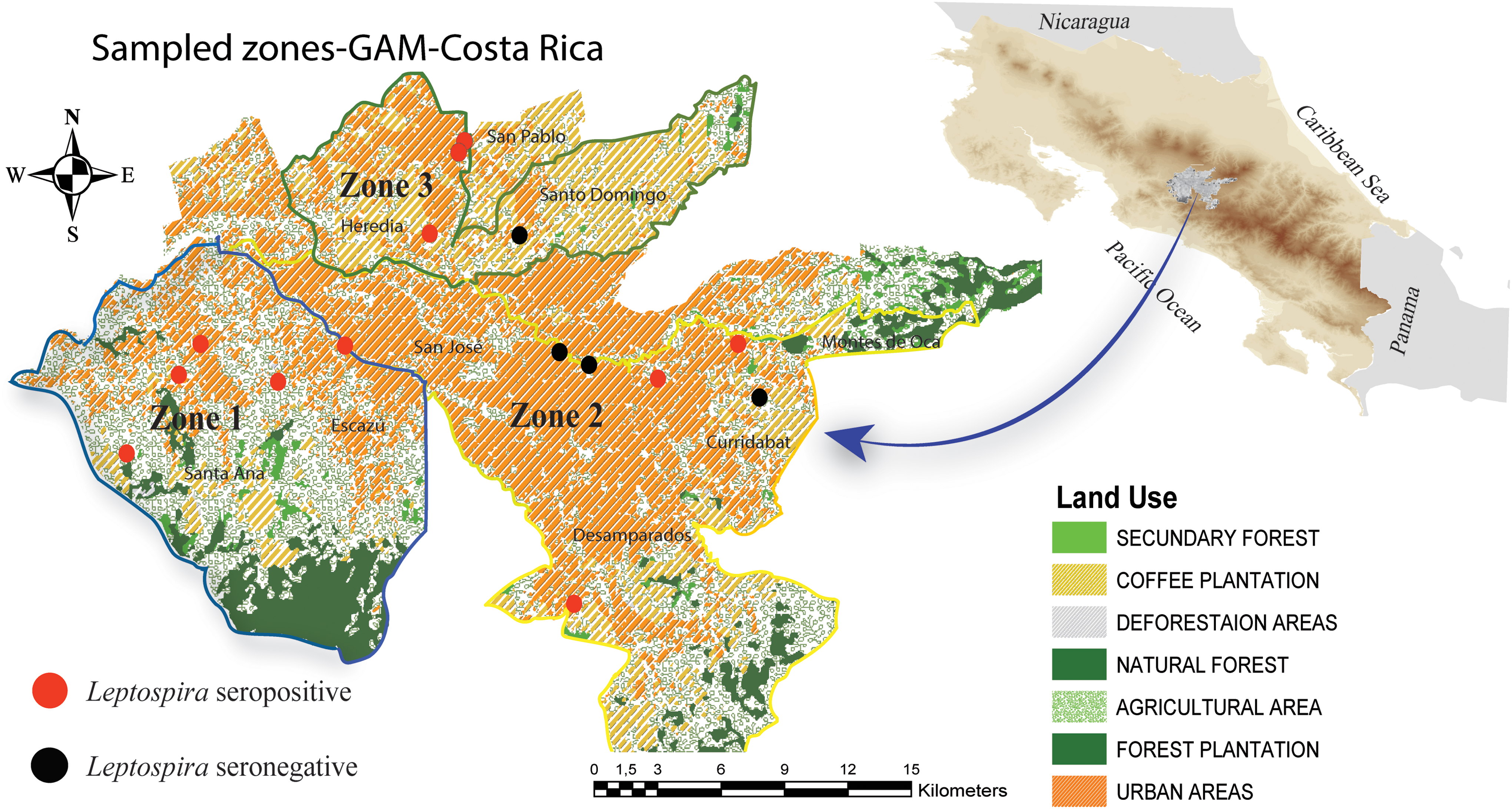
Shows the areas in the GAM within three sectors where the raccoon samples were taken. GAM, Great Metropolitan Area. Color images are available online.
We set up traps in 15 locations in three defined zones (Zone 1: Santa Ana-Escazu, Zone 2: Heredia, and Zone 3: San Jose). We captured and removed individuals at locations where raccoon conflicts had been reported to the local wildlife service authorities. The remaining animals were part of a mark–catch–recapture study to estimate the population density of raccoons in the GAM. Permissions from the Research Ethics Board, FCSA-EMV-CBA-007-2013, and Institutional Review Board, ACCVC-OH-512, were obtained before sampling.
Samples and laboratory analysis
Twenty raccoons were euthanized with established anesthesia and euthanasia protocols (Baldi et al., 2016). Standard necropsies were performed at the National University pathology laboratory (UNA-Costa Rica) to determine the presence of Leptospira and rabies lesions (Fiette and Slaoui, 2011, Leary and Golab, 2013). From these 20 animals, kidney tissues and urine samples were aseptically collected to cultivate, isolate, and identify Leptospira. In addition, serum samples were assessed for the presence of Leptospira antibodies. Evaluation of internal organs was performed according to standard protocols. Tissue samples from internal organs (liver, kidney, lung, and spleen), including brain, were fixed in 10% neutral buffered formalin for histopathological examination. Additionally, a brain aliquot was frozen at −20°C for further analysis. Location, weight, length, and tooth eruption were recorded for each individual.
Seventy-seven raccoons were anesthetized using a combination of ketamine–medetomidine 2 mg/kg/4 mg/kg (Aveco Co., Fort Dodge, IA., Domitor® 1 mg/mL; Zoetis, Kalamazoo, MI) and reversed 45 min later with atipamezole (Antisedan® 5 mg/mL; Zoetis) (West et al., 2014). Approximately 5 mL of blood was collected by jugular vein puncture. All biological samples were stored at ∼4°C in a cooler and transported to the National Veterinary Laboratory in less than 3 h. Location and biological measurements, sex, age, tooth eruption, and weight were recorded for each captured raccoon (Table 1).
Number of Captured Raccoons (Procyon lotor) by Age, Sex, and Weight in KG (Average)
We classified individuals as either juvenile or adult based on their weight, size, and dentition (Grau et al., 1970). The collected blood was allowed to clot and then centrifuged at 2000 rpm for 15 min. All serum samples were transferred into microcentrifuge tubes, and two aliquots were stored at −20°C and −70°C for further analysis. Kidney tissue was stained with hematoxylin and eosin (H&E), following standard procedures (Aughey and Frye, 2001).
Leptospira
A total of 95 serum samples were analyzed against Leptospira serogroups at the Laboratorio Nacional de Servicios Veterinarios (LANASEVE) using microscopic agglutination testing (MAT) with anti-Leptospira rabbit serum samples, following OIE recommendations (OIE, 2013). The Leptospira serovar battery of reference strains consisted of the following serovars: Arenal, Australis, Autumnalis, Batabiae, Canicola, Castellonis, Copenhageni CR, Copenhageni, Corredores, Costa Rica, Grippotyphosa, Hardjo, Hebdomadis, Icterohaemorrhagiae RGA, Icterohaemorrhagiae Kantrowics, Patoc, Pomona, Panama, Pyrogenes, Tarassovi, Alexi, Rama, Rio, and Shermani. Results were reported as seropositive if the titer was >1:100, as previously stated (Balows, 1988).
For isolation of Leptospira bacteria, aseptically macerated kidneys and urine samples were accordingly inoculated into Ellinghausen, McCullough, Johnson, and Harris (EMJH) media (Forbes et al., 2007). All cultures were incubated aerobically and protected from light at room temperature (25–30°C) for 6 months. Kidney tissue samples from all seropositive raccoons were also stained with Warthin–Starry to determine the inflammatory response and the presence of spirochetes (Colvin, 2016).
Rabies virus
Twenty brains were stained with H&E and analyzed by a veterinary pathologist for signs of encephalitis and cytoplasmic inclusions in Negri bodies (Rupprecht et al., 2002, Jackson, 2013). Additionally, we used frozen brain material from the same raccoons to perform virus antigen detection using a commercial immunochromatographic antigen detection test (rapid immunoassay assay or RIDT (Rabies Ag test Kit, Bionote®, Inc., Korea), with a reported sensitivity of 95% and specificity of 98.5% (Yang et al., 2012). RIDT is a validated test for domestic animals and has also been used in wild carnivores (Léchenne et al., 2016). The producer's instructions and interpretation recommendations were adhered to (Eggerbauer et al., 2016).
Statistical analyses
Descriptive statistics were performed to establish Leptospira seroprevalence by the zone where they were captured, age, and sex of the animals. We then examined the association between potential risk factors (age, sex, and weight) and Leptospira-positive outcomes through a conditional, backward step, binary logistic regression model. Analyses and generation of confidence intervals were performed using Excel (Microsoft) and SPSS Statistics, version 24 (IBM, Armonk, NY).
Geocoding and spatial analysis
A map was generated with georeferenced points for each sampling point in each of the three zones. Information from 19 Leptospira serovars identified from a total of 31 seropositive animals was geocoded, we assigned a site where the serovar was collected and identified. The map was generated using ArcGis 10.1 software (ERSI, Redlands, CA).
Results
Information and samples from 97 raccoons, 47% (n = 46) males and 54% (n = 52) females, 15% juveniles (15) and 84% (82) adults, collected over the study period were included. None of the raccoons exhibited clinical disease, and all necropsied animals appeared to be in good health at gross examination. The mean body weight was 4.87 (95% CI: 4.50–5.26) kilograms. Table 1 shows the weight ranges per sex and age class.
Leptospira
Thirty-one of 95 raccoon (32.6%, 95% CI: 23.6–43.1) samples were positive for Leptospira antibodies. The seroprevalence by zone is presented in Table 2. Of these 31 positive samples, we identified 19 serovars implicated in morbidity of both humans and domestic animals in Costa Rica (Hutter and Sandi, 2012, INCIENSA, 2015). Only six animals were seropositive to a unique serovar (with titers of 1:100 to 1:1600), which included Pyrogenes, Hebdomadis, Icterohemorrhagic, Icterohaemorrhagiae Kantrowics and Travossi. The other 25 samples comprised multiple Leptospira serovars with titers ranging from 1:100 to 1:6400. Eleven raccoons presented with one high titer (for example, one raccoon yielded Pyrogenes titers of 1600) among the other titers. Fourteen samples had multiple similarly high titers. Details of the titers and their corresponding serovars can be found in Tables 3 and 4.
Proportion of Samples (with 95% Confidence Intervals) Testing Positive for Leptospira Antibodies by Location Zone
CI: 95% confidence interval
Raccoons with Elevated Titers of Only One Leptospira Serovar Detected
= single elevated serovar
Raccoons with Elevated Titers of Multiple Leptospira Serovars
A, Alexi; Ast, Australis; C, Canicola; Co, Copenhageni; CoCr, Copenhageni CR; CR, Costa Rica; Hb, Hebdomadis; HbCR, Hebdomadis CR*; IcRGA, Icterohaemorrhagiae RGA; IcKw, Icterohaemorrhagiae Kantrowics; P, Panama; Py, Pyrogenes; Ri, Rio; Sh, Shermani; T, Tarassovi; Bt, Batavia; Ra, Rama.
Bold font implies that all of these serovars had equal levels of titers on the same animal.
Three (15%) of the 20 animals, which were analyzed histopathologically, presented interstitial nephritis and spirochetes in E&H and Warthin–Starry stains. The stained positives were also positive in the MAT. There were other minor histopathological findings in kidneys and other tissues, but none of them were associated directly with leptospirosis.
In the binary logistic regression model, we could not find any statistical evidence of association between seropositivity and sex (p = 0.971896) or age (p = 0.395133). Although weight had a significant p-value (p = 0.04414), the odds ratio was exactly 1 (95% CI: 1.000–1.001), which shows that age was a confounder for weight. Therefore, neither sex and age nor weight influenced the presence of Leptospira antibodies.
Rabies
The 20 brain samples tested for the presence of virus antigen were negative in the tissue stain (H&E) and RIDT (no positive bands observed). All the RIDT diagnostic kits yielded a clear and well-defined, single, blue negative band.
Discussion
This study establishes the presence of a zoonotic bacterium, Leptospira, and suggests the absence of the raccoon rabies virus variant or any other rabies variants in this raccoon population in a tropical urban zone in Costa Rica. Both Leptospira and rabies are important zoonotic pathogens and are considered endemic in Costa Rica (INCIENSA, 2015). Leptospira and rabies virus have been reported in raccoons in temperate zones in the past, and raccoons are pointed out as effective hosts and reservoirs for both diseases (Reilly, 1970, Bigler et al., 1974, Rupprecht et al., 2002, Compton et al., 2008, Jardine et al., 2011b, Lee et al., 2011, Shearer et al., 2014). However, these disease agents have never been reported in urban, tropical raccoon populations before. With this study, we contribute toward filling the knowledge gap of these agents in raccoon populations in endemic neotropical zones.
Leptospira
Our results show that raccoons are competent, peridomestic wildlife hosts for Leptospira in urban areas of Costa Rica. The identified Leptospira prevalence (32.6%) in Costa Rica is consistent with studies in temperate zones performed by Jardine et al. (2011a) and Duncan et al. (2012), who reported similar findings (30%), and is consistent with other studies, where prevalence ranged from 10% to 70% (Mitchell et al., 1999, Hamir et al., 2001, Richardson and Gauthier, 2003, Bischof and Rogers, 2005, Junge et al., 2007, Koizumi et al., 2009, Raizman et al., 2009, Jardine et al., 2011b, Tan et al., 2014). However, to date, no data were available for the prevalence of Leptospira in raccoons in a Mesoamerican country.
The frequency of Leptospira varies widely across different sampling locations. Similarly, the serovars recognized in raccoons as causing human disease in Costa Rica are widely heterogeneous between individuals, locations, and within locations (INCIENSA, 2015). In Costa Rica, no information on the presence of Leptospira serovars in raccoons was previously available. This study demonstrates the great diversity of serovars in raccoons and highlights the potential public health risk. The MAT assay is the gold standard to assess evidence of leptospirosis exposure. Nevertheless, paired samples are needed in patients to clarify the infectious serovar. MAT constraints include cross-reactivity when two or more serovars are simultaneously infecting one individual at a point in time (Picardeau, 2017).
The results of this study demonstrate this constraint as we could collect only a single sample per animal. It is well known that raccoons can be reinfected with different serovars over time (Jardine et al., 2011a). Since a second sampling event was not possible, we were unable to identify serovars or active infections in our raccoons.
All 19 serovars identified in raccoons have also been reported in humans in Costa Rica (INCIENSA, 2015). Leptospirosis can persist in the environment not only in reservoir hosts such as rats and mice but also Leptospira-competent hosts such as raccoons (Picardeau, 2017). Raccoons have been classified as competent host mammals and are able to shed bacteria in urine (Junge et al., 2007, Jardine et al., 2011a). Being asymptomatic or chronically infected, raccoons express little or no significant renal pathology (Hamir et al., 2001, Duncan et al., 2012). The findings in this study also suggest that such pathological changes are limited or absent in our sampled raccoons. This supports our hypothesis that urban raccoons in Costa Rica are able to maintain Leptospira and contaminate tropical urban environments in a regular manner.
Outbreaks of Leptospira are often associated with the rainy season and flooding in Costa Rica. However, an analysis over a 5-year period (Carvajal and Fagerstrom, 2017) showed that there were more cases during the dry season than the rainy season, contrary to what was expected. The authors point out that environmental factors, reservoir animals, and other wild animals could be responsible for this seasonality. Constant environmental contamination is fundamental in maintaining the infection cycle and raccoons could play a direct role in environmental contamination during the dry season. Raccoons use water bodies as pathways and break into houses and/or use ceilings as both shelter and latrines regularly in Costa Rica (Ramírez Vargas et al., 2012, Navaez Viviana, 2014). Due to the wide distribution of raccoons in urban areas in Costa Rica and common feeding places, they can constantly contaminate each other and spread Leptospira serovars into the GAM and beyond.
In Costa Rica, the most prevalent serogroups in human cases during the first trimester of 2015 were Pyrogenes, Icterohaemorrhagiae, Tarassovi, and Sejroc (INCIENSA, 2015). In this study, the first three serovars were also circulating in the urban raccoon population. Using raccoons as sentinels can help to monitor important known serovars and contribute toward identifying new virulent strains and novel circulating serovars in urban areas (Bigler et al., 1974, Hamir et al., 2001). The high diversity of serovars encountered in the GAM suggests that raccoons are good candidates for this task.
Rabies
Raccoons are well recognized as natural reservoirs of the type 1 North American raccoon variant (RRV) in temperate zones, especially in southeastern United States (Rosatte, 1988, Rupprecht et al., 2002, Jackson, op. 2013, Eggerbauer et al., 2016), but there have also been other variants of rabies and hosts implicated as vectors to humans in Central and South America, including two cases, one in El Salvador and the other in Brazil linked with rabid raccoons (Belotto et al., 2005). In Latin America, including Costa Rica, the common vampire bat (Desmodus rotundus) is classified as the most important reservoir for paralytic rabies (genotype 1 antigen variant AgV3) (Vigilato et al., 2013, Escobar et al., 2015, Hutter et al., 2016), while dog-transmitted rabies incidence has seen a significant reduction over the last four decades due to compulsory vaccination programs of dogs (Freire de Carvalho et al., 2018).
Most rabies outbreaks in Costa Rica during the last 30 years and to date have been reported in cattle at altitudes below 500 meters above sea level, with the main vector (transmitter) being the common vampire bat (Hutter et al., 2018). Although the raccoon's natural altitudinal range overlaps with this altitude (0–2300 meters), in Costa Rica, raccoons (five individuals in 30 years) have not been diagnosed with any variant of rabies so far under the passive rabies surveillance program by the national veterinary authority (Hutter et al., 2016). The GAM is situated at around 1200 meters above sea level, making vampire-transmitted rabies outbreaks in raccoons in this area highly unlikely since neither the reservoir nor its preferred prey (cattle) is present in this area and even more the fact that raccoons are unable to be reservoirs for the bat rabies strain.
The RIDT results plus the negative central nervous system histology findings give us confidence that none of our tested animals were positive to this virus. Nevertheless, due to the small sample size of healthy animals in only one zone of Costa Rica, it is impossible to be assured that raccoons might not be infected with any rabies virus strain in other areas of the country.
Therefore, any raccoon (or other wild mammals, in particular carnivores) with neurological signs reported to the veterinary authority or before the national wildlife service (SINAC) should be tested for rabies virus on a regular basis, to identify spillover risk event toward humans or domestic animals, and particular attention should be paid to animals that come from lower lying areas closer to sea level. Evidence of this type of bat rabid spillover has been recorded in Costa Rica before when rabies cases in humans were due to other rabid mammal contact (squirrel, opossum, and cat) (Badilla et al., 2003, Hutter et al., 2018).
Conclusions
This study provides first evidence of the presence of Leptospira in urban raccoons in Costa Rica and also some limited evidence of the absence of rabies in these populations. Raccoons, due to their natural and evolutionary history, can interlink air, aquatic, and terrestrial environments. As a result, they can be used as sentinels for pathogens such as Leptospira in disease monitoring and surveillance programs (Martinez, 2009, Rosenblatt-Farrell, 2009). This is especially important in endemic tropical areas, as in the GAM of Costa Rica where raccoons are peridomestic. Due to the increasing contact between raccoons and domestic pets (dogs–cats) (Baldi et al., 2016), rabies should not be neglected in raccoons in the tropics and should be considered in the national rabies surveillance system.
Moreover, ecological studies should be performed in tropical urban areas to understand the variables associated with the risk of infection with zoonotic agents. This will improve knowledge of the eco-epidemiology of these disease agents and their natural hosts in urban ecosystems.
Footnotes
Acknowledgments
The authors would like to thank Mario Romero and Martha Piche for fieldwork assistance. This work was funded by a grant from the Fondo Institucional de Desarrollo Académico-2013-Universidad Nacional-Universidad de Costa Rica, Fondo del Sistema-Consejo Nacional de Rectores (ACUERDO-VI-167-2013).
Author Disclosure Statement
The authors have no competing interests.