
Abstract
Presence of Leishmania spp. was evaluated in the blood of nine red howler monkeys (Alouatta seniculus) from a specific area of French Guiana, located in the northeast of the Amazon. The molecular detection was performed based on PCR targeting the markers 18S rRNA, kDNA and ITS2 genes, as well as rapid immunomigration tests. Two monkeys were positive for Leishmania infantum and one for Leishmania guyanensis. While L. guyanensis cutaneous leishmaniasis is common, visceral leishmaniasis (human and canine) caused by L. infantum has never been described in this area. The howler monkey proved to be a sentinel and a potential reservoir of a serious zoonosis. These results must be carefully considered by public health officials and veterinarians in the future.
Introduction
French Guiana is an overseas department located between Brazil and Suriname, and whose surface (84,000 km2) is equivalent to about one seventh of the territory of metropolitan France. Its climate is equatorial (hot and humid) and the Amazonian forest covers 90% of its territory. Its ecosystem is characterized by the exceptional richness of fauna and flora (5500 plant species, 700 species of birds, and 177 mammalian species) and a very dense hydrographic network. This department, like the entire Amazon, is an environment conducive to the emergence of infectious diseases, including various zoonoses. Many wild animals in the forest act as reservoirs of pathogens. To evaluate the impact of leishmanial infections, we have conducted an epidemiological investigation in one place on a few howler monkeys.
The red howler monkey (Alouatta seniculus) (Gargominy, 2018) belongs to the family of Atelidae and is found throughout the Amazon. Its cries are piercing and can be heard several kilometers away. It weighs 4.5–6.5 kg and can live up to 12 years. It feeds on fruits, seeds, small reptiles, small birds, and small mammals. They live in small groups of four to seven individuals, perched in the trees, and they move skillfully but relatively slowly. They are good jumpers because they use their prehensile tail. They sometimes go down to the ground but never spend the night there (Eisenberg and Redford 1999). Red howler monkeys are familiar to the local population and are hunted by native populations, making them a potential source of zoonotic pathogens.
The Leishmania spp. known in Guiana are Leishmania guyanensis, Leishmania braziliensis, Leishmania amazonensis, Leishmania naiffi, and Leishmania lainsoni (Rotureau 2006). Recently, a case of human leishmaniasis caused by Leishmania martiniquensis has been described in Guyana (Desbois et al. 2014, Depaquit et al. 2018, Polley et al. 2018). In this area, the majority of human leishmaniasis cases are caused by L. guyanensis. The 2017 activity report of the French Reference Center for Leishmaniasis indicates an average of 180 cases of cutaneous leishmaniasis in French Guiana per year: 85% are caused by L. guyanensis and 10% are caused by L. braziliensis. No cases of autochthonous visceral human leishmaniasis caused by Leishmania infantum (synonym with Leishmania chagasi) have been observed in this area so far (Bastien 2018).
Materials and Methods
In January 2016, we were able to obtain samples from howler monkeys that were hunted by two Amerindian hunters and intended for consumption by the family. This wild animal species is huntable because its International Union for Conservation of Nature conservation status is a “least concern” (Boubli et al. 2008). The hunters applied the provisions of the prefectural decree regulating the quotas of species that can be taken by a person in the department of Guiana (No. 583/DEAL of April 12, 2011). The hunt took place in the deep forest (4°01′39.5″N 52°31′32.5″W), near the Approuague River, 50 km from the village of Regina. Nine howler monkeys (five females and four males) were hunted. Blood of the heart was collected in EDTA tubes and kept cold in a cooler before being frozen at −20°C until analysis.
DNA extraction
In our laboratory (IHU-Méditerranée Infection, Marseille, France), the DNA was extracted from 200 μL of blood using a commercial DNA extraction kit (QIAamp DNA Mini Kit®; Qiagen, Courtaboeuf, France) and performed on BioRobot EZ1 (Qiagen) as per the manufacturer's instructions. To achieve more yield, digestion with proteinase K was performed at +56°C overnight in a Qiagen lysis buffer before the QIAamp DNA Mini Kit was used. DNA was eluted in 200 μL of distilled water.
Molecular and serological analysis
All the DNA samples were screened for Leishmania spp. using two real-time quantitative PCR (qPCR) assays. The first one used primers Leish.F (GGTTTAGTGCGTCCGGTG) and Leish.R (CGGCCCATAAGATCCCCAA) and probe Leish.P (FAM-CGGCCGTAACGCCTTTTCAACTCA-TAMRA) to target the conserved region of encoding ribosomal RNA gene 18S. It was used for the initial screening and the detection of DNA from all Leishmania spp. (Medkour et al., submitted). The second one, the specific L. infantum qPCR assay, uses primers RV1 (CTTTTCTGGTCCTCCGGGTAGG) and RV2 (CCACCCGGCCCTATTTTACACCAA) and probe (FAM-TTTTCGCAGAACGCCCCTACCCGC-TAMRA), based on the amplification of kinetoplast minicircle DNA (kDNA), recognized for its high sensitivity, which allows the quantification of the parasite load as previously shown (Mary et al. 2004). Samples were considered positive when the cycle threshold (Ct) was <38 for the 18S qPCR and <35 for the kDNA qPCR. PCR assays were performed in a CFX96 Real-Time system (Bio-Rad Laboratories, Foster City, CA) with the following Roche protocol: first, an incubation step at 50°C for 2 min and an initial denaturation step at 95°C for 5 min, followed by 40 cycles of denaturation at 95°C for 5 s and annealing–extension at 60°C for 30 s.
Positive samples for both 18S and/or kDNA qPCRs were amplified using conventional PCR targeting 370–450 bp fragment of ITS2 gene and sequenced to identify Leishmania spp. using the generic primers LGITSF2 (GCATGCCATATTCTCAGTGTC) and LGITSR2 (GGCCAACGCGAAGTTGAATTC) (de Almeida et al. 2011). PCR amplifications were performed in a Peltier PTC-200 model thermal cycler (MJ Research, Inc., Watertown, MA) and visualized in electrophoresis on 2% agarose gels. For a second time, amplicons were purified using NucleoFast 96 PCR plates (Macherey Nagel EURL, Hoerdt, France) as per the manufacturer's instructions and sequenced using the Big Dye Terminator Cycle Sequencing Kit (Perkin Elmer Applied Biosystems, Foster City, CA) with an ABI automated sequencer (Applied Biosystems). The obtained electropherograms were assembled and edited using the ChromasPro software (ChromasPro 1.7; Technelysium Pty Ltd., Tewantin, Australia) and compared with those available in the GenBank database by NCBI BLAST (
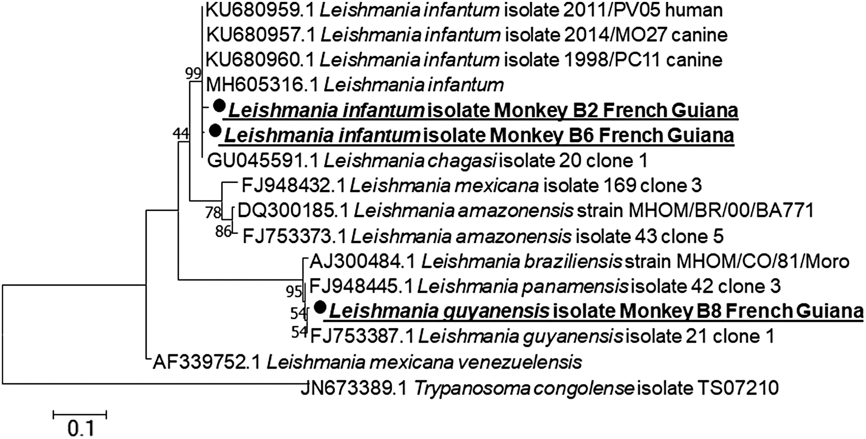
Phylogenetic tree showing the position of Leishmania infantum and Leishmania guyanensis compared with other Leishmania species. The sequences of the ITS2 gene amplified in this study with other ITS2 sequences available in the GenBank were aligned using CLUSTAL W implemented on BioEdit v3. The evolutionary history was inferred by using the maximum likelihood method based on the Tamura 3-parameter model. The tree with the highest log-likelihood (−1159.66) is shown. The percentage of trees in which the associated taxa clustered together is shown next to the branches. Initial tree(s) for the heuristic search was obtained automatically by applying the neighbor-join and BioNJ algorithms to a matrix of pairwise distances estimated using the maximum composite likelihood approach and then selecting the topology with superior log-likelihood value. The tree is drawn to scale, with branch lengths measured in the number of substitutions per site. The analysis involved 16 nucleotide sequences. All positions containing gaps and missing data were eliminated. There were a total of 246 positions in the final data set. Evolutionary analyses were conducted in MEGA7. Statistical support for internal branches of the trees was evaluated by bootstrapping with 1000 iterations.
A serological test was also carried out on blood samples from these nine monkeys after thawing. Rapid immuno-migration (RIM), also known as immunochromatography, is one of the fastest and most practical techniques for detecting antibody–antigen interactions. In the most common iteration of this method, an antibody specific to a given antigen is fixed to colloidal gold molecules, which are dried as a stripe on a chromatography paper (nitrocellulose) strip encased in a plastic holder. The Witness® Leishmania test (Zoetis, Lyon, France) is based on the RIM technology and uses an antigen from L. infantum to quickly identify antibodies in Leishmania-infected animals. Sensitized colloidal gold particles, bound to anti-Leishmania antibodies present within the sample of blood, are allowed to migrate along a strip. The complex is then captured on a sensitized reaction line where its accumulation causes the formation of a clearly visible pink band. A control band, located at the end of the reading window, ensures that the test was performed correctly.
Results
Screening with Leishmania genus-specific 18S-based qPCR resulted positive in 33.3% (3/9) of howler monkeys (Table 1). L. infantum-specific kDNA-based qPCR was also positive, but for two of three specimens only. Sequencing of 360 bp length ITS2 gene amplicons, obtained from these two monkeys (B2 and B6), had 99% concordance with L. infantum sequences in the GenBank (i.e., KU680960, KU680959, KU680957); a third monkey (B8) resulted positive for Leishmania genus-specific qPCR, but negative for L. infantum-specific qPCR (Table 1). A 315 bp length sequence was also obtained from this monkey; BLAST search showed 99% identity with L. guyanensis (FJ753387) (Fig. 1). L. infantum parasite load quantified by kDNA qPCR was medium to high, as previously described by Martínez et al. (2011), around 100 for B2 monkey and 5200 parasites/mL of blood for B6 monkey. L. guyanensis-infected monkey was weakly positive by the serological Witness Leishmania test. This may be related to the specificity and sensitivity of diagnostic tests that use L. infantum as antigens to reveal antibodies, so that in B8 monkeys, the positive serological test was probably due to a cross-reaction between L. infantum and L. guyanensis.
Molecular Detection and Serology of Leishmania spp. in the Blood of Howler Monkeys in French Guiana
Based on k
−, negative result; qPCR, real-time quantitative PCR.
Discussion
Human leishmaniasis caused by L. guyanensis occurs in French Guiana as well as in neighboring countries. L. guyanensis is widespread in the east of the western Andes mountain range in Colombia, south of Bolivia, and east of Guyana. It is found in a range of habitats from dry deciduous forest to multistrata tropical rainforest. L. guyanensis causes a simple skin form of the disease characterized by multiple ulcerous lesions (“pian bois”) (Simon et al. 2017). The main reservoir of this Leishmania is the two-toed sloth (Choloepus didactylus) and also the small anteater (Tamandua tetradactyla) and the common opossum (Didelphis marsupialis) (Dedet et al. 1989). Our results showed that the howler monkey can also be considered a potential reservoir of L. guyanensis. Our study is the first to report the presence of this infection in macroscopically nonaffected howler monkey. L. guyanensis leishmaniasis is a zoonosis of dense moist forest. Infection remains occasional, directly related to human entry into the forest. The cycle takes place at the level of the forest canopy, where we consider that the three-toed sloth is the reservoir. The main vector for L. guyanensis is Lutzomyia umbratilis, whose habitat is also the forest canopy (Pajot et al. 1982). In November, at the beginning of the rainy season, sand flies are present near the ground (old tree stumps) and are a source of human infection. The howler monkeys and sand flies live in the same ecosystem located in trees over 15 meters high. Humans are not usually exposed to the bites of infected sand flies. However, with environmental changes (deforestation, forest fragmentation, and anthropization), the interface between mammalian reservoirs, such as howler monkeys, vectors, and humans, is made possible.
We report here for the first time the presence of a potential wild reservoir for human visceral leishmaniasis caused by L. infantum in French Guiana, where no indigenous human cases were reported. L. infantum has been found to cause disease in neighboring countries such as Brazil, Suriname, Guyana, and Venezuela. Lutzomyia longipalpis is the main vector of L. infantum in Brazil (Lainson and Rangel 2005, Bauzer et al. 2007, Lara-Silva Fde O et al. 2015). In northern Brazil, where this type of leishmaniasis is endemic, the dog acts as a reservoir. In French Guiana, no publication reported autochthonous canine leishmaniasis caused by L. infantum. However, one observation was reported concerning an infected dog imported from Spain that caused a secondary case (Rotureau et al. 2006). In the Colombian forest, an infection of the common opossum (D. marsupialis) has been confirmed by culture of L. infantum from the spleen (Travi et al. 1994). In northern Brazil, the crab-eating fox (Cerdocyon thous) is also suspected to be a wild reservoir of L. infantum (Gomes et al. 2007). Spleen culture also revealed that 42% (11/26) of these foxes were carriers of parasites. All were apparently healthy (Lainson et al. 1987). It has been shown that the synanthropic rodents can play a role as leishmaniasis reservoirs (Oliveira et al. 2005, Lara-Silva et al. 2014). In Brazil, the blood of one synanthropic bat (Pteronotus parnelli) was PCR positive for L. infantum (da Costa et al. 2015). In the country of São Paulo, the presence of L. infantum DNA has been found in three specimens belonging to the species Molossus molossus and Glossophaga soricina (Savani et al. 2010). In a serological Brazilian study, mammals of the species Callithrix jacchus, Lepus europaeus, Sphiggurus villosus, Nasua nasua, Eira barbara, and Galictis cuja were found positive with high titers for antibodies to anti-L. infantum (≥1280), for the last three (Paiz et al. 2015). In 2013, de Araújo et al. (2013) isolated L. infantum in a bone marrow sample from an anteater (T. tetradactyla) of the Brazilian Amazon basin. Therefore, it is clear that there may be parasitic circulation in various wild mammals. However, very few studies have been conducted on primates for the infection by Leishmania spp. in America. Some of them showed that primate species harboring Leishmania parasites (Table 2). Howler monkeys have previously been suspected to be vectors of vector-borne zoonotic protozoa. A serological study conducted in French Guiana on the blood of 81 monkeys (A. seniculus) showed that 97% of these monkeys were positive for Plasmodium malariae/brasilianum, a human malaria agent (Volney et al. 2002). In addition, Vasconcelos dos Santos et al. (2018) showed that there was a great diversity of phlebotomines 80 km from the village of Oiapoque, where the monkeys were hunted. Some are carriers of Leishmania spp. They make bloodmeals on various animals including human, but the origin of bloodmeals was not searched for the howler monkeys. The study by Chavy et al. (2019) shows that there are also many potential sand flies vectors of Leishmania spp. 20 km from our study area (Nouragues reserve). So, it seems that there is or has been a parasitic cycle in the Guyana forest with wild animals in the past.
Leishmania Detection in Primates from America
Had signs of visceral leishmaniasis.
DAT, direct agglutination test.
Our results show that three of the nine howler monkeys were carriers of leishmaniasis, in particular L. infantum, the agent of visceral leishmaniasis unknown in this area. Although our results are preliminary, because only a few monkeys were tested, they nevertheless confirmed the importance of the red howler monkeys in potential zoonosis. The role of primates as sentinel and/or potential reservoir of a serious zoonosis should be considered by public health officials and veterinarians in all the future investigations.
Footnotes
Acknowledgments
We especially thank Christophe B., Amélie V., and Coarasi S. for their help in providing samples and Magdalen L. for reviewing English. This study was supported by the Institut Hospitalo-Universitaire (IHU) Méditerranée Infection, the National Research Agency under the program “Investissementsd'avenir,” reference ANR-10-IAHU-03, the Region Provence-Alpes-Côte d'Azur, and European funding FEDER PRIMI.
Author Disclosure Statement
No conflicting financial interests exist.