
Abstract
Leptospirosis is a globally important, fatal disease of humans, and over 160 species of animals are associated with more than 250 bacterial serovars in 64 species, but its ecology varies regionally and has changed over time with expansion of human development on previously agricultural and wild land. Sporadic human cases and clusters of canine leptospirosis, primarily attributable to Leptospira interrogans serogroup Pomona, have been detected in northern California. Small mesocarnivores such as raccoons and skunks frequent peridomestic space across much of the western United States and could serve as reservoirs for human and canine leptospirosis. We aimed to summarize the prevalence of infection with pathogenic leptospires in skunk and raccoon renal and urinary samples across broad geographic zones in California, and to determine whether prevalence changed during wet and dry seasons, and as functions of host species and demographic characters. Overall, 25.6% (22/86 tested) of raccoons and 28.5% (39/137 tested) of skunks were PCR-positive for Leptospira spp. in either renal tissue or urine, with leptospiral DNA in 22.0% of kidney samples and 18.8% of urine samples from raccoons and 27.8% and 14.5% of kidney and urine samples from skunks, respectively. Raccoons from the Central California and skunks from the San Francisco Bay Area had the highest overall PCR-prevalence (35.7% and 44.4%), respectively, and adults were more likely to be PCR-positive for Leptospira spp. than juveniles. There was moderate agreement between urine and renal tissue Leptospira spp. PCR with sensitivity for both host species in renal tissue of 0.86–0.97 and 0.42–0.64 in urine. Cases of human leptospirosis are thought to be underrecognized in the continental United States and possibly increasing in some states, including California. Our data document regionally high rates of infection in common mesocarnivores, which can pose a threat to humans and dogs, revealing an important periurban epidemiological cycle.
Introduction
Leptospirosis is a globally important, common, and potentially fatal bacterial disease of humans and animals (World Health Organization 1999, 2011, Bharti et al. 2003, Pappas et al. 2008, Ko et al. 2009). Leptospira spp. are motile spirochetes comprising more than 250 serovars distributed among 64 species divided into two clades—“Saprophytes” (S) and “Pathogens” (P) (Vincent et al. 2019). The P clade is further subdivided into two subclades—P1 and P2, with P1 corresponding to those species previously classified as “pathogenic” and P2 corresponding to species previously classified as “intermediate.”
In the P1 subclade, there are now 17 species and P2 contains 21 species. Recent advances in Leptospira spp. genomics have resulted in a dramatic increase in the number of named species from just a few years ago when nine pathogenic, five intermediately pathogenic, and seven nonpathogenic species were recognized (Levett 2015). Pathogenic Leptospira spp. infects over 160 domestic and wild animal species worldwide (Babudieri 1958, Matthias et al. 2008, Ko et al. 2009). Clinical leptospirosis in humans and dogs ranges from subclinical to fatal, with a mortality rate up to 20% (World Health Organization 1999, Levett 2001, van de Maele et al. 2008). Clinical sequelae most commonly include renal and hepatic failure, but meningitis, uveitis, myocarditis, and severe respiratory disease are also seen (Faine et al. 2000, Greene et al. 2006, Gouveia et al. 2008, Lo et al. 2011).
Infection occurs through bacterial invasion across mucous membranes and abraded skin, often after contact with urine, or water contaminated by urine, from infected animals or less commonly via bite wounds, ingestion, and venereal and placental transfer. Recent estimates put the annual number of human cases of leptospirosis worldwide at 1.03 million, of which ∼59,000 were fatal (Costa et al. 2015); in California, human leptospirosis has been a reportable disease since 1922, and there were 18 cases reported between 2013 and 2017 (California Department of Public Health 2017, 2018).
Data regarding the infecting Leptospira species and/or serogroups are not available for the majority of recent human cases in California but Leptospira interrogans serovars Mini and Australis were identified as being the serogroups to which samples reacted most strongly on the MAT in two cases and one case, respectively, from a human cluster in California in 2000, (Meites et al. 2004).
Despite decades of documentation of domestic and wildlife leptospirosis in continental North America, any serious risk for dogs or people is often dismissed. Defining that risk remains highly challenging, in part, because the ecology of leptospirosis is incredibly complex, with many animals that are reservoir-competent for several of the many species and strains of the bacteria. With extensive serological cross-reactivity across serogroups, serology has limitations for detecting epidemiological cycles, whereas more recent DNA-based approaches suffer from lack of extensive reference databases, relatively poor sensitivity for detecting active infection in many individual animals, and unclear correspondence of the significance of distinct sequence types with particular serovars.
Moreover, understanding modern risk of leptospirosis informed by data from older studies must be approached with caution because of differing diagnostic modalities and the changing landscape of the infection as much land has been converted from wild lands or agriculture to suburban and urban centers. Very little research has addressed leptospirosis in North America (except Hawaii) in the last decade, and the western continental states have received even more limited attention.
Recent clusters of canine leptospirosis have been detected in northern California (Gautam et al. 2010, Hennebelle et al. 2013), and the majority of these cases (Ghneim et al. 2007, Hennebelle et al. 2013) and infections in wildlife species (Gulland et al. 1996, Lloyd-Smith et al. 2007, Roug et al. 2012) are associated with L. interrogans serogroup Pomona. This serogroup has also been implicated in disease in humans (Heath et al. 1965).
A possible source of human and canine leptospirosis in California may be mesocarnivores, including raccoons (Procyon lotor) and skunks (Mephitis mephitis). Raccoons that seroreacted to serogroups Australis, Autumnalis, Bratislava, Canicola, Grippotyphosa, Hardjo, Icterohaemorrhagiae, and Pomona, and skunks reacting to Autumnalis, Ballum, Grippotyphosa, and Icterohaemorrhagiae were detected in the Midwest and eastern North America (Alexander et al. 1972, Ferguson and Heidt 1981, Mikaelian et al. 1997, Mitchell et al. 1999, Richardson and Gauthier 2003, Bischof and Rogers 2005, Raizman et al. 2009, Allen et al. 2014), and active infection was reported in these regions as well (Roth et al. 1963, Shearer et al. 2014, Tan et al. 2014). However, their roles in leptospirosis in western North America were poorly documented (Duncan et al. 2012, Britton et al. 2017).
These species might be considered reservoirs if they can permanently maintain infection within their populations and serve as a source to a target population (Haydon et al. 2002), in this case dogs and people. Active infection in kidneys in a large number of individuals across broad geographic regions would support a hypothesis that these species may serve as reservoirs, and finding of shedding of leptospires into urine would underscore that these peridomestic animals could be important sources of risk for humans and dogs.
In this study, we sought to estimate the prevalence of infection with pathogenic Leptospira spp. in renal and urinary samples in raccoons and skunks from several regions of California. We also evaluated whether infection was more common in dry vs. wet seasons, which are particularly distinct in most of California, and whether host demographic characteristics (sex and age) altered risk of being infected. Our last objective was to compare the agreement between PCR results from renal tissue and urine.
Materials and Methods
Trapping and sample collection
Convenience samples were assessed by PCR in this study when carcasses were available from collaborators and wildlife management agencies (USDA Wildlife Services and the California Department of Fish and Wildlife), and during live-trapping studies performed concurrently by members of the laboratory for other unrelated studies. Availability of samples was not under the control of the authors of this study. Collection took place in 20 counties in California from March 2011 to November 2016.
Trapping and sampling of live raccoons and skunks were covered under California Department of Fish and Wildlife scientific collecting permit #SC-854 and approved by the University of California, Davis Institutional Animal Care and Use Committee. Live-trapping was done with Tomahawk (Tomahawk Live Trap, Hazlehurst, WI) traps baited with canned cat food. Traps were opened in the evening and checked in the early morning. Skunks were exposed to isoflurane on a cotton ball until they became uncoordinated and then given 20 mg/kg ketamine and 0.5 mg/kg midazolam intramuscularly. Raccoons were anesthetized only with 10 mg/kg ketamine and 0.5 mg/kg midazolam administered intramuscularly.
Animals were aged (adult vs. juvenile) and sexed and uniquely ear-tagged before release. Urine was collected aseptically by cystocentesis from animals with a palpable urinary bladder and maintained cool for transfer to UC Davis. Urine was centrifuged at 4000 g for 15 min and the pellet resuspended in 200 μL sterile PBS. Samples were then frozen at −20°C until further use. Donated, previously frozen, carcasses in good postmortem condition were necropsied, and urine was collected and processed as described for samples from live-trapped animals. Kidney tissue was also collected during necropsy and frozen at −20°C until DNA extraction was performed. Donated carcasses were aged, sexed, and evaluated for overall body condition. The county of collection was recorded for donated carcasses.
DNA extraction and PCR
DNA was extracted from urine and kidneys using a kit (Qiagen DNAeasy Blood and Tissue Kit, Qiagen, Valencia, CA). DNA extraction was performed between 2 weeks and 4 years from initial sample collection. Renal tissue was collected in a thin section encompassing the corticomedullary junction and extending equally into the cortex and medulla, and 25 mg were extracted according to manufacturer's instructions. DNA was extracted from 100 μL of the resuspended urine sediment following the manufacturer's protocol for whole blood extraction.
Real-time PCR using an in-house assay (Stoddard et al. 2009) targeting the LipL32 gene of pathogenic Leptospira spp. was performed on extracted DNA. The target codes for a major outer membrane lipoprotein, which is an important virulence factor and occurs only in pathogenic species of Leptospira (Yang et al. 2002, Levett et al. 2005, Stoddard et al. 2009). A Ct of less than 45 was considered positive. A higher Ct than in the original reference (Stoddard et al. 2009) was used because repeated sampling (three replicates) of tissues from seven skunks with initial Ct's of between 40 and 42.7 (the highest initial Ct obtained from any sample with a characteristic amplification curve) resulted in Ct values that were either below 40 or between 40 and 42, indicating that Leptospira spp. DNA was indeed present in the tissues. One positive control (DNA extracted from cultured L. interrogans serovar Pomona (serogroup Pomona)) and three negative controls of PCR grade water were included in each batch of PCRs.
Statistical analyses
Data were maintained in Excel (Microsoft, Redmond, WA) and analyzed in R (version 3.3.2, R-Development Core Team,
The presence of leptospires in either the urine or kidneys of raccoons and skunks was evaluated for associations with sex, age (adult or juvenile), region, and season of sampling (dry or wet) using logistic regression analysis. Each variable was first assessed using univariable logistic regression. Predictor variables with p-values of ≤0.2 were included in the multivariable analysis, as were potential confounding variables. Confounding was defined as a change in the odds ratio of greater than 10%. Two-way interactions were also evaluated for significance. Individuals that were PCR-positive in either renal tissue or urine, or both, were considered overall PCR-positive for Leptospira spp. The dry season was defined as May through October and the wet season was defined as November through April.
Cohen's kappa coefficient (k) (Cohen 1960) was calculated to assess the agreement between renal and urinary Leptospira spp. PCR assays for both raccoons and skunks. The sensitivity of both renal and urinary PCR for each species was also calculated. Individuals that were positive on either or both renal and urinary PCR were considered infected with Leptospira spp. and were used as the denominator in sensitivity calculations.
Results
A total of 86 raccoons (four of which were live-trapped, the remaining 82 were donated carcasses) and 137 skunks (four live-trapped and 133 donated) were sampled (Table 1). Of the raccoons, kidney tissue was available from 82 individuals and urine was collected from 48. Kidney tissue was available from 133 skunks and urine was available from 76. Raccoon and skunk samples were obtained from the Bay Area, Central Valley, and Northern Coast of California. In addition, skunks from Southern California were sampled. No significant differences were found in the sex distributions between different geographic regions for either species. The seasonal (p < 0.001) and age (p = 0.004) distributions of samples were different between geographic regions for skunks, but not for raccoons (Table 1).
Sex and Age Class Distribution of Raccoons (Procyon lotor) and Striped Skunks (Mephitis mephitis) from Regions of California Sampled for Leptospira spp. Infection Between 2011 and 2016
Bay Area includes Alameda, Contra Costa, San Francisco, San Mateo and Santa Cruz counties; Central Valley includes Butte, Kern, Lake, Nevada, Placer, Sacramento, Solano, and Yolo counties; Northern Coastal includes Humboldt, Marin, Mendocino, and Sonoma counties; Southern California includes Los Angeles, San Luis Obispo, and Ventura counties.
A significant difference was found between regions for skunks for age (p = 0.004) and for season (p < 0.0001).
Dry = May through October, Wet = November through April.
Overall, 25.6% (22/86 tested) of raccoons and 28.5% (39/137 tested) of skunks were PCR-positive for Leptospira spp. in either renal tissue or urine (Table 2). Leptospiral DNA was detected in 22.0% (18/82 tested) of kidney samples and 18.8% (9/48 tested) of urine samples from raccoons and in 27.8% (37/133 tested) and 14.5% (11/76 tested) of kidney and urine samples from skunks, respectively. No significant differences were found between raccoons and skunks for renal, urinary, or overall Leptospira spp. PCR-prevalence.
PCR-Prevalence of Renal and Urinary Leptospires in Raccoons (Procyon lotor) and Striped Skunks (Mephitis mephitis) in California Sampled Between 2011 and 2016
PCR prevalence was determined using a real-time PCR assay targeting the LipL32 gene of pathogenic Leptospira spp. performed on extracted DNA from renal tissue or urine. A Ct of less than 45 was considered positive.
Significant differences were found in the overall PCR-prevalence for skunks by region and by age.
Bay Area includes Alameda, Contra Costa, San Francisco, San Mateo, and Santa Cruz counties; Central Valley includes Butte, Kern, Lake, Nevada, Placer, Sacramento, Solano, and Yolo counties; Northern Coastal includes Humboldt, Marin, Mendocino, and Sonoma counties; Southern California includes Los Angeles, San Luis Obispo, and Ventura counties.
Dry = May through October, Wet = November through April.
CI, confidence interval.
Risk factor analysis
Raccoons from the Central Valley region of California had the highest overall PCR-prevalence (35.7%), although that prevalence was not statistically significantly different than that found in Bay Area (11.1%) or North Coastal (27.8%) raccoons (Fig. 1 and Tables 2, 3). Adult raccoons were more likely to be PCR-positive for Leptospira spp. than juveniles (OR = 5.50), although not significantly so (p = 0.12). No significant difference was found between the overall PCR-prevalence of Leptospira spp. between male and female raccoons nor between raccoons sampled during the wet season or during the dry season (Table 3). Two-way interactions were not significant.
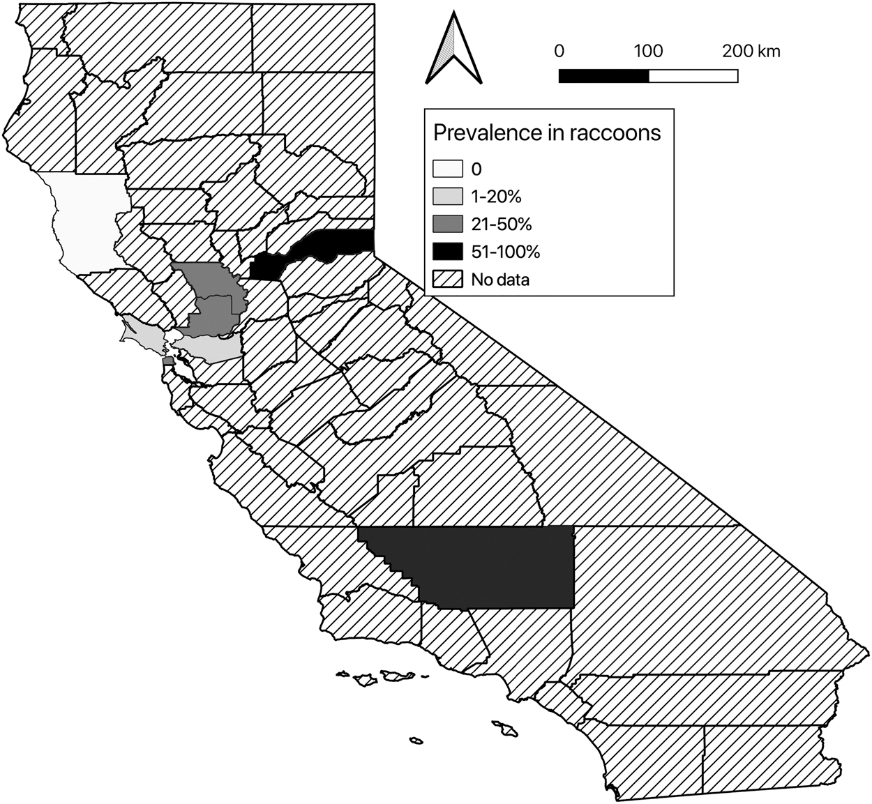
Map of California counties showing prevalence of Leptospira sp. infection in raccoons in counties from which samples were available.
Results of Logistic Regression Analyses of Potential Risk Factors for PCR Detection of Renal or Urinary Leptospires in Raccoons (Procyon lotor) and Striped Skunks (Mephitis mephitis) in California Sampled Between 2011 and 2016
A multivariable model was constructed for skunks on the basis of multiple significant univariable risk factors.
Bay Area includes Alameda, Contra Costa, San Francisco, San Mateo, and Santa Cruz counties; Central Valley includes Butte, Kern, Lake, Nevada, Placer, Sacramento, Solano, and Yolo counties; Northern Coastal includes Humboldt, Marin, Mendocino, and Sonoma counties; Southern California includes Los Angeles, San Luis Obispo, and Ventura counties.
Dry = May through October, Wet = November through April.
ref = reference category.
Skunks from the Bay Area had the highest overall PCR-prevalence (44.4%) compared to skunks from the Central Valley (32.9%), Southern California (25.0%), and the Northern Coastal region (9.7%) (Fig. 2 and Table 3). On univariable analysis, skunks from the Bay Area (OR = 7.47, p = 0.009) and Central Valley region (OR = 4.57, p = 0.020) were more likely to be PCR-positive than were skunks from the Northern Coastal region of California (Table 3). Adult skunks were more likely to be PCR-positive than were juvenile skunks (OR = 8.82, p = 0.004).

Map of California counties showing prevalence of Leptospira sp. infection in skunks in counties from which samples were available.
No significant association was found between sex or the season of sampling and overall PCR-prevalence of Leptospira spp. in skunks. Two-way interactions between potential risk factors were not significant. Age confounded the association between region and overall PCR status, but age was already to be included in the multivariable model due to its significant association with overall PCR status. When region and age were assessed by multivariable logistic regression analysis, both remained significantly associated with overall PCR-prevalence in skunks; however, the increased likelihood of skunks in the Central Valley being PCR-positive for Leptospira spp. compared to skunks in the Northern Coastal region of California became statistically nonsignificant with the inclusion of age in the model (Table 3). Two-way interactions were not significant for either skunks or raccoons.
Urine versus renal tissue PCR
Cohen's kappa coefficient was 0.443 (95% CI: 0.0939–0.7921) indicating moderate agreement between urine and renal tissue Leptospira spp. PCR in raccoons was 0.415 (95% CI: 0.1616–0.6676), also indicating moderate agreement between the assays, in skunks. The sensitivity of renal tissue PCR was 0.86 (95% CI: 0.626–0.962) in raccoons and 0.97 (95% CI: 0.846–0.997) in skunks, while the sensitivity of urine PCR was 0.64 (95% CI: 0.356–0.860) for raccoons and 0.42 (95% CI: 0.240–0.628) for skunks. Of the 10 raccoons that were PCR-positive in renal tissue and that had urine available to test, five were also positive by urine PCR, and of the 24 skunks that were positive by renal PCR and had urine available for testing, nine were positive by urine PCR.
Discussion
While living among nature and wildlife can have numerous mental and physical health benefits (Wells 2000, de Vries et al. 2003, Bratman et al. 2012, Cox et al. 2017), it can also lead to exposure to zoonotic pathogens, including Leptospira spp. We documented common, active infection by pathogenic Leptospira spp. in raccoons and skunks in all regions of California included in our study. While the duration of infection in these hosts is not known, we presume that infection is likely chronic in order that so many apparently healthy individuals would have active infection during a cross sectional sampling. Although more commonly detected in renal tissue, leptospiral DNA was also found in urine, underscoring the potential for environmental contamination with pathogenic leptospires (Levett 2001).
Given that leptospires can survive for months outside of their hosts under favorable conditions (Levett 2001), there is risk to both human and animal health from contact with urine or areas contaminated by urine of these common peridomestic mesocarnivores. Our data suggest that these species may serve as reservoirs, although additional experimental infection with strains derived from patients, including dogs, humans, or wildlife, would be valuable to clarify infection dynamics.
Previous studies have also found evidence of Leptospira spp. infection in raccoons and skunks in North America. Both of these species are widespread across much of the continent and, while often found in wild areas, have high tolerance for human development as well and occur frequently in peridomestic parks, yards, and other vegetated areas (Chupp et al. 2013, Rosatte et al. 2019). Both species may be crepuscular and will eat unguarded pet food, edible garden vegetation, and other attractants near humans. We acknowledge that our sampling strategy limits our ability to conclude prevalence of infection at ecologically meaningful spatial scales, for example parks or towns where many individual skunks, raccoons, dogs, and people may be in contact with each other and contaminated fomites.
In addition, we likely underestimate the true prevalence, because leptospires may circulate in blood during acute infection, before establishing in renal tissue. However, there is no reason that the sampling would introduce bias for or against finding infected animals as they were collected in both remote regions under study of multiple wildlife diseases as well as periurban areas as part of pest management programs. However, statewide and regional prevalence values surely lump together high- and low-risk regions, and thus, we recommend additional targeted studies of individual areas of concern.
Shearer et al. (2014) detected leptospires by IHC in renal tissue from 42% of 33 skunks and 33% of 245 raccoons in Ontario, Canada and Duncan et al. (2012) found 30% of 65 raccoons in Colorado to be positive for renal leptospires by IHC. We found a higher renal PCR-prevalence in California skunks than the 18% (n = 49) that was detected recently in British Columbia, Canada (Britton et al. 2017) but a substantially lower rate of infection than was found in raccoons in Indiana where 100% of 34 raccoons from two sites were culture positive for renal leptospires (Tan et al. 2014).
The lack of seasonal variation in either species was interesting as summers in California are very dry and leptospires survive longer in moist environments, which we hypothesized would lead to a greater prevalence during the wet season. However, Leptospira spp. can remain in the kidneys of naturally infected skunks for more than 2 years (Roth et al. 1963), more than 6 months in experimentally infected skunks (Tabel and Karstad 1967), and more than 3 months in experimentally infected raccoons (McGowan and Karstad 1965). In this regard, the significant increased risk of being PCR-positive in older skunks could be attributable to chronic infection, accumulated risk over time, and exposure to contaminated environments.
Although the sensitivity of renal tissue PCR was higher than that of urine PCR in both raccoons and skunks, each method failed to detect some animals that were carrying pathogenic leptospires. Urine and renal tissue vary in the presence of PCR inhibitory characteristics as well as homogeneity of distribution of Leptospira DNA within the sample and these characteristics can change over the course of infection, underlying the less than full agreement of kidney and urine PCR for each animal. The failure of renal tissue PCR to detect infection in individuals who were PCR positive in the urine may be explained by the multifocal, rather than diffuse, distribution of leptospires in kidney tissue (Meites et al. 2004, Ajayi et al. 2017). The lower sensitivity of urine PCR compared to renal tissue PCR may be due to the intermittent shedding of leptospires in the urine, which has been documented in both skunks (Roth et al. 1963) and raccoons (McGowan and Karstad 1965) or inadequate sample volume from some individuals.
In addition, PCR inhibitors present in urine may have contributed to the lower sensitivity of urine PCR (Chernesky et al. 1997) as could the DNA degradation that can take place in stored urine (Cannas et al. 2009). Use of a housekeeping or other calibrator gene such as 18S in a quantitative PCR could help confirm that inhibitors in a sample might be a cause of false-negative results for Leptospira spp. PCR; such assays are straightforward for samples such as tissue, but problematic for samples such as urine where host DNA might be expected to be minimal, and our funding did not allow for such calibration to be performed on all of our samples. However, our findings clearly show that surveillance by either urine or kidney tissue alone may lead to an underestimation of the prevalence of Leptospira spp. infection in raccoons and skunks. We therefore recommend that both samples be tested when possible, and that animals where only one sample is positive not be ruled out completely as possibly infected with Leptospira spp.
The brown rat (Rattus norvegicus) is the most important source of human infections in much of the world (Haake and Levett 2015), and rats are often infected by L. interrogans serovars within serogroup Icterohaemorrhagiae (Ido et al. 1917, Hartskeerl and Terpstra 1996, Faine et al. 2000). In general, human leptospirosis is thought to be underrecognized in the continental US and possibly increasing in some states, including California (Meites et al. 2004) where rat-Leptospira epidemiological cycles appear far less important than cycles involving the serovar Pomona.
Further work is needed to explore the genetic relatedness of the Leptospira spp. circulating in California peridomestic wildlife species and domestic animals to better understand the ecology and epidemiology of the disease in the state. In addition, further sampling of raccoons and skunks in areas that were not well represented in our study (e.g., southern California, the Sierra Nevada) would also address gaps in our knowledge of the disease in these reservoir species in California. More comprehensive understanding of the risk of leptospirosis will require studying numerous species that can serve as reservoirs and evaluating the risk of contamination of the environment as well.
Conclusions
Leptospira spp. infection is common and widespread in raccoons and skunks in California and a substantial proportion of these species are shedding leptospires into the environment. Our findings highlight the potential for these peridomestic mesocarnivores to act as sources of infection for humans as well as domestic animals.
Footnotes
Acknowledgments
We thank Deana Clifford, Shannon Chandler, Rebecca Mihalco, and Jane Reiner for their invaluable assistance.
Author Disclosure Statement
All authors declare that no competing financial interests exist.
Funding Information
This study was partially funded by Zoetis.