
Abstract
As one of the important tick-borne zoonotic pathogens, Anaplasma has both veterinary and public health significance. Here, we performed a survey of Anaplasma infection in the goats from a farm in Beijing, China, and found 44.6% (41/92) were infected with Anaplasma capra, and 22.8% (21/92) were infected with Anaplasma sp. This Anaplasma sp. bacterium was close to a recently emerging Anaplasma platys strain based on gltA and groEL gene phylogenetic analysis. As to further understand the characteristics of Anaplasma sp., we raised a couple of positive goats (n = 2) in the laboratory with tick-free settings. We observed inappetence, vomiting, high fever, and weakness of limbs in the goat's offspring (n = 3). In addition, the blood samples from all offspring were all positive of this Anaplasma spp. We did not see any intracellular morulae in neutrophils, monocytes, and erythrocytes, but we identified some in the platelets of the blood smears from the positive goats by light microscopy. We named it A. platys-like and suggested it may infect platelets and be transmitted vertically through the placenta of goats. These findings deserve further evaluation.
Introduction
Tick-borne diseases (TBDs) have been an increasing threat on animal production, welfare, and human health worldwide (Torina et al. 2007, Perronne 2014). The Anaplasma spp. are tick-borne pathogens with mammalian hosts in temperate, subtropical, and tropical regions of the world (Geurden et al. 2008, Tay et al. 2014, Yang et al. 2018). Anaplasma is an intracellular gram-negative bacterium, which belongs to the Anaplasmataceae family and Rickettsiales order (Dumler et al. 2001). In recent years, the public health significance of Anaplasma as emerging pathogens has also been realized (Ismail et al. 2010, Botelho-Nevers et al. 2012, Stuen et al. 2013). Anaplasma ovis, Anaplasma centrale, Anaplasma bovis, Anaplasma platys, Anaplasma phagocytophilum, and Anaplasma marginale are recognized species contained in the genus Anaplasma (Dumler et al. 2001). Recently, Anaplasma capra (Li et al. 2015a) and Anaplasma odocoilei (Tate et al. 2013) species have been included in the Anaplasma genus. The different species have diverse blood cell tropisms (Silaghi et al. 2017).
Anaplasmosis is one of the common tick-borne zoonotic diseases in domestic animals, such as sheep and goats on pastures. Clinical characteristics of infected animals depend on animal welfare and immune status (Čobádiová et al. 2013). The clinical symptoms of infected animals include inappetence, weight loss, sudden high fever, reduced milk production, abortions, and even death (Mathieson 1991, Stuen 2007, Liu et al. 2012, Yasini et al. 2012). Furthermore, infected animals are more susceptible to be coinfected by other pathogens (Čobádiová et al. 2013). For example, A. phagocytophilum is not only an aborticide but also associated with various bacterial opportunistic infections in sheep farms in northern Spain (Garcia-Perez et al. 2003). In addition, the human NY-18 A. phagocytophilum isolate can infect sheep and transmit to the lamb with some clinical symptoms (Reppert et al. 2013). Vertical transmission of A. ovis can cause death of neonatal elk calf (Hendrix et al. 2019).
The previous study has indicated that there was a high incidence of anaplasmosis in the goats in the suburban areas of Beijing, China (Zhang et al. 2012). In 2012–2013, we performed a survey and found A. capra infections among the goats from a farm in Fangshan District, another suburban region of Beijing. The goats in this farm were usually grazing on the mountains, where Haemaphysalis longicornis had a high density in the peak season (Li et al. 2015a). In this study, we continued the survey in this farm and identified A. capra and a new Anaplasma spp. To understand the characteristics of this new species, we brought a couple of positives from this farm to the laboratory for further investigation.
Materials and Methods
Investigation and sample collection
The investigation was conducted on a herd of goats (n = 92) from a farm in Dayuan village, Qinglonghu Town, Fangshan District, Beijing (E116°3′ N39°45′36″), in October 2016. To assess the Anaplasma infection on the goats, 92 ethylenediaminetetraacetic acid (EDTA)-anticoagulant blood samples of the goats were collected and detected by PCR targeting 16S rRNA and gltA genes. After initial screening, two adult positive goats (G1 [male], G2 [female]) were further raised in the laboratory with tick-free settings. Soon after, the couple naturally bore their offspring (G3 [female], G4 [male]), and G1 and G3 gave birth to G5 [male]). Their relationship is described in Figure 1. Those five goats were raised in the laboratory of Beijing Institute of Microbiology and Epidemiology, following the animal ethics authority regulations (IACUC-13-2016-004).
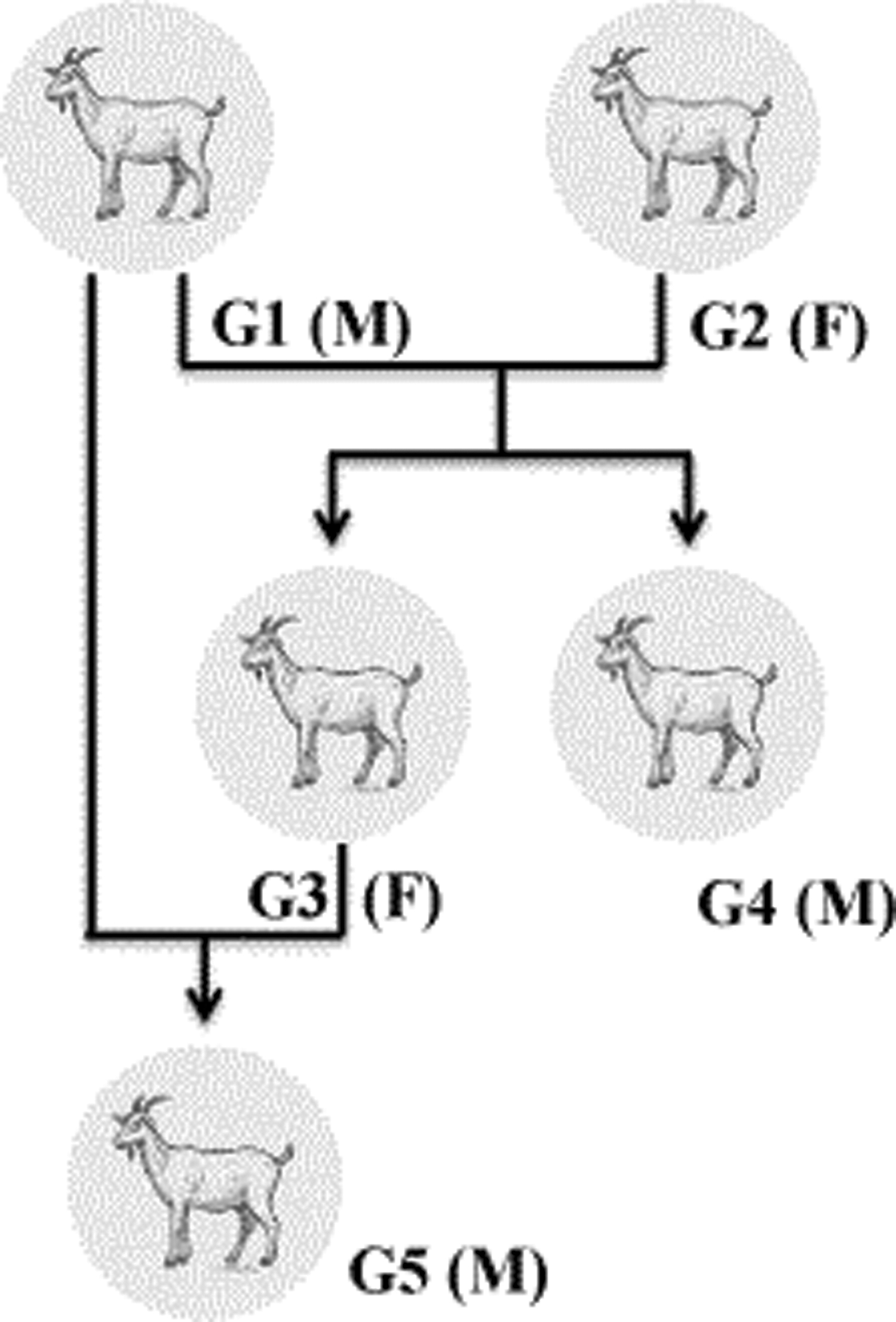
G1 and G2 gave birth to G3 and G4, G1 and G3 gave birth to G5. F, female; M, male.
Molecular detection and amplification of Anaplasma sp. genes
Total DNA was extracted from all samples using the TIANamp Genomic DNA Kit (DP304; TIANGEN (Beijing) Biotech, Beijing, China) according to the kit recommended protocol. The DNA specimens were diluted into 80 μL nucleic acid eluant and stored at −20°C. All DNA samples were tested by nested PCR using the primer pairs, targeting the 16S rRNA and citrate synthase (gltA) genes for first screening of Anaplasma. The identification of A. capra was followed by our previously described methods (Li et al. 2015a). To obtain the Anaplasma sp. gene sequences, DNAs of the goats' family (goat 1–goat 5, G1–G5) were amplified for the near-complete 16S rRNA, and partial gltA and groEL genes. To obtain the 16S rRNA gene, the primer pair Eh-out1/Eh-3-17 was used in the first round, and the primer pairs Eh-out1/Eh-out2/Eh-out2/Eh-3-17 were used in the second round reaction. Seminested PCR was used to amplify the gltA and groEL genes, and the primers are shown in Table 1. Nuclease-free water was used as a negative control, and 2 × DreamTaq Master Mix (Thermo Scientific, USA) including DNA polymerase was used in the PCR. To avoid cross-contamination, the experimental operations of DNA extraction, PCR preparation, DNA amplification, and agarose gel electrophoresis were in separate rooms; in addition, the template isolation and amplification were performed with the dedicated pipettes.
Primer Sequences Used in This Study
Sequencing of PCR products and phylogenetic analysis
All PCR products were sent to Sangon Biotech (Shanghai, China) for sequencing. The sequences obtained were checked and assembled by CLC Main Workbench 5, and were compared with the registered sequences in GenBank using the BLAST program (NCBI, Bethesda, MD) (
Morphological examination
The blood smears of the goats' family (G1–G5) were prepared by air-drying and were further fixed in absolute formaldehyde and acetone 1:1 for 10 min. Wright–Giemsa staining was then performed. The slides were rinsed with water slowly, dried, examined under the ordinary light microscope (Olympus BX41, Japan).
Results
We have tested the whole-blood samples from the 92 goats by PCR, and found that the infection rate of A. capra was 44.6% (41/92), and the positive rate of Anaplasma sp. was 22.8% (21/92). Anaplasma sp. infection was found in the goat couples (G1, G2) and goats G3–G5. We have obtained the sequences of near-complete 16S rRNA gene (∼1470 bp) and partial gltA (∼920 bp) and groEL genes (∼850 bp) for genetic identification of this Anaplasma sp. Based on the evolutionary analysis in the 16S rRNA phylogenetic tree, the Anaplasma sp. in our study and the Anaplasma sp. in the Wen-Ping Guo study formed a cluster, and close to A. phagocytophilum, A. platys, and A. platys-like strains identified in Ankang (Fig. 2a) (Guo et al. 2019). The partial gltA gene sequence of our Anaplasma sp. formed a cluster with the Anaplasma sp. identified in Xi'an, and this cluster was close to A. platys-like strain identified in Rhipicephalus microplus in Ankang, northwest China (Fig. 2b) (Guo et al. 2018, 2019). For the groEL gene, Anaplasma sp. found in our research was also close to the Anaplasma spp. identified in Xi'an, and it was genetically related to the A. platys and A. platys-like strains in Ankang (Fig. 2c) (Guo et al. 2018, 2019).
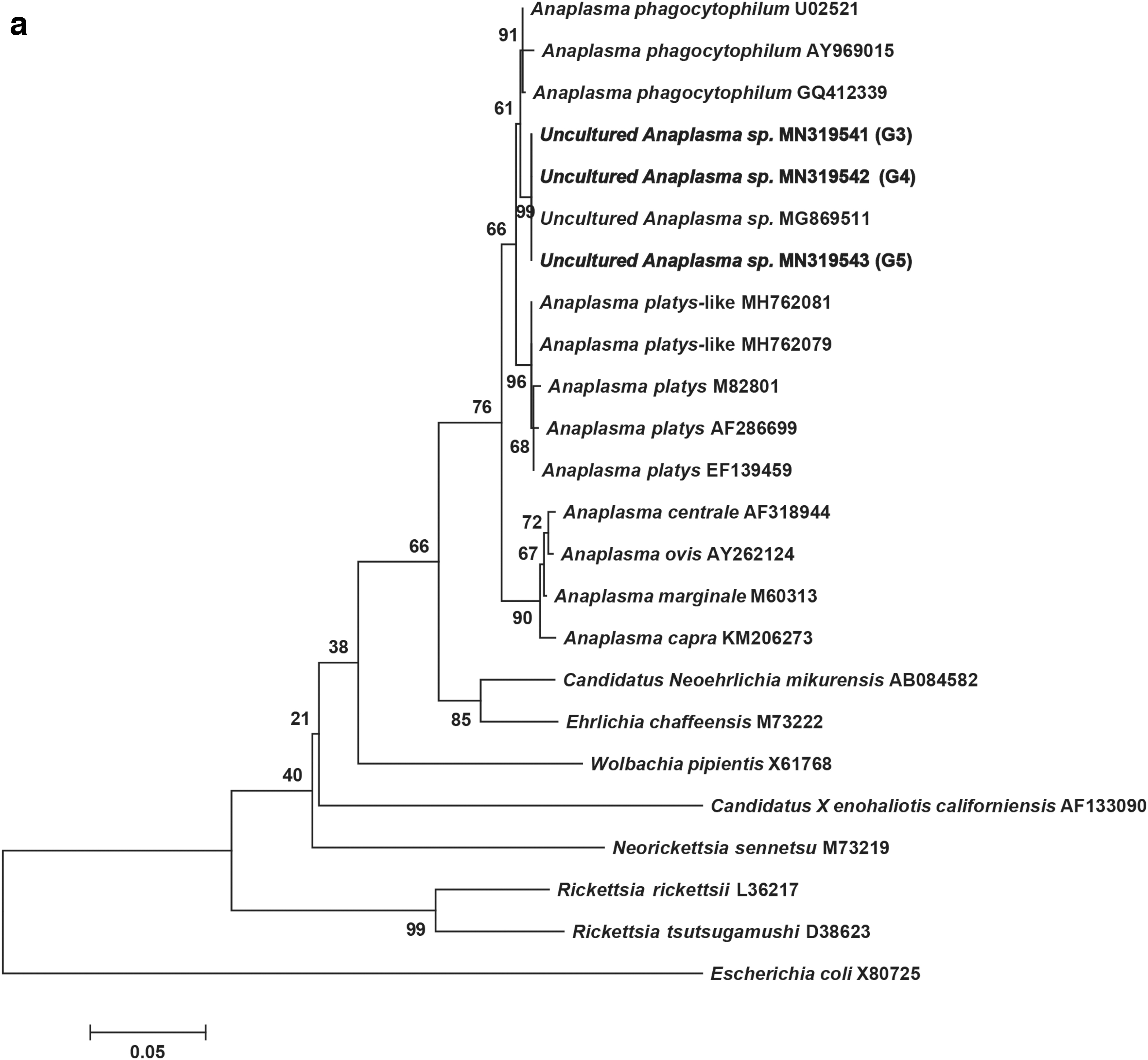
Phylogenetic analyses based on nucleotide sequences of the 16S rRNA (1170 bp)


The couple of positive goats infected with Anaplasma sp. gave birth in our laboratory settings. We found that all the family members of the goats (G1–G5) were infected with this Anaplasma unexpectedly (Fig. 2), although there were some gene variations. The almost full-length 16S rRNA sequences shared 100% nucleotide identities with each other and 99.93% identities with the Anaplasma strain detected in the goats from Xi'an, China (Guo et al. 2018) (Fig. 2a). The sequences of the partial gltA gene showed 98.5–100% similarity with each other and exhibited 99.07–99.87% identities with Anaplasma sp. found in Xi'an (Fig. 2b). For the partial sequences of groEL gene, they showed 98.9–100% nucleotide similarity with each other and presented 98.03–99.64% identities with Anaplasma spp. identified in Xi'an (Fig. 2c). We only got the short-length gene sequence of 16S rRNA of G1 and G2 but not the complete one; thus, we did not include them in the phylogenetic tree. We observed the offspring G3–G5 with some symptoms of anaplasmosis, including inappetence, high fever, vomiting, and weakness of limbs. During the examination, morulae were found in the platelet of the goats' family (G1–G5) in the blood smears by Wright–Giemsa staining, but not in monocytes, neutrophils, and red blood cells (Fig. 3). Consequently, we named this bacterium A. platys-like.

The blood smear of Anaplasma platys-like-infected goat was stained with the Wright–Giemsa staining method, and the inclusion is identified with arrowhead as blue-purple stained bodies within the platelet cytoplasm.
Discussion
In this study, the goat couples (G1, G2) were fed in a tick-free environment, and the goats G3–G5 were found to be all infected with A. platys-like bacterium. This suggests the vertical transmission of A. platys-like in goats. We also observed some clinical symptoms in the offspring, which might be caused by A. platys-like. By morphological examination of blood smears from the infected goats, we suggest that this Anaplasma might infect platelets.
In previous research, there are six species of the genus Anaplasma, which infect mammals and particular cell types of hosts such as erythrocytes, neutrophils, platelets, and monocytes (Dumler et al. 2001). Currently, A. platys is the only classified rickettsial species known to infect platelets, and caused cyclic thrombocytopenia in dogs (Dumler et al. 2001); this pathogen has also been found in cats (Lima et al. 2010), cattle, Camelus bactrianus, goats, Cervus elaphus, and African buffalo (Chochlakis et al. 2009, Dahmani et al. 2015, Li et al. 2015b, Lorusso et al. 2016a, 2016b, Machado et al. 2016). The investigation from Italy indicated that the prevalence of A. platys-like infection was higher in the goats than in the calves and sheep (Zobba et al. 2014), and it was also lower in the cattle in Algeria than in the calves in Italy (Dahmani et al. 2015). Through pathogen detection and symptom observation of the goat offspring (G3–G5), we suggest that there is a high probability that A. platys-like strain might impact goat husbandry, which deserves further epidemiological investigations and surveillance.
Up to date, there are two Anaplasma spp. that are recognized as human pathogens. For instance, A. phagocytophilum was the agent of human granulocytic anaplasmosis (Chen et al. 1994). A. capra was identified as a human pathogen in China recently (Li et al. 2015a). A. platys and A. ovis have been identified as the potential human pathogens (Chochlakis et al. 2010, Arraga-Alvarado et al. 2014). Whether A. platys-like identified in our study could infect human is an important issue to be clarified, however, we did not successfully isolate the bacterium. Further studies should be carried out to determine the pathogenicity of Anaplasma infected with platelets to humans.
To date, molecular and pathogenic identifications are still the significant methods of detecting Anaplasma, and serological testing is also commonly used as a supplementary detection. In our study, we identified the infection with Anaplasma by PCR and blood smear, but not by the antibody detection because A. platys-like strain isolate is not available. Based on the previous comparison on diagnosis methods, PCR was highly sensitive and endorsed for blood parasite diagnosis (Rucksaken et al. 2019, Wardrop et al. 2016). In addition, for PCR detection, it is worth noting that only targeting the 16S rRNA gene is not sufficient for the identification of Anaplasma. Phylogenetic analysis based on the sequences of 16S rRNA, gltA, and groEL indicated that A. platys-like in our study is similar to Anaplasma sp. identified from goats in Xi'an and is related to A. platys strains and A. platys-like in R. microplus in Ankang. With the phylogenetic analysis, the gltA and groEL sequences from the five goats (G1–G5) were not completely identical, which suggested that the G3–G5 might be coinfected with other Anaplasma sp. However, we performed molecular detection and blood smear observation, and found no other Anaplasma infection.
The previous reports also identified Anaplasma infection in domestic animals in northern China including Beijing, Inner Mongolia, Shanxi, and Hebei. The investigations in rural areas of Beijing showed that 48.9% (44/90) of goats, 23.9% (17/71) of cattle, and 0% (0/2) of dogs were infected with A. phagocytophilum by PCR amplification (Zhang et al. 2012). In Shanxi Province, 78.1% (25/32) of sheep were infected with Anaplasma spp., and 8.8% (30/338) of goats were identified as A. capra positive (Li et al. 2012, Zhang et al. 2016, Peng et al. 2018). The study from Hebei indicated that A. capra was detected in 51.1% (23/45) of sheep (Yang et al. 2017). The infection rate of A. capra in the goats in Inner Mongolia was 10% (4/40) (Peng et al. 2018). The seroprevalence of A. phagocytophilum was higher in rural residents from Tianjin (41.8%) than from Beijing (13.6%) (Zhang et al. 2014). According to the data described above, the infection rate of A. capra in the goats in our study was 44.6% (41/92), which was lower than in the sheep in Hebei (51.1%). Until now, there has not an A. platys infection report in northern China.
Ticks play an important role in the transmission of Anaplasma (Rar et al. 2011). Rhipicephalus sanguineus is considered to be the primary vector of A. platys transmission (Simpson et al. 1991, Cicuttin et al. 2015, Li et al. 2015b). Rhipicephalus turanicus and Rhipicephalus microplus were reported to be infected with A. platys-like strains (Harrus et al. 2011, Lu et al. 2017), and A. capra (Guo et al. 2019).
The limitations of this study were as follows. Although we have observed that A. platys-like-infected goats had some symptoms, the veterinary significance of this bacterium should be further evaluated with a larger sample size. In addition, we found the possibility of vertical transmission, however, this observation was beyond our initial plan, and thus, no detailed procedure was designed to describe this transmission. Many critical characteristics were not available, such as what time was the offspring found positive after its birth. This should be evaluated in further animal models. Finally, the infection rate of A. platys-like in ticks is unknown, specially around the farm where we got goat samples.
Conclusions
In recent years, due to frequent contact between the rural and urban populations, the threat of TBDs to human health is not limited to rural areas. In our study, we detected A. platys-like in goats in Fangshan district, Beijing, which might infect platelets of goats and be a risk of vertical transmission although its pathogenicity to human was uncertain.
Footnotes
Author Disclosure Statement
No competing financial interests exist.
Funding Information
This work was supported by the Natural Science Foundation of China (81773492), State Key Research Development Program of China (2019YFC1200401, 2019YFC1200202, 2019YFC1200501), Guangxi scientific and technological research (2020AB39264), and Special Program for Prevention and Control of Infectious Diseases in China (2018ZX10712001-018-001).