
Abstract
Background and Objectives:
Chlamydia spp. are potential zoonotic pathogens that can infect a wide range of animal hosts. In reptiles, Chlamydia can cause hepatitis, pneumonitis, and conjunctivitis and it can cause high mortality in young animals. The objectives of this study were to estimate the pooled prevalence of chlamydial infections in reptiles and to assess the trend of these infections over time.
Materials and Methods:
The study followed the Preferred Reporting Items for Systematic Reviews and Meta-Analyses (PRISMA) guidelines. Relevant studies were retrieved from PubMed, Scopus, and Web of Science. The retrieved studies were screened for eligibility. Then, important data were extracted from the included studies. A random effects model was used for all analyses. Subgroup analysis was used to assess heterogeneity for orders of reptiles, continents where the studies were conducted, and types of specimens. Cumulative meta-analysis and meta-regression were used to determine the trend of the prevalence over time. The quality of each included study was evaluated.
Results:
Of 106 studies (with a total of 2607 samples), 20 met the inclusion criteria and were included in the meta-analysis. The pooled prevalence of chlamydial infections in reptiles was 23.5% (95% confidence interval [CI]: 15.4–34.0). The trend of chlamydial infections increased from 1990 to 2008; thereafter, it was almost stable at slightly over 20%. The most commonly reported Chlamydia spp. were Chlamydia psittaci, Chlamydia pneumoniae, Chlamydia pecorum, and Chlamydia caviae. Among reptiles, the prevalence of chlamydial infections was highest in crocodiles (57.3% [95% CI: 32.5–78.9]). Among continents, the prevalence of chlamydial infections was highest in Australia (68.6% [95% CI: 36.8–89.1]).
Conclusions:
Based on the included studies, the prevalence of chlamydial infections in reptiles was high, especially in crocodiles. Because C. psittaci and C. pneumoniae are commonly found in reptiles and are well-known zoonotic pathogens, they should be of concern for human health.
Introduction
C
Several Chlamydia spp. are well-known pathogens that cause particular infections or diseases in human and animal hosts; moreover, some Chlamydial spp. exhibit the possibility of zoonotic transmission. In humans, Chlamydia trachomatis and Chlamydia pneumoniae are well recognized to cause oculogenital infections (e.g., blinding trachoma, cervicitis in females, and prostatitis in males) and respiratory infections (e.g., community-acquired pneumonia and pharyngitis), respectively (Monno et al. 2002, Mackern-Obertiet et al. 2013, Malhotra et al. 2013).
In animals, some Chlamydia spp. tend to have specific primary animal hosts (Cheong et al. 2019). For example, Chlamydia abortus can cause abortion in late gestation among small ruminants (sheep and goats), Chlamydia avium and Chlamydia gallinacea are responsible for diseases in birds, Chlamydia felis causes conjunctivitis in cats and dogs, and Chlamydia suis causes respiratory disorders in pigs. In contrast, some Chlamydia spp. tend to have a wide range of animal hosts. For example, C. pneumoniae can infect a wide range of mammals and reptiles. Some Chlamydia spp. are zoonotic pathogens. Chlamydia psittaci is a well-known zoonotic pathogen that causes psittacosis in birds (primary host) and humans (Cheong et al. 2019). Other potential zoonotic Chlamydia spp. include C. abortus, Chlamydia caviae, C. gallinacea, and C. suis (Cheong et al. 2019).
Reptiles are categorized into four orders: Crocodilia (crocodiles and alligators), Sphenodontia (tuataras), Squamata (lizards and snakes), and Testudines (turtles and tortoises). Although most reptiles are wild animals, some species are popularly raised as pets, such as iguanas and snakes. Moreover, crocodiles are commercially raised for their skin and meat and are popularly used for tourism in some regions (Cohen et al. 2019). These circumstances allow such animals to come in close contact with humans and may provide an opportunity for zoonotic pathogens, such as Chlamydia, to be transmitted to humans. Several studies have reported the prevalence of chlamydial infections in different reptile species and in different regions of the world (Frutos et al. 2014, Sariya et al. 2015, Mitura et al. 2017, Staub et al. 2018). However, these data have not yet been combined to estimate the overall prevalence. The objective of this study was to determine the pooled prevalence and the trend of chlamydial infections in reptiles over time.
Materials and Methods
Search strategy
This study was conducted in accordance with the Preferred Reporting Items for Systematic Reviews and Meta-Analyses (PRISMA) guidelines (
Inclusion and exclusion criteria
The inclusion criteria for study eligibility were as follows: (1) the study must be a full-text article published in English, (2) the study must be a cross-sectional (survey) study, and (3) the study must report the prevalence of chlamydial infections in reptiles (number of positive and total samples). Studies were excluded if the title and abstract were not relevant to or did not fulfill the inclusion criteria.
Quality of the studies
The publications were evaluated for their risk of bias with a quality assessment checklist adapted from the study by Ding et al. (2017). The following items were used to calculate a score based on a simple scale system (2 for yes, 0 for no, or 1 for unsure). Therefore, the possible total score for each study ranged from 0 to 10.
Was the research objective clearly described and stated?
Were the period and location of the study clearly stated?
Was the sample categorized into different species or orders?
Was the sampling method described in detail?
Were the examination method and procedure clearly described?
Data extraction
The following data were extracted and entered into a Microsoft Excel form: study identification (the first author and year of publication), location of the study, order of reptiles, type of specimen, laboratory method, number of positive and total samples, Chlamydia species, and quality score.
Data analysis
Prevalence was calculated as the number of positive samples divided by the number of total samples. The point estimate of the pooled prevalence and its 95% confidence interval (CI) were calculated using a random effects model. The heterogeneity of results from the included studies was determined by Cochrane's Q test with a significant value (p < 0.05). In addition, the I 2 statistic was used to describe the percentage of variation between studies, with values of 25%, 50%, and 75% indicating low, moderate, and high degrees of heterogeneity, respectively (Higgins et al. 2003). Publication bias was assessed using Egger's test and a funnel plot (Egger's et al. 1997). A sensitivity analysis was conducted to evaluate the robustness of the pooled prevalence due to (1) model selection (random vs. fixed) and (2) the influence of an individual study.
Subgroup analysis was conducted using a random effects model for four factors: order of reptiles (four subgroups: Crocodilia, Squamata, Testudines, or unknown orders), continent where the study was conducted (six subgroups: Africa, Asia, Australia, Europe, North America, or South America), type of specimen (two subgroups: carcass or life), and diagnostic technique (two subgroups: PCR or non-PCR). Some studies reported multiple subgroups for particular characteristics. In this circumstance, each subgroup was considered an independent unit of analysis. Cumulative meta-analysis and meta-regression were conducted to assess the trend of the prevalence over time. Data were analyzed using Comprehensive Meta-Analysis software, version 3 (Biostat, Englewood, NJ, USA).
Results
Search results and characteristics of the included studies
A total of 106 articles published between 1989 and 2018 were identified from PubMed (44), Scopus (31), Web of Science (28), and Google Scholar (3). Of these, 81 were excluded (20 duplicated publications and 61 irrelevant titles or abstracts). The full text of the remaining 25 articles was assessed for their eligibility. Of these 25 articles, 5 were case reports or review articles. As a result, 20 studies reporting the prevalence of chlamydial infections in reptiles between 1990 and 2018 were included for this systematic review and meta-analysis (Jacobson and Telford 1990, Homer et al. 1994, Huchzermeyer et al. 1994, 2008, Jacobson et al. 2002, 2004, Soldati et al. 2004, Hotzel et al. 2005, Jerrett et al. 2008, Robertson et al. 2010, Frutos et al. 2014, Di Ianni et al. 2015, Kabeya et al. 2015, Ruegg et al. 2015, Sariya et al. 2015, Taylor-Brown et al. 2015, Lukac et al. 2017, Mitura et al. 2017, Rostami et al. 2017, Staub et al. 2018). A flow chart of the selection process is shown in Fig. 1. The characteristics of the included studies are presented in Table 1. Across the 20 studies, 646 samples (from a total of 2607 samples) were positive for chlamydial infections. Most studies were conducted in Europe (n = 8) and on members of the order Squamata (lizards and snakes) (n = 9). PCR methods were most commonly used (n = 14). C. pneumoniae and C. psittaci were commonly reported. The mean quality score was 9.15 (of 10).
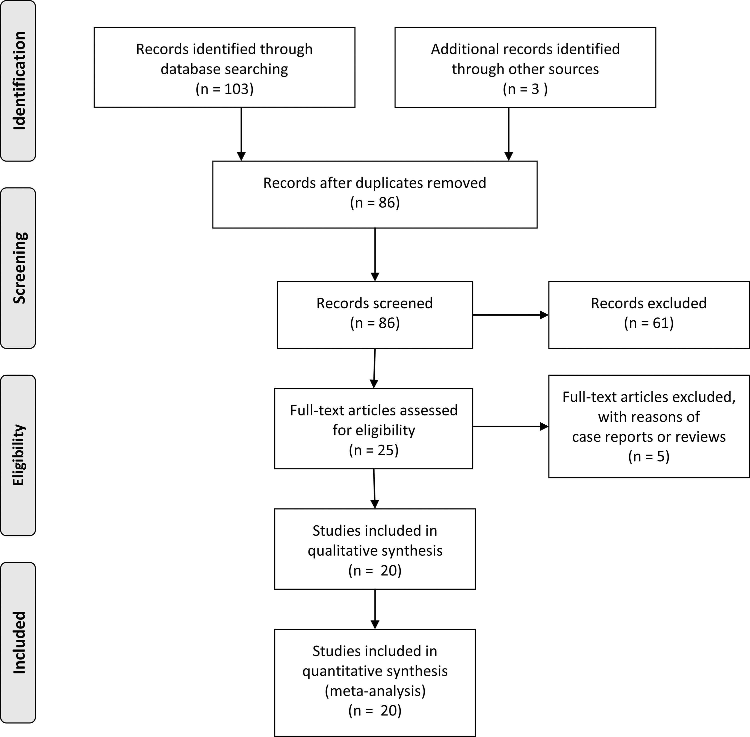
PRISMA flow diagram depicting the selection process of the studies included in the analysis. PRISMA, Preferred Reporting Items for Systematic Reviews and Meta-Analyses.
Characteristics of the Included Studies (n = 20 Studies)
Non-PCR represents other methods such as enzyme-linked immunosorbent assay, scanning electron microscopy, immunohistochemistry, and cell culture.
Several included studies did not classify bacteria at the species level.
Two included studies did not separately report the total number of samples for each animal species.
Pooled prevalence
The pooled prevalence of chlamydial infections was 23.5% (95% CI: 15.4–34.0). High heterogeneity was observed among the included studies (Q-value = 308.2, df = 19, p < 0.001; I 2 = 93.8%) (Table 2).
The Overall Pooled Prevalence and Subgroup Analysis for Chlamydial Infections in Reptiles
95% CI; Q; I 2 represents the inconsistency index describing the percentage of variability due to heterogeneity rather than sampling error; Non-PCR represents other methods such as enzyme-linked immunosorbent assay, scanning electron microscopy, immunohistochemistry, and cell culture.
95% CI, 95% confidence interval; I 2, I-squared; Q, Cochran's Q-tests for heterogeneity.
Subgroup analysis
Significant differences among subgroups were found for three characteristics (continents, orders of reptiles, and types of specimens) (Table 2). For continents, the prevalence was highest in Australia (68.6% [95% CI: 36.8–89.1]) and lowest in Africa (5.8% [95% CI: 1.4–20.8]). For orders of reptiles, the prevalence was highest in Crocodilia (crocodiles and alligators) (57.3% [95% CI: 32.5–78.9]) and lowest in Testudines (turtles and tortoises) (8.9% [95% CI: 3.2–22.2]). For types of samples, the pooled prevalence of chlamydial infections was higher in carcass specimens than in live specimens (38.5%, [95% CI: 22.2–57.7] vs. 15.6%, [95% CI: 8.3–27.4]). For diagnostic techniques, the prevalence was higher for PCR studies (28.4% [95% CI: 18.6–40.9]), but was not significantly different (p value = 0.375) from that of non-PCR studies (17.4% [95% CI: 5.5–43.3]).
Cumulative meta-analysis and meta-regression
Cumulative evidence of the pooled prevalence over time is presented in Fig. 2. The pooled prevalence of chlamydial infections in reptiles gradually increased from 1.8% in 1990 to 22.9% in 2010. Then, the pooled prevalence was relatively stable at ∼21.6–25.4%, with the latest cumulative prevalence in 2018 at ∼23.5% (Fig. 2). A result from the meta-regression is shown in Fig. 3. The rate of prevalence gradually increased over time (regression coefficient = 0.07 [95% CI 0.00–0.13], p = 0.049).
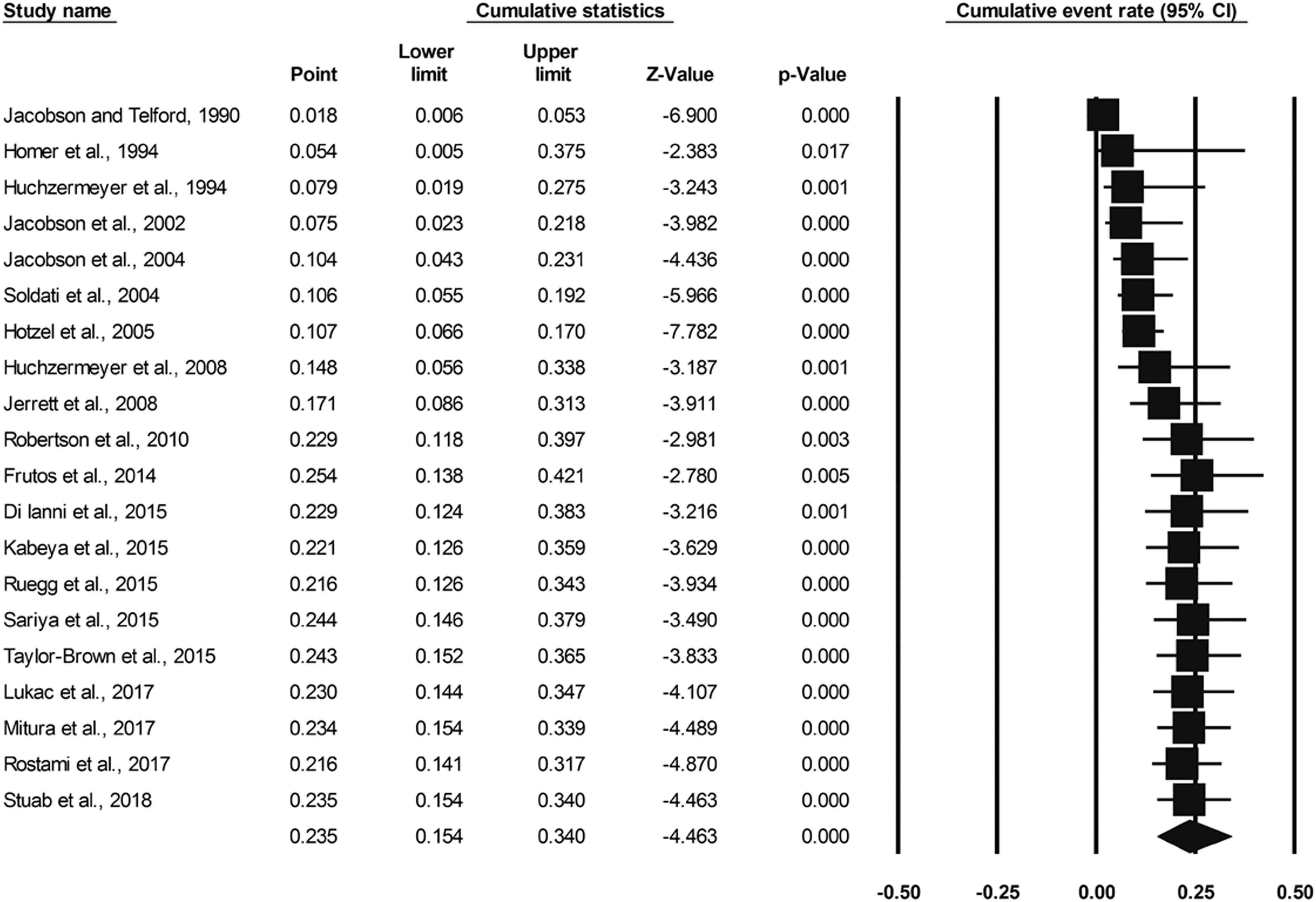
A forest plot depicting the cumulative prevalence of chlamydial infections in reptiles. The cumulative prevalence gradually increased from 1.8% in 1990 to 22.9% in 2010; then, it was relatively stable at ∼21.6% to 25.4%, with the latest cumulative prevalence in 2018 at ∼23.5%.
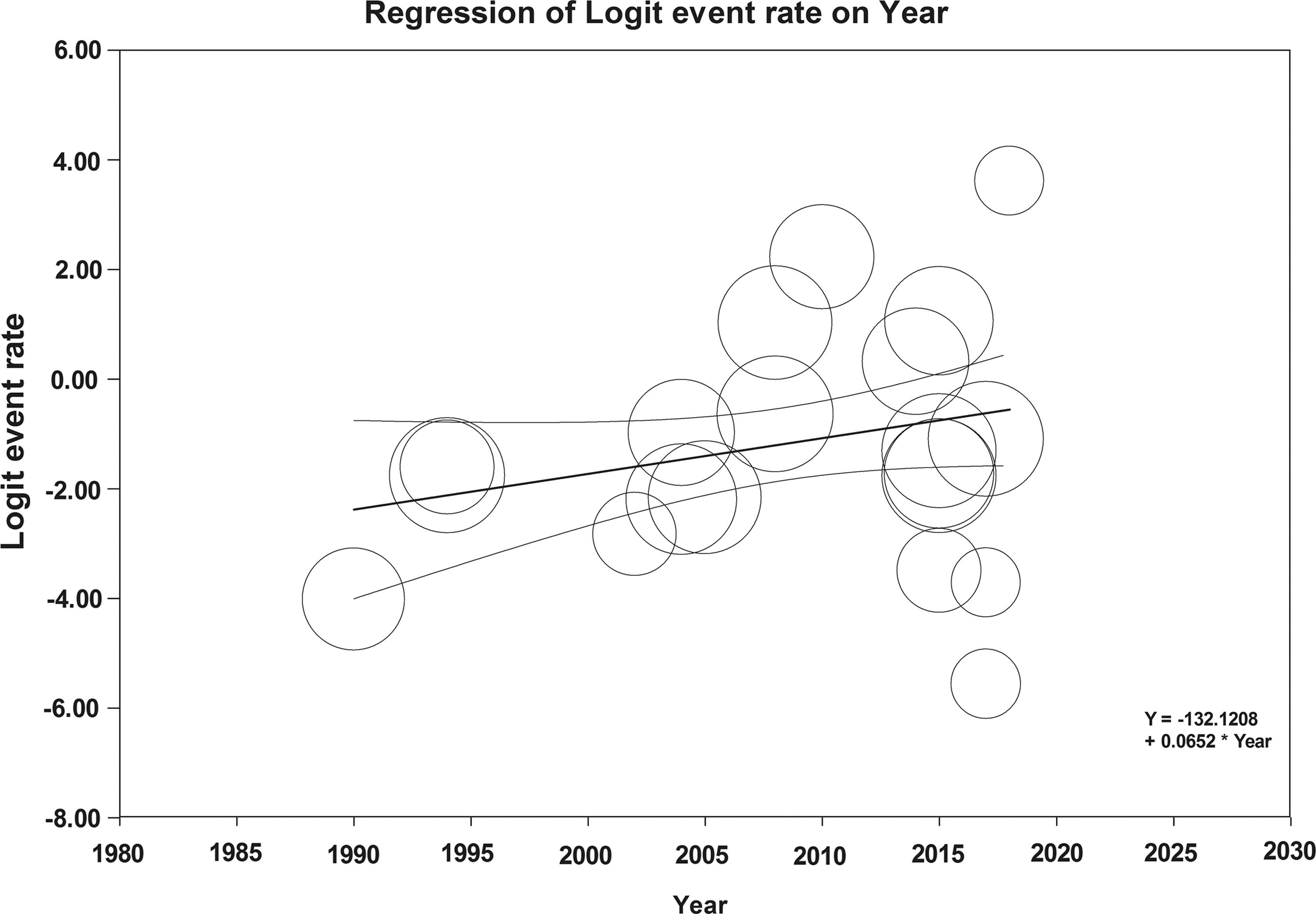
A scatter plot for the regression analysis. The Y-axis represents the logit event rate, and the X-axis shows years. A circle represents each included study (n = 20). The straight line in the middle is a regression line braced with its 95% CI lines. CI, confidence interval.
Publication bias and sensitivity test
No evidence of publication bias was observed. The funnel plot appeared approximately symmetrical. Egger's test for publication bias was not significant (p = 0.722). For the sensitivity analysis, a random effects model yielded a slightly lower pooled prevalence than a fixed effects model (23.5% [95% CI 15.4 − 34.0] vs. 27.1% [95% CI 25.0 − 29.2]). The influence of an individual study on the point estimate of pooled prevalence was minimal, ranging from 20.5% to 26.4%.
Discussion
This study showed that the cumulative prevalence (based on 20 studies analyzed) of chlamydial infections in reptiles is quite high at ∼21.6% to 25.4% over recent years. The cumulative prevalence in our study was calculated as aggregated values of prevalence over time. Therefore, it represents accumulative evidence of prevalence over time. This should be interpreted in a different way from results of the meta-regression that used a single point of data (not aggregate) to determine the trend of prevalence over time. A high cumulative prevalence indicated that reptiles are at risk for chlamydiosis (a disease caused by chlamydial infections). Infected reptiles may have depression, anorexia, hepatitis, conjunctivitis, fibrinous pharyngitis, pneumonia, and granulomatous lesions in the heart, lung, liver, spleen, and intestine with a high mortality rate in young infected animals (Soldati et al. 2004, Ebani 2017). Therefore, an outbreak of chlamydiosis in reptiles may result in an economic loss for farms and biodiversity loss of rare species of reptiles in nature. The high prevalence of chlamydial infections in reptiles also indicated that these animals may be potential sources of transmission of pathogens to other animal classes. In this study, we found four studies reporting C. psittaci infections in reptiles (Huchzermeyer et al. 1994, Jerrett et al. 2008, Robertson et al. 2010, Kabeya et al. 2015). This pathogen is a well-known zoonotic pathogen that causes psittacosis in humans. However, there is still lack of evidence of chlamydial transmission from reptiles to humans. We also found that C. pneumoniae was the most commonly reported species in reptiles. Although C. pneumoniae is the most common cause of chlamydial infection in humans, it is unclear whether this pathogen can be transmitted from animals to humans (Cheong et al. 2019). However, the finding of animal genotypes of C. pneumoniae in humans suggests a possible zoonotic transmission (Kutlin et al. 2007).
For reptile species, we found that the prevalence of chlamydial infections was highest in crocodiles (57.3%) and lowest in turtles (8.9%) (Table 2). In juvenile crocodiles, chlamydial infections can result in sudden death (Thongkamkoon et al. 2018). The prevalence of chlamydial infections in crocodiles is still high when compared with other classes of animals. Several studies have shown that the prevalence in birds is ∼20% (Aaziz et al. 2015, Ebani et al. 2016, Feng et al. 2016). The prevalence in ruminants was also ∼20% (Talafha et al. 2012, Benkirane et al. 2015, Bhardwaj et al. 2017). However, this pooled prevalence in crocodiles was only estimated from five studies. The pooled estimate may be subject to change when new studies of prevalence of chlamydial infections in crocodiles are published. In lizards and snakes, the pooled prevalence of chlamydial infections was estimated to be ∼12.5% (Table 2). This estimate was far lower than that of crocodiles. However, Chlamydia species among snakes are also recognized as emerging pathogens; indeed, a recent study found that novel Chlamydia species isolated from snakes exhibit decreased susceptibility to azithromycin (Staub et al. 2018). The reptiles with the lowest prevalence of chlamydial infections were turtles and tortoises (the point estimate was ∼8.9%). It is interesting that chlamydial infections were found both in wild and captive turtles and tortoises (Mitura et al. 2017). This indicated the ubiquitous presence of Chlamydia in turtles and tortoises.
For continents, surprisingly, we found that the prevalence of chlamydial infections was highest in Australia (68.6%). This can possibly be explained by the fact that three studies (Huchzermeyer et al. 2008, Jerrett et al. 2008, Robertson et al. 2010) from Australia reported a high prevalence of chlamydial infections in crocodiles, and no other orders of reptiles were reported from Australia (Table 2). Most studies (n = 8) reported chlamydial infections in reptiles from Europe, and the point estimate was ∼16.1% (Table 2). However, in Europe, the reptiles under study are members of the orders Squamata (lizards and snakes) and Testudines (turtles and tortoises), but not Crocodilia (crocodiles and alligators). It can be concluded that differences between the prevalence levels of chlamydial infections among continents may result from differences between the reptile species under study in each continent. This may reflect the differences in the reptile species of interest across continents.
For specimen types, we found that the prevalence of chlamydial infections was higher in carcasses (38.5%) than in live specimens (15.6%) (Table 2). These findings are not surprising and can be explained as follows. Most carcass samples were collected from internal organs such as the lung, liver, and spleen (Thongkamkoon et al. 2018). Chlamydia can infect and multiply in these organs; as a result, the chance of detecting pathogens from these organs is higher. On the other hand, the swabbing technique used for live animals (using a cotton tip to gently touch the surface of the conjunctiva, choana, and cloaca for sample collection) has a lower chance of yielding a sample with a high density of pathogens. Thus, the samples from internal organs are more likely to yield positive results than swab samples from live animals.
For diagnostic techniques, our study found no significant difference in the prevalence between PCR and non-PCR techniques. This result, however, must be interpreted carefully because of certain confounders. First, only six studies were included for non-PCR techniques; as a result, the prevalence estimate is not precise (because of its wider CI). This reduces the power of the statistical test, resulting in a high chance of false negative errors. Second, different diagnostic techniques have different sensitivity and specificity and possess different pros and cons. For example, PCR techniques have high specificity and enable fast identification, but work best only in the acute phase of chlamydial infection (Nieuwenhuizen et al. 2018).
This study has some limitations. The meta-analysis technique relies solely on available data from published original studies. The number of included studies reporting particular subgroups of some characteristics is small. This circumstance can result in a low power for statistical tests, probably leading to false negatives (type 2 error). Additional relevant studies that will be published in the future may change the results of the pooled prevalence analysis and subgroup analysis.
Conclusions
Few studies have reported the prevalence of chlamydial infections in reptiles. Based on available data, the prevalence of chlamydial infections in reptiles was quite high (∼23.5%). The cumulative evidence has shown an almost stable prevalence of slightly over 20% since 2010. Therefore, reptiles could be potential reservoirs of Chlamydia and may be a risk for humans or other animal species. However, the prevalence varied among different reptile species (with the highest prevalence in crocodiles and the lowest prevalence in turtles and tortoises). More studies of the prevalence of chlamydial infections in reptiles are required to better estimate the global prevalence.
Footnotes
Acknowledgments
The authors would like to thank the Faculty of Veterinary Medicine, Khon Kaen University, for their support in this study.
Author Disclosure Statement
No conflicting financial interests exist.
Funding Information
This study was supported by the Faculty of Veterinary Medicine, Khon Kaen University.