
Abstract
Fleas are carriers for many largely understudied zoonotic, endemic, emerging, and re-emerging infectious disease agents, but little is known about their prevalence and role as a vector in Africa. The aim of this study was to determine the diversity of fleas and the prevalence of infectious agents in them collected from human dwellings in western Kenya. A total of 306 fleas were collected using light traps from 33 human dwellings; 170 (55.56%) were identified as Ctenocephalides spp., 121 (39.54%) as Echidnophaga gallinacea, 13 (4.25%) as Pulex irritans, and 2 (0.65%) as Xenopsylla cheopis. Of the 306 individual fleas tested, 168 (54.9%) tested positive for rickettsial DNA by a genus-specific quantitative real-time PCR (qPCR) assay based on the 17-kDa antigen gene. Species-specific qPCR assays and sequencing revealed presence of Rickettsia asembonensis in 166 (54.2%) and Rickettsia felis in 2 (0.7%) fleas. Borrelia burgdorferi, normally known to be carried by ticks, was detected in four (1.3%) flea DNA preparations. We found no evidence of Yersinia pestis, Bartonella spp., or Orientia spp. Not only were Ctenocephalides spp. the most predominant flea species in the human dwellings, but also almost all of them were harboring R. asembonensis.
Introduction
Fleas (Order: Siphonaptera) are holometabolous insects, and thus progress through four life stages: egg, larva, pupa, and adult. The adults survive as obligate ectoparasites on mammalian or avian hosts (Bitam et al. 2010). Flea-borne infections have been increasingly prevalent with chances of re-emerging in epidemic forms (Eisen and Gage 2012). They are known to be carriers for many largely understudied zoonotic, endemic, emerging, and re-emerging infectious disease agents such as Rickettsia typhi, Rickettsia felis, Yersinia pestis, Bartonella spp., Erhlichia spp., and Anaplasma spp. (Bitam et al. 2010, Eisen and Gage 2012, Torina et al. 2013, Leulmi et al. 2014). Transmission occurs through biting and/or inoculation of feces into pruritic bite lesions (Bitam et al. 2010, Eisen and Gage 2012). Flea bites are not only a nuisance, but may also cause hypersensitivities in humans and animals (Bitam et al. 2010). In addition, they may act as intermediate hosts for zoonotic tapeworms, Dipylidium caninum (dog tapeworm), and Hymenolepis diminuta (rat tapeworm), which are parasites for dogs and rats, respectively (Mane and Sangwan 2016, Jiang et al. 2017). In most developing countries, domestic companion animals such as cats and dogs roam freely and, therefore, serve as potential sources of disease transmission (Haydon et al. 2002). Of the pathogens transmitted by fleas, Y. pestis causes a life-threatening disease and is still a major health problem in some parts of Africa (Andrianaivoarimanana et al. 2013, Butler 2013). R. felis is a common cause of fever in Africa (Richards et al. 2010, Socolovschi et al. 2010, Sokhna et al. 2013).
In addition, louse-borne relapsing fever is caused by Borrelia recurrentis and transmitted by the human body louse (Pediculus humanus), and tick-borne relapsing fever due to Borrelia duttoni and transmitted by argasid ticks in the Ornithodorus moubata complex are still common in Africa (Trape et al. 2013, Diatta et al. 2016). Conversely, Lyme disease caused by Borrelia burgdorferi is transmitted by ticks in genus Ixodes (Ixodes scapularis, I. pacificus, I. ricinus) is rarely reported in Africa (Elhelw et al. 2014, Ben Said et al. 2016). The literature concerning Lyme disease in Kenya is limited to two cases reports (Jowi and Gathua 2005).
Previous studies in Kenya have documented the presence of Rickettsia species in febrile patients (Maina et al. 2012), and in fleas (Jiang et al. 2013, Luce-Fedrow et al. 2015) from western Kenya, but none have documented the diversity of other flea-borne pathogens in fleas from this region. There is only limited data concerning the presence and prevalence of other vector-borne pathogens in fleas in Kenya.
Materials and Methods
Ethical statement
The study was approved by the Kenya Medical Research Institute's (KEMRI's) Ethical Review and Animal Care and Use Committees (SSC no. 2412) and was embedded on Population-Based Infectious Disease Surveillance (PBIDS) (SSC Protocol no. 1899) conducted by the Kenyan International Emerging Infections Programme (Feikin et al. 2011).
Study site
This study was conducted in Rarieda subcounty, western Kenya; rural site located on the eastern shores of Lake Victoria falling within a health and demographic surveillance system run by the KEMRI and the United States Centers for Disease Control and Prevention (CDC) since 2001 (Adazu et al. 2005). KEMRI/CDC has conducted PBIDS of the human population in this site since 2005 (Oria et al. 2013). The PBIDS population is ∼25,000 people living in 33 villages in 6800 households within a 5.5 km radius. The community lives in dispersed settlements, and houses are made of mud, cement, or brick, with roofs of iron sheet or thatch (Bigogo et al. 2010) with animal ownership at >90% (including dogs and cats) (Thumbi et al. 2015) (Fig. 1).
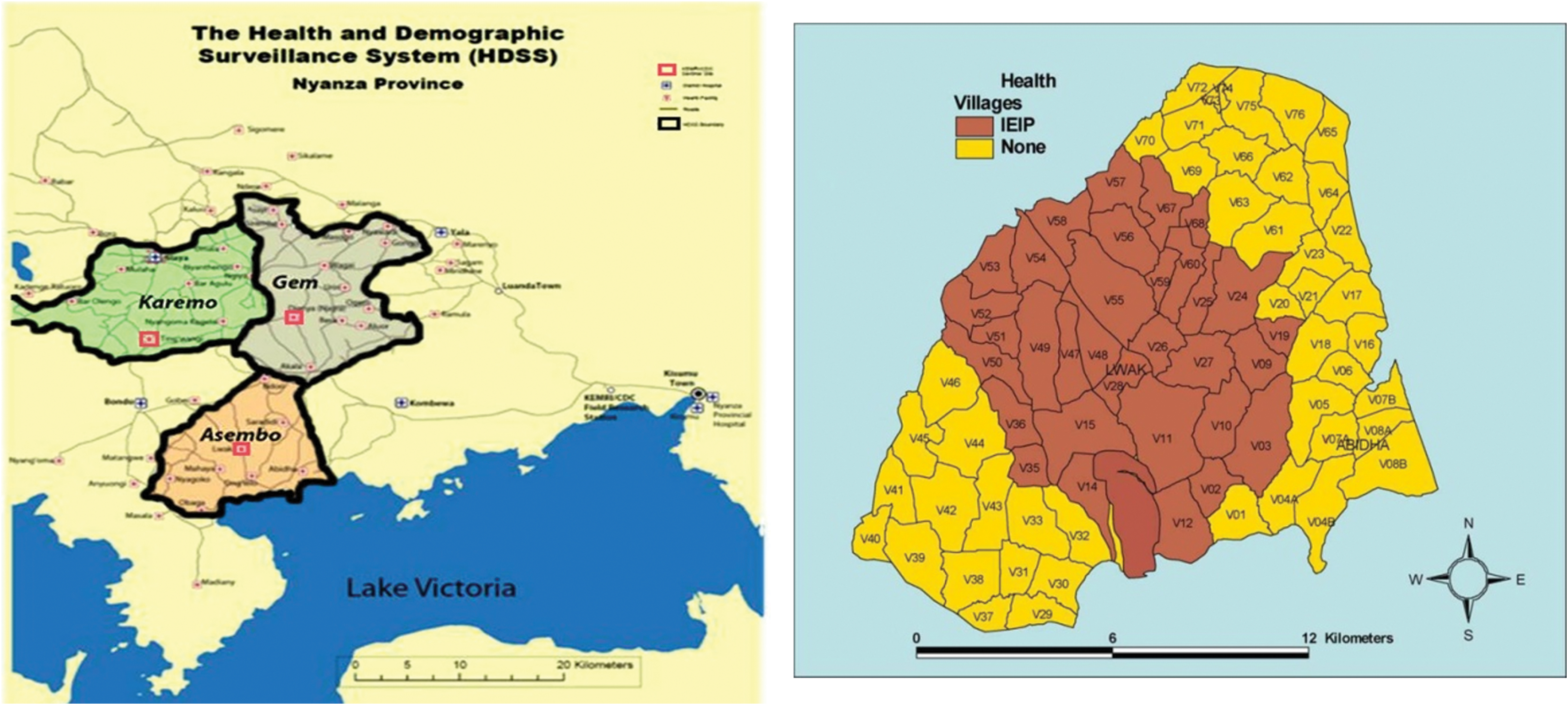
Study area map displaying the locations of the villages in Asembo area of western Kenya where the study was conducted. Color images are available online.
Sampling and laboratory procedures
Flea collections occurred in 2012 during the months of February (hot dry season) and March (long rains season). Fleas were collected from 33 randomly selected houses using light traps that consisted of a hurricane lamp 3–13 cm above a tray containing water with dish soap and petroleum jelly (Vaseline) smeared on the sides to prevent fleas from crawling out. The fleas were then pooled and preserved in 70% ethanol until delivery to the KEMRI/CDC laboratory in Kisumu, where they were stored at −80°C. Identification was done using standard entomological taxonomic keys, and then separated into individual tubes. The fleas were then individually washed three times in molecular-grade water and mechanically disrupted using disposable pellet pestles (Fisher Scientific, Waltham, MA). Genomic DNA was extracted from individual fleas using Prepman Ultra sample preparation kits (Applied Biosystems, Foster City, CA) according to the manufacturer's instructions. Nuclease-free/molecular-grade water was used a negative extraction control.
Quantitative real-time PCR assays
A quantitative real-time PCR (qPCR) assay that amplifies and detects a 111-bp segment of the 17-kDa antigen gene (Rick17b) (Jiang et al. 2012) was used to screen the individual flea DNA preparations for the presence of rickettsial DNA. Flea DNA preparations that tested positive by Rick17b assay were subsequently tested using three species-specific qPCR assays: (i) RFel_Phosp_MB, which targets the membrane phosphatase gene from R. felis; (ii) Rasem, which targets a 112-bp of the ompB gene specific to R. asembonensis; (iii) Rtyph, which targets a 122-bp fragment of the ompB gene of R. typhi (Henry et al. 2007, Jiang et al. 2013, Leulmi et al. 2014).
The DNA preparations were also screened for the presence of Bartonella spp. using a genus-specific assay targeting the ssrA gene (Diaz et al. 2012); Y. pestis using a species-specific (Yper_PLA) assay (Leulmi et al. 2014); and B. burgdorferi and Orientia spp. using multiplex TaqMan species-specific assays (Courtney et al. 2004, Jiang et al. 2004) (Table 1). In addition to negative extraction controls, nuclease-free/molecular-grade water (no-template controls) were included as negative controls in all qPCR runs. Plasmid DNA preparations for the aforementioned microbes were used as positive controls in each qPCR run.
Oligos Used in the Study
PCR.
Nested PCR.
Sequencing.
To confirm the identity of fleas morphologically identified as Pulex irritans or Xenopsylla cheopis, a P. irritans-specific assay was employed (Mediannikov et al. 2012).
To confirm the identity of the pathogens detected by the various qPCR assays in the fleas, standard and nested PCR (nPCR) together with sequencing of the rickettsial 16S rRNA (Jiang et al. 2013) and ompB genes (Roux and Raoult 2000, Jiang et al. 2005, 2013) were performed. Two primers (FlaBd and FlaBrc) were used for PCR to generate a 278 bp fragment of the Borrelia flagellin (flaB) gene (Bugrysheva et al. 2011). A subset of 13 individual flea DNA samples, including four that were positive by the Rick17b and Rasem qPCR assays, two that were Rick17b and RFel_Phosp_MB positive, three that had a late cycle threshold (Ct) by Rick17b but were negative in all species-specific assays, and four that were positive by the Bburg qPCR assay were selected. No positive controls were included in PCR and nPCR reactions, in an effort to reduce contamination potential. Nuclease-free/molecular-grade water was used as negative controls. Sequencing reactions were performed utilizing both DNA strands with the Big Dye Terminator v3.1 Ready Reaction Cycle Sequencing Kit (Life Technologies, Foster City, CA), according to the manufacturer's instructions on an ABI 3500 genetic analyzer (Applied Biosystems). Sequences were assembled using CodonCode Aligner version 5.0.1 (CodonCode Corporation, Centerville, MA) and exported to MEGA version 7 software (CEMI, Tempe, AZ). BLAST searches were performed using the National Center for Biotechnology Information website (
Results
A total of 306 fleas were collected and identified morphologically using standard entomological keys and confirmed by molecular assays. Of these, 170 (55.56%) were Ctenocephalides spp., 121 (39.54%) Echidnophaga gallinacea, 13 (4.25%) P. irritans, and 2 (0.65%) X. cheopis (Table 2).
Rickettsia spp. Detected in Five Flea Species
Of the 306 fleas, 168 (54.9%) tested positive for rickettsial DNA by a genus-specific qPCR assay based on the 17-kDa antigen gene. Three species-specific Rickettsia qPCR assays were then conducted: (i) using the R. asembonensis-specific assay, 166 (54.25%) tested positive; (ii) using the R. felis-specific assay, 2 (0.65%) tested positive; and (iii) using the R. typhi-specific assay, none tested positive. Nearly all the Ctenocephalides fleas (167 fleas, 98.24%) were positive for Rickettsia spp., of which 97.1% were positive for R. asembonensis, whereas the remaining 1.18% were positive for R. felis. One of two X. cheopis was positive for R. asembonensis. There were no coinfections and no rickettsiae detected in the other flea species (P. irritans and E. gallinacea).
Four of the individual flea DNA preparations (1.31%) were positive using the B. burgdorferi-specific qPCR assay, all of which belonged to the genus Ctenocephalides. Three other qPCR assays were also performed, which screened for Y. pestis, Bartonella, and Orientia, but no samples returned positive results. Male P. irritans and X. cheopis are difficult to distinguish by their physical traits, so a qPCR assay (Pirr) specific for P. irritans was utilized for species differentiation. Of the 15 fleas morphologically classified as P. irritans and X. cheopis, 13 out of the 15 DNA samples (86.7%) tested positive for P. irritans (Table 2).
The 16S rRNA and ompB sequence from the four individual flea DNA samples that were positive using Rick17b and Rasem qPCR assays were 100% identical to R. asembonensis (NZ_JWSW00000000). The sequences from the two samples that were Rick17b and RFel_Phosp_MB positive were confirmed to be 100% identical to R. felis (CP000053). No amplicons were obtained from the three samples that had a late cycle Ct by Rick17b and tested negative using all species-specific assays. The flaB sequences from the four samples that were positive by Bburg qPCR were identical to each other and were determined to be 100% (278/278-bp) identical to B. burgdorferi (CP002228).
Discussion
Flea species in the genera Ctenocephalides spp. (C. felis and C. canis) E. gallinaecea, P. irritans, and X. cheopis were identified in human dwellings. Our findings are corroborated by recent studies that described an abundance of human-associated fleas (P. irritans, C. felis, and X. cheopis) in human dwellings in plague-endemic regions of Africa (Laudisoit et al. 2007, Eisen et al. 2008, Miarinjara et al. 2016). C. felis and C. canis are extremely common in many temperate and tropical regions, and are known to infest domestic animals such as dogs and cats, as well as wild animals (Pung et al. 1994). The Ctenocephalides genus represents the majority (94%) of fleas found in human homes in Uganda (Eisen et al. 2008); similarly, they were the most prevalent in this study (55.56%). This is in contrast to the findings from previous studies that reported the human flea (P. irritans) to be the most predominant flea (72.4%) in houses in Usambara of western Tanzania (Laudisoit et al. 2007) and Madagascar (Miarinjara et al. 2016). P. irritans is mostly referred to as the human flea, has a cosmopolitan distribution, and attacks a wide range of mammals with potential infestation of humans sharing dwellings or living near habitations for animals (He et al. 1997, Loftis et al. 2006). Although this species was reportedly among the most predominant in many homes in sub-Saharan Africa, it was among the least (4.25%) abundant flea species encountered in this study.
E. gallinacea (stick tight flea) was the second most prevalent flea collected in the human houses (39.54%). It is found in free-living avian species in south central Florida (Boughton et al. 2006), and commonly infests fowl, but may also infest a range of mammals such as rodents, opossums, raccoons, feral cats, and domestic ruminants (Pfaffenberger and Valencia 1988, Pung et al. 1994, Akucewich et al. 2002, Boughton et al. 2006, Loftis et al. 2006). A recent study demonstrated that 83.6% of the people in rural Kenya shared houses with livestock at night (Ndenga et al. 2016). Correspondingly, chickens are the most commonly owned domestic animals in this region (Thumbi et al. 2015), and are known to be highly associated with human habitations; this may explain the high prevalence of E. gallinacea found in human dwellings in this study.
X. cheopis (Oriental rat flea) was the least abundant flea species encountered in this study (0.65%). It is distributed throughout Africa and central and southern Asia, coinciding with the distribution of gerbils or rats; it is the primary vector for plague, murine (endemic) typhus, and parasitic helminths as well as Bartonella (Bitam et al. 2010).
Rickettsiae were the most common (54.2%) bacterial agents detected in these fleas. Not only were the Ctenocephalides spp. the most common flea species found in this study, but also almost all of them (98.2%) were carrying R. asembonensis. Species-specific qPCR assays and sequencing confirmed that 54.2% of rickettsiae identified in this study were R. asembonensis and a small proportion (0.7%) were R. felis. R. asembonensis was predominantly detected in Ctenocephalides spp. (97.1%) and to a much lesser extent (1/2 fleas) in X. cheopis. As such, there were too few X. cheopis fleas for meaningful comparison. R. felis was only detected in Ctenocephalides spp. Although E. gallinacea and P. irritans were negative for rickettsial DNA, previous studies have detected Rickettsia species Rf2125 (Loftis et al. 2006, Jiang et al. 2013) and R. asembonensis (Jiang et al. 2013) in those flea species.
Ticks in the genus Ixodes are historically known to play a major role in the transmission of B. burgdorferi; therefore, it was intriguing to detect Borrelia in fleas collected. Fleas are not known vectors of Borrelia, but four of the samples tested positive at the late Ct values. Sequencing of the flaB gene from the four positive samples revealed 100% identity with B. burgdorferi. Our finding corresponds with earlier data demonstrating the isolation of the agent from C. felis (Teltow et al. 1991). In addition, previous studies have shown that B. burgdorferi can be detected in other invertebrates such as mosquitoes (Culex pipiens) (Zakovska et al. 2002), deer flies, horse flies (Magnarelli et al., 1986), and fleas (Ctenophthalmus spp., Histrichopsylla talpae) (Netusil et al. 2013). However, the vectorial importance of these different types of invertebrates has not yet been demonstrated.
Although Bartonella spp. have previously been detected in small mammals and in bats in Kenya (Kosoy et al. 2010, Young et al. 2014, Halliday et al. 2015), none of these fleas were positive for Bartonella.
Y. pestis has been associated with several species of fleas and mammals, and X. cheopis is considered as the primary vector for plague (He et al. 1997, Bitam et al. 2006). The vectorial competence of C. felis in transmitting Y. pestis has been previously demonstrated experimentally (Eisen et al. 2008). Plague is endemic to sub-Saharan Africa and has been identified in fleas as well as humans during outbreaks (Davis 1953, Bitam et al. 2006, Laudisoit et al. 2007, Leulmi et al. 2014). Despite this apparent endemicity of Y. pestis in Africa, including in countries neighboring Kenya, none of the fleas tested in the study was positive for the pathogen. In one study (Laudisoit et al. 2007), P. irritans was not only the most abundant flea species found in the human dwellings, but the density of P. irritans was positively correlated with high plague frequency. None of the fleas tested in our study were found to carry Y. pestis. This result is likely due to the few numbers of fleas studied/collected, or may also reflect a decline of plague carriers in western Kenya due to the extensive use of insecticides and rodenticides.
Conclusion
Consequently, in this study we have demonstrated that rickettsiae are the predominant agents present in fleas collected from the human dwellings in western Kenya. R. felis, the causative agent for flea-borne spotted fever is sympatric with R. asembonensis. Both of these agents are genetically related and belong to the larger genetic group referred to as the transition group of Rickettsia. Conclusions concerning the finding of B. burgdorferi in Ctenocephalides spp. will require additional studies to better understand the role of fleas associated with this traditionally tick-borne pathogen. The presence of R. felis (causative agent of flea-borne spotted fever in humans) and R. asembonensis (unknown/unconfirmed pathogenicity in humans) warrants additional studies to better detail/expand the knowledge of pathogen carriage by fleas in human dwellings in Kenya.
Footnotes
Disclaimer
The views expressed in this article are those of the author and do not necessarily reflect the official policy or position of the Department of the Navy, Department of Defense, nor the U.S. Government. A.L.R. was and C.M.F. is an employee of the U.S. Government and their work was prepared as part of their official duties. Title 17 U.S.C. §105 provides that “Copyright protection under this title is not available for any work of the United States Government.” Title 17 U.S.C. §101 defines a U.S. Government work as a work prepared by a military service member or employee of the U.S. Government as part of that person's official duties.
Author Disclosure Statement
No conflicting financial interests exist.
Funding Information
This study was supported by the Global Disease Detection program of the United States Centers for Disease Control and Prevention and the Armed Forces Health Surveillance Branch and its Global Emerging Infections Surveillance and Response (GEIS) Section (funding year 2015, ProMIS ID P0043_15_NM); NMRC work unit A0074.