




Abstract
Background:
Q fever is a worldwide zoonosis caused by Coxiella burnetii. This study was carried out to investigate the occurrence of C. burnetii among apparently healthy pregnant, parturient, and postparturient dogs and cats to highlight their role in the transmission of such disease to humans.
Materials and Methods:
A total of 88 apparently healthy pet animals (48 dogs and 40 cats) were enrolled in this study, vaginal swabs were obtained from pregnant and postparturient animals while birth fluids were collected from parturient ones. All samples were subjected to DNA extraction followed by nested PCR for molecular detection of C. burnetii.
Results:
Out of 40 cats, 3 were positive for C. burnetii with an overall prevalence of 7.5%, all positive samples were birth fluids of parturient queens with a prevalence of 15.8% (3/19) while all pregnant and postparturient animals were negative. In contrast, none of 48 dogs yielded positive result. Moreover, the phylogenetic analysis and sequence identity matrix of the obtained sequence from a parturient cat showed high genetic relatedness to strains derived from human cases rather than those of ruminants to indicate the public health burden of such strain.
Conclusion:
This study underscores the occurrence of C. burnetii among parturient cats to point out the possible zoonotic transmission to human contacts.
Introduction
Q (Query) fever is a re-emerging disease of human and animals with worldwide distribution. Coxiella burnetii, etiology of Q fever, is an obligate intracellular Gram-negative bacterium with a complicated life cycle (Angelakis and Raoult 2010). Owing to its very low infective dose and its ability to disperse great distances, U.S. Centers for Disease Control and Prevention classified this pathogen as a category B bioterrorism agent (Roest et al. 2013a). The disease may be asymptomatic among humans, nonetheless, some patients show febrile illness, pneumonia, or hepatitis in the acute form of the disease and endocarditis in chronic form (Terheggen and Leggat 2007).
The main primary reservoirs for human Q fever infections are ruminants, particularly cattle, sheep, and goats, whereas wild animals, ticks, and birds have been considered as potential sources (Angelakis and Raoult 2010, Ghoneim et al. 2020). The disease in farm animals is subclinical but the main clinical outcomes are reproductive disorders encompassing abortion, premature birth, weak newborns, and stillbirths from which massive number of bacteria are excreted contaminating the environment (Abdel-Moein and Hamza 2017, Plummer et al. 2018). Human infection results mainly through inhalation of contaminated aerosols of birth fluids either from abortions or from normal parturitions (Roest et al. 2013a).
Although zoonotic transmission is usually associated with livestock, pet animals have been included in urban Q fever outbreaks as well (Greene 2012). In Australia, it was noted that there were raising number of human cases without a history of occupational exposure to ruminant animals, a matter that warranted looking for another potential source of infection, which may be companion animals (Archer et al. 2017). Despite there were many human Q fever outbreaks linked with exposure to infected parturient cats (Kosatsky 1984, Marrie et al. 1988, Malo et al. 2018) and to a lesser extent dogs (Buhariwalla et al. 1996), researchers paid more attention to investigate association of C. burnetii with reproductive disorders in pet animals rather than apparently healthy ones (Agerholm 2013, Fujishiro et al. 2016, Stefanetti et al. 2018).
Therefore, this study was conducted to investigate the presence of C. burnetii among apparently healthy pregnant, parturient, and postparturient dogs and cats to provide more knowledge about their potential role in the epidemiology of such pathogen.
Materials and Methods
Collection of samples
Birth fluids as well as vaginal swabs were collected from 88 apparently healthy pet animals (48 dogs and 40 cats) from veterinary hospitals and private pet clinics. Birth fluids were gathered in sterile cups from apparently healthy parturient animals (without any reproductive disorders or fetal anomalies) admitted to pet clinics to undergo cesarean section whereas vaginal swabs were obtained from pregnant and postparturient (1–4 weeks after parturition) animals using sterile swabs after disinfection of the vulva. All swabs were inserted in sterile screw capped tubes containing 1 ml of sterile phosphate buffer saline (Sheikh et al. 2020). All samples were transported in an ice box to the laboratory where they were stored at −20°C till further processing. The examined number of pregnant, parturient, and postparturient dogs and cats is given in Table 1.
The Number of Examined Pregnant, Parturient, and Postparturient Dogs and Cats
Molecular detection of C. burnetii in animal samples
DNA extraction
The vaginal swab samples were thawed and vortexed for 10 s to drive out any material from the tip and side of the tube. Two hundred microliters of each vaginal swab elute and birth fluids were subjected for DNA extraction using the QIAamp DNA mini kit (Qiagen, Germany) according to manufacturer's instructions, then extracted DNA was stored at −20°C for further use in PCR.
Nested PCR assay
The nested PCR was conducted using two sets of primers amplifying the repetitive element IS1111, which can vary dramatically in copy number between strains. PCR assays were carried out with EmeraldAmp GT PCR master mix (Takara, Japan) in T100™ Thermal Cycler (Bio-Rad, USA). The initial set of primers IS111 F1 (5′-TACTGGGTGTTGATATTGC-3′) and IS111 R1 (5′-CCGTTTCATCCGCGGTG-3′) was designed to amplify a 485 bp fragment, whereas the second set IS111 F2 (5′-GTAAAGTGATCTACACGA-3′) and IS111 R2 (5′-TTAACAGCGCTTGAACGT-3′) was used for nested PCR step that targets a 260 bp fragment (Fournier and Raoult 2003).
The first PCR was carried out with a temperature profile of initial denaturation at 95°C for 3 min then 40 cycles of denaturation (95°C for 30 s), annealing (52°C for 30 s), and extension (72°C for 1 min) followed by 4 min of final extension at 72°C. Thereafter, nested PCR step of PCR products was carried out as follows: after initial denaturation at 95°C for 3 min, 30 cycles of denaturation, annealing, and extension at 95°C, 52°C, and 72°C for 30 s in each step, respectively, were conducted, then final extension at 72°C for 4 min and subsequently specific band was obtained at 260 bp after ethidium bromide staining of PCR products (Fig. 1).
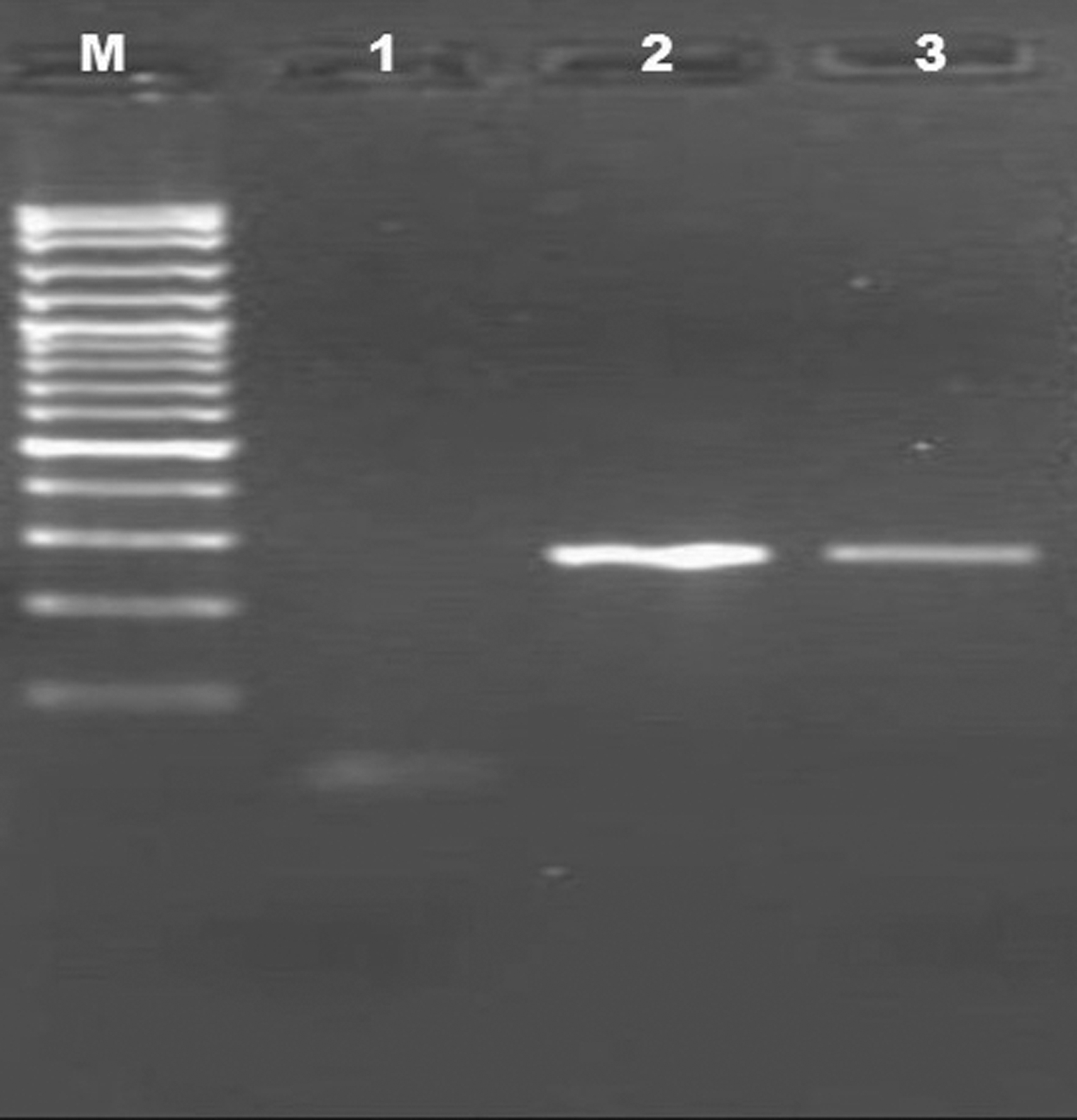
Molecular detection of Coxiella burnetii in birth fluids of parturient cats. Lane M: DNA ladder (100 bp); lane 1: negative control; lanes 2 and 3: positive samples with specific bands at 260 bp.
Sequencing step
PCR amplicon of one positive parturient cat from the first PCR reaction was purified through Qiaquick purification kit (Qiagen) following the manufacturer's protocol. Then, the purified product was subjected to sequencing using Big Dye Terminator V3.1 kit (Applied Biosystems, USA) in ABI 3500 Genetic Analyzer (Applied Biosystems).
Nucleotide sequence accession number
The obtained sequence from a parturient cat was deposited in the GenBank under accession number MT472649.
Sequence identity matrix and phylogenetic analysis
The obtained nucleotide sequence from a parturient cat was blasted on the NCBI website (
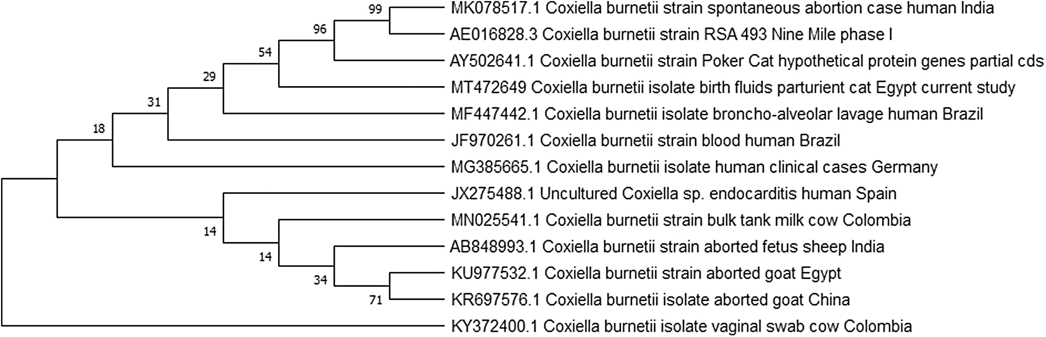
Neighbor-joining phylogenetic tree was inferred to show evolutionary history and genetic relationship of Coxiella burnetii parturient cat sequence and those from GenBank records. The bootstrap consensus tree was constructed based on C. burnetii partial sequence of htpAB-associated repetitive element using Mega 7 software.
Results
Three out of 40 apparently healthy cat samples were positive with an overall prevalence of 7.5%, whereas all of them were obtained from birth fluids of parturient cats with a prevalence 15.8%, and all vaginal swabs of the examined pregnant and postparturient queens yielded negative results. In contrast, none of the investigated 48 apparently healthy dogs was positive (Table 2). The results of sequence identity matrix are displayed in Table 3.
Occurrence of Coxiella burnetii DNA Among Examined Dogs and Cats
Sequence Identity Matrix of the Obtained Parturient Cat Sequence and Some Coxiella burnetii Strains Retrieved from GenBank
Discussion
Pet animals, especially dogs and cats, are important companions, elsewhere, they are common households in many settings and usually kept in close contact with their owners. In the meantime, they may harbor some emerging pathogens with a great public health implication (Abdel-Moein and Samir 2014).
In this study, our results revealed that C. burnetii was detected in the birth fluids of apparently healthy parturient cats with a prevalence of 15.8% (3/19). Such result is higher than that recorded in the Netherlands, where the examined placentas that were collected during parturition from 15 cats after the 2007–2010 Q fever outbreak were negative for C. burnetii (Roest et al. 2013b). The potential role of healthy parturient cats in the transmission of such zoonotic pathogen to human was proposed by several reported outbreaks in Canada (Kosatsky 1984, Langley et al. 1988) and the United States (Pinsky et al. 1991).
Seriously, healthy parturient queens are cause for concern because they shed huge number (109) of bacteria into the environment during parturition, while cats are asymptomatic (Greene 2012). What is more, they usually go through normal delivery inside the house and thereby birth fluids may be considered an important source of infection for pet owners through direct contact with birth fluids or indirectly through contamination of households mentioning that C. burnetii could survive for long periods in the contaminated environment along with its resistance to several disinfectants (Maurin and Raoult 1999), making the contaminated object is very hard to be decontaminated, likewise, the reported Q fever outbreak was traced to exposure to contaminated clothing of cat owners having newborn kittens (Marrie et al. 1989).
Noteworthy, the vaginal swabs of all pregnant and postparturient queens in this study were negative. This result is similar to the findings of Kopecny et al. (2013) who found that reproductive tissues of 11 weeks postparturient cats incriminated in a human Q fever outbreak in Australia were negative as well as to the findings of Ma et al. (2020) who did not detect C. burnetii in six pregnant queens. In this context, Ma et al. (2020) concluded that shedding of such pathogen may be low outside parturient cats. On the contrary, C. burnetii was found in feline uterus at 3 weeks postparturition during outbreaks in Canada (Marrie et al. 1988).
Of note, the sequence identity matrix was done to include sequences from humans, Nine Mile phase I strain, Poker cat strain and others from ruminants, which are the main reservoir for human infection and the most probable source for environmental contamination. Unexpectedly, the obtained cat strain showed a higher level of identity (90.8–91.2%) to strains from human cases in different countries than those retrieved from cattle, sheep, and goat, including sequence obtained from an aborted goat in Egypt (88.2–90.8%) (Table 3). Furthermore, the phylogenetic tree demonstrated two clusters: the first cluster comprised sequences retrieved from human cases, Nine Mile phase I strain and cat strains (our strain and Poker cat strain), whereas the second cluster encompassed the vast majority of C. burnetii strains obtained from ruminants.
The results of sequence identity matrix coupled with that of phylogenetic analysis may draw a conclusion that points out to the high similarity between the cat strain and those circulated among humans elsewhere, a matter that bears a public health implication and may elucidate why there were many human Q fever outbreaks related to parturient cats, especially among humans without history of contact with farm environment.
In contrast, none of the 48 apparently healthy dog samples comprising birth fluids and vaginal swabs was positive. Such a result is consistent with that obtained by Ma et al. (2020), but conflicted with that of Roest et al. (2013b) who reported C. burnetii in 4 (7%) out of 54 placentas from parturient bitches after the 2007–2010 Q fever outbreak in the Netherlands. Nevertheless, several studies concluded that the role of dogs in human Q fever infection is still controversial (Porter et al. 2011, Stefanetti et al. 2018).
Conclusion
This study elucidates the potential role of parturient cats in transmitting C. burnetii to humans, especially cat owners or those who live in the vicinity of cats. Accordingly, strict hygienic measures should be implemented during the delivery of pregnant cats either inside the house or in the veterinary hospitals to tackle the transmission of such deadly pathogen.
Footnotes
Author Disclosure Statement
No competing financial interests exist.
Funding Information
No funding was received for this work.