




Abstract
We conducted serologic surveillance for flaviviruses and orthobunyaviruses in vertebrate animals in Mexico in 2018–2019. Sera were collected from 856 vertebrate animals, including 323 dogs, 223 horses, and 121 cows, from 16 species. The animals were from 3 states: Chihuahua in northwest Mexico (704 animals) and Guerrero and Michoacán on the Pacific Coast (27 and 125 animals, respectively). Sera were assayed by plaque reduction neutralization test using four flaviviruses (dengue type 2, St. Louis encephalitis, West Nile, and Zika viruses) and six orthobunyaviruses from the Bunyamwera (BUN) serogroup (Cache Valley, Lokern, Main Drain, Northway, Potosi, and Tensaw viruses). Antibodies to West Nile virus (WNV) were detected in 154 animals of 9 species, including 89 (39.9%) horses, 3 (21.4%) Indian peafowl, and 41 (12.7%) dogs. Antibodies to St. Louis encephalitis virus (SLEV) were detected in seven animals, including three (0.9%) dogs. Antibodies to Lokern virus (LOKV) were detected in 22 animals: 19 (8.5%) horses, 2 (1.7%) cows, and a dog (0.3%). Antibodies to Main Drain virus (MDV) were detected in three (1.3%) horses. WNV and LOKV activity was detected in all three states, SLEV activity was detected in Chihuahua and Michoacán, and MDV activity was detected in Chihuahua. None of the animals was seropositive for Cache Valley virus, the most common and widely distributed BUN serogroup virus in North America. In conclusion, we provide serologic evidence that select flaviviruses and BUN serogroup viruses infect vertebrate animals in Chihuahua, Guerrero, and Michoacán. We also provide the first evidence of LOKV and MDV activity in Mexico.
Introduction
Many arthropod-borne viruses (arboviruses) of veterinary significance occur in North America. Some of these viruses are classified in the genus Flavivirus and others are classified in the Bunyamwera (BUN) serogroup of the genus Orthobunyavirus. The most prevalent and widely distributed flavivirus of veterinary importance in North America is West Nile virus (WNV), which occurs throughout Mexico, the contiguous United States and southern Canada (Elizondo-Quiroga and Elizondo-Quiroga 2013, Zheng et al. 2014, Petersen 2019). The most prevalent and widely distributed BUN serogroup virus of veterinary importance in North America is Cache Valley virus (CVV), with isolations made as far north as Canada and as far south as the state of Yucatan, Mexico (Thorsen et al. 1980, Calisher et al. 1986, Blitvich et al. 2012a).
The introduction of WNV into the Americas in 1999 was associated with extensive morbidity and mortality in horses in the United States and Canada (Castillo-Olivares and Wood 2004). Surprisingly, there have been few cases of WNV disease in horses in Mexico, despite serological evidence of widespread WNV activity (Blitvich 2008, Elizondo-Quiroga and Elizondo-Quiroga 2013). Several licensed WNV vaccines are now available for horses (Bosco-Lauth and Bowen 2019, Saiz 2020). These vaccines are routinely used in the United States and Canada, but their use in Mexico is sporadic. Domestic ruminants are also susceptible to WNV infection and there have been occasional reports of encephalitis in alpacas and sheep (Dunkel et al. 2004, Rimoldi et al. 2017). WNV infections in companion animals are usually subclinical but fatal cases have occurred (Read et al. 2005, Schwab et al. 2007). WNV also infects poultry, often progressing to clinical disease in geese (Komar et al. 2001, Swayne et al. 2001).
Other flaviviruses that occur in North America include St. Louis encephalitis virus (SLEV), dengue virus (DENV), and Zika virus (ZIKV). SLEV is maintained in a bird/mosquito transmission cycle, but other vertebrates can become infected (Reisen 2003, Diaz et al. 2018). Vertebrate animals susceptible to SLEV infection include chickens, cows, horses, pheasants, and turkeys (Morris et al. 1994, Ulloa et al. 2003, Marlenee et al. 2004). There have been no reports of fatal SLEV infections in vertebrate animals in North America, but the virus was isolated from brain tissue of a horse that died of encephalitis in Brazil (Rosa et al. 2013). DENV and ZIKV are not usually associated with infections of domestic animals, but antibodies to these viruses were recently identified in horses on islands in the South Pacific (Beck et al. 2019).
CVV infections in sheep are common and can result in embryonic and fetal death, stillbirths, and congenital defects (Noronha and Wilson 2017, Waddell et al. 2019). The first documented outbreak of CVV occurred in a sheep flock in Texas in 1987 (Chung et al. 1990). Nineteen percent of the 360 lambs born during the outbreak had musculoskeletal and central nervous system defects and the total neonatal loss was 26%. Other vertebrate animals susceptible to CVV infection include cows, goats, horses, poultry, and rabbits (Waddell et al. 2019). Additional BUN serogroup viruses that occur in North America are Lokern virus (LOKV), Main Drain virus (MDV), Potosi virus (POTV), Northway virus (NORV), and Tensaw virus (TENV) (Calisher et al. 1986, Heard et al. 1991). MDV has been isolated from brain tissue of a horse that died of encephalitis and caused musculoskeletal and nervous system malformations and death in ovine fetuses after experimental challenge (Emmons et al. 1983, Edwards et al. 1997, Wilson et al. 2015). LOKV, POTV, NORV, and TENV are not recognized pathogens of vertebrate animals but their ability to cause disease has not been widely investigated.
Many studies have been performed to estimate the seroprevalence of flaviviruses and BUN serogroup viruses in vertebrate animals in Canada and the United States, but relatively few studies have been performed in Mexico. BUN serogroup virus surveillance has been especially limited. In the last 20 years, there have been two serological investigations performed in Mexico where vertebrate animals were tested for antibodies to BUN serogroup viruses and these were performed in the states of Chiapas, Quintana Roo, and Yucatan (Ulloa et al. 2003, Blitvich et al. 2012b). To increase our understanding of the host range and seroprevalence of BUN serogroup viruses and flaviviruses in Mexico, we serologically assayed vertebrate animals in Chihuahua, Guerrero, and Michoacán for select BUN serogroup viruses and flaviviruses known to occur in North America.
Methods
Study sites and sample population
The sample population consisted of vertebrate animals in three states of Mexico: Chihuahua in northwest Mexico and Guerrero and Michoacán on the Pacific Coast (Fig. 1). Study sites were located in 13 of the 67 municipalities of Chihuahua: Ahumada, Ascención, Batopilas de Manuel Gómez Morín, Buenaventura, Casas Grandes, Chihuahua, Dr. Belisario Domínguez, Galeana, Juárez, Namiquipa, Nuevo Casas Grandes, Ocampo, and Riva Palacio. In Michoacán and Guerrero, study sites were located in the municipalities of Lázaro Cárdenas and La Unión, respectively.
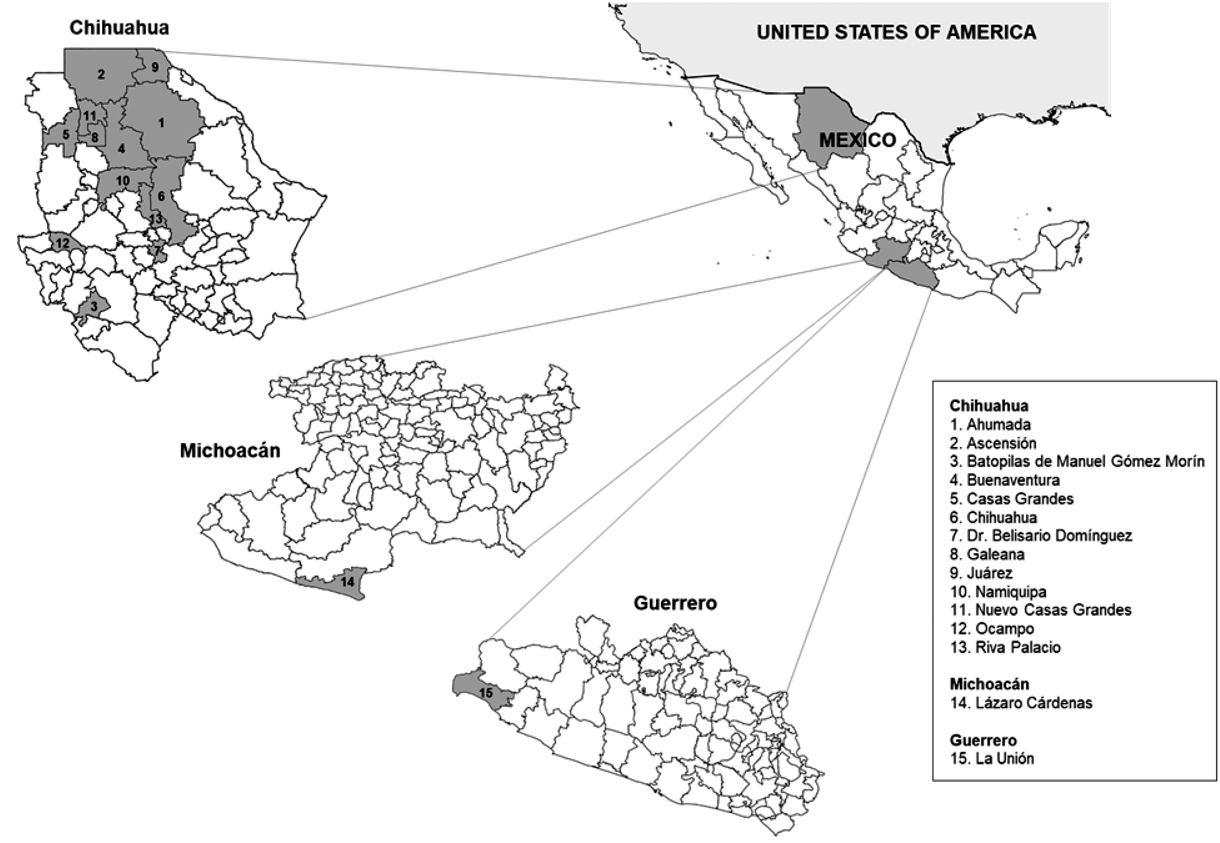
Geographic location of the study sites. Municipalities in Chihuahua, Guerrero, and Michoacán from where animals were sampled are shaded.
Sera were collected from 856 vertebrate animals of 16 species and 1 nothospecies and they are as follows: cat (Felis silvestris catus), cow (Bos taurus), dog (Canis lupus familiaris), domestic goose (Anser anser domesticus), goat (Capra aegagrus hircus), golden pheasant (Chrysolophus pictus), helmeted guinea fowl (Numida meleagris), horse (Equus ferus caballus), hybrid duck (interspecies breeding between Anas platyrhynchos and Anser anser), Indian peafowl (Pavo cristatus), mallard duck (A. platyrhynchos), pig (Sus scrofa domesticus), pigeon (Columba livia), sheep (Ovis aries), silver pheasant (Lophura nycthemera), white-sided jackrabbit (Lepus callotis), and wild turkey (Meleagris gallopavo) (Table 1). All of the cats, cows, dogs, goats, horses, pigs, rabbits, and sheep were from a convenience selection of ranches, farms and houses. All ducks, pigeons and geese were from the Parque Central Poniente Hermanos Escobar, Ciudad Juárez (Central Park, Juárez City). All peafowl, pheasants, guinea fowl and turkeys were from the Recreativo Zoológico San Jorge, Ciudad Juárez (St. Jorge Recreational Zoo, Juárez City). All animals were asymptomatic at the time of sampling, except for one horse in Ciudad Juárez (Juárez City) with ataxia and vision loss that eventually recovered. None of the animals had ever left the state or been vaccinated against WNV. Sera were collected from animals with the approval of the Animal Ethics Committees from each institution that participated in the study (Log No. A3236-01, 11-14-7897-G, 19-072, and 18-173).
Number of Vertebrate Animals in the Study Population, Mexico, 2018–2019
Hybrid ducks were not included in the species total.
Cell culture and viruses
African Green Monkey kidney (Vero) cells were cultured at 37°C with 5% CO2 in Dulbecco's modified Eagle's medium (Thermo Fisher Scientific, Waltham, MA) supplemented with 10% fetal bovine serum, 2 mM
Plaque reduction neutralization test for flaviviruses
Plaque reduction neutralization tests (PRNTs) were performed in six-well plates containing confluent monolayers of Vero cells following standard methods (Beaty et al. 1995). Initially, all sera were screened at a single dilution of 1:20 using WNV. The initial screen was performed using WNV because it is the most widespread flavivirus in North America. These assays are not WNV specific; flaviviruses are antigenically similar and therefore, antibodies to other flaviviruses are also detected (Calisher et al. 1989). All sera with antibodies that neutralized WNV were further diluted and tested by comparative PRNT using DENV2, SLEV, WNV, and ZIKV to identify the flavivirus responsible for these infections. Titers were expressed as the reciprocal of highest serum dilutions yielding ≥90% reduction in the number of plaques (PRNT90). For etiologic diagnosis, the PRNT90 antibody titer to the flavivirus was required to be at least fourfold greater than that to the other flaviviruses tested. If neutralizing antibodies were detected, but there was not at least a fourfold difference in PRNT90 antibody titers, the animal was considered to have antibodies to an undetermined flavivirus.
PRNTs for BUN serogroup viruses
PRNTs were performed in six-well plates containing confluent monolayers of Vero cells (Beaty et al. 1995). Initially, all sera were screened at a single dilution of 1:20 using CVV, the most prevalent and widespread BUN serogroup virus in North America. These assays are not CVV specific; BUN serogroup viruses are antigenically similar and therefore, antibodies to other BUN serogroup viruses are also detected (Hunt and Calisher 1979). All sera with antibodies that neutralized CVV were further diluted and tested by PRNT using CVV, LOKV, MDV, NORV, POTV, and TENV. For etiologic diagnosis, the PRNT90 antibody titer to the BUN serogroup virus was required to be at least fourfold greater than that to the other BUN serogroup viruses tested. If neutralizing antibodies were detected but there was not at least a fourfold difference in PRNT90 antibody titers, the animal was considered to have antibodies to an undetermined BUN serogroup virus.
RNA extraction and RT-PCR
Total RNA was extracted from the serum of the symptomatic horse using the QIAamp Viral RNA Mini Kit (Qiagen) according to the manufacturer's instructions and tested by RT-PCR using flavivirus-specific primers (Kuno et al. 1998). The IRB at each participating university waived the requirement that signatures be acquired from the owners and caregivers of animals.
Results
Sera were collected from 856 vertebrate animals of 16 species and 1 nothospecies in Mexico in 2018–2019 (Table 1). The majority (82.3%) of the animals were from Chihuahua, with the remainder from Michoacán (14.6%) and Guerrero (3.2%). The most commonly sampled animals were dogs (37.7%), horses (26.0%), and cows (14.1%). The municipality that comprised most (65.3%) of the sample population was Juárez in Chihuahua with 559 animals, followed by Lázaro Cárdenas in Michoacán (125 animals; 14.6%) and Buenaventura in Chihuahua (74 animals; 8.6%). All of the sheep, 112 of the 197 (56.9%) horses and 44 of the 117 (37.6%) cows were from the municipality of Juárez.
Sera were tested at a dilution of 1:20 by PRNT using WNV. Antibodies that neutralized WNV were detected in 296 animals: 19 (15.7%) cows, 80 (24.8%) dogs, 12 (27.3%) domestic geese, 1 (50.0%) goat, 1 (100%) golden pheasant, 1 (16.7%) helmeted guinea fowl, 153 (68.6%) horses, 5 (16.1%) hybrid ducks produced from crossbreeding, 12 (85.7%) Indian peafowl, 3 (25.0%) mallard ducks, 1 (11.1%) pigeon, 4 (11.4%) sheep, 1 (100%) silver pheasant, 1 (25.0%) white-sided jackrabbit, and 2 (100%) wild turkeys. All sera that neutralized WNV in the initial screen were further diluted and tested by PRNT using DENV2, SLEV, WNV, and ZIKV. The comparative PRNTs revealed that 154 animals were seropositive for WNV, 7 animals were seropositive for SLEV, and 135 animals were seropositive for an undetermined flavivirus. Antibodies to WNV were detected in animals from nine species and the crossbreed (Table 2). Of the species/crossbreed where at least 10 individuals were sampled, seroprevalence for WNV was highest for horses (39.9%), Indian peafowl (21.4%), and hybrid ducks (16.1%). One horse seropositive for WNV developed symptoms several days before serum collection. Serum of the symptomatic horse was tested for flavivirus RNA by RT-PCR and shown to be negative. WNV activity was detected in all three states. Antibodies to SLEV were detected in five species (Table 3). Of the species where at least 10 individuals were sampled, seroprevalence for SLEV was highest for domestic ducks (2.6%). SLEV activity was detected in Chihuahua and Michoacán. Antibodies to DENV2 and ZIKV were not detected in any animals.
Vertebrate Animals Seropositive for West Nile Virus in Mexico, 2018–2019
Animals are not listed if none was seropositive.
Vertebrate Animals Seropositive for St. Louis Encephalitis Virus in Mexico, 2018–2019
Animals are not listed if none was seropositive.
Sera were tested at a dilution of 1:20 by PRNT using CVV. Antibodies that neutralized CVV were detected in 42 animals: 2 (1.7%) cows, 5 (1.5%) dogs, 33 (14.2%) horses, 1 (2.9%) sheep, and 1 (25%) white-sided jackrabbit. All sera that neutralized CVV were further diluted and tested by PRNT using CVV, LOKV, MDV, NORV, POTV, and TENV. The comparative PRNTs revealed that 22 animals were seropositive for LOKV, 3 animals were seropositive for MDV, and 17 animals were seropositive for an undetermined BUN serogroup virus. Antibodies to LOKV were detected in animals from three species (Table 4). Seroprevalence for LOKV was highest for horses (8.5%) and cows (1.7%). LOKV activity was detected in all three states. All three animals seropositive for MDV were horses in Chihuahua. The overall seroprevalence for MDV in horses was 1.3%. Antibodies to CVV, NORV, POTV, and TENV were not detected in any animals.
Vertebrate Animals Seropositive for Lokern Virus in Mexico, 2018–2019
Animals are not listed if none was seropositive.
Twenty-five animals were seropositive for two viruses. Two horses from Chihuahua were seropositive for MDV and WNV, a horse from Guerrero was seropositive for LOKV and SLEV, 11 horses (4 from Chihuahua, 2 from Guerrero, and 5 from Michoacán) were seropositive for LOKV and WNV, and a cow from Michoacán was seropositive for LOKV and WNV. Five animals (four horses and one sheep) were seropositive for WNV and an undetermined BUN serogroup virus and five other animals (three dogs, one horse, and one rabbit) were seropositive for an undetermined BUN serogroup virus and an undetermined flavivirus.
Discussion
We provide serologic evidence of LOKV, MDV, SLEV, and WNV infections in vertebrate animals in Mexico. LOKV and MDV have never before been reported outside of the United States. LOKV has been isolated from arthropods and vertebrate animals in five states of the United States, including three (California, New Mexico, and Texas) that border Mexico (Crane et al. 1983, Calisher et al. 1986, Kramer et al. 1990). Isolations of MDV have been made from arthropods and vertebrate animals in six states, including three (Arizona, California, and Texas) than border Mexico (Calisher et al. 1986). Serological data suggest that LOKV and MDV also occur elsewhere in the United States (Sahu et al. 2002, Johnson et al. 2014, Meyers et al. 2015). In this study, we provide evidence that the geographic distributions of LOKV and MDV extend beyond the United States. The detection of LOKV activity as far north as Chihuahua and as far south as Guerrero indicates that this virus is widely distributed across Mexico.
All viruses in the BUN serogroup have tripartite, single-stranded, negative-sense RNA genomes and the three genome segments are designated as small, (S), medium (M), and large (L) (Elliott et al. 1991, Horne and Vanlandingham 2014). The viral envelope glycoproteins are encoded by the M segment. Recent evidence suggests that LOKV is a reassortant that acquired its S and L segments from MDV and its M segment from an undiscovered, possibility extinct, virus (Tangudu et al. 2018). Because LOKV and its apparent unidentified donor share the same M segment, antibodies to these viruses cannot be differentiated by PRNT. Therefore, if the M segment donor still occurs in nature, some or all of the animals considered seropositive for LOKV could have instead been infected with the undiscovered donor virus.
CVV is the most common and widely distributed BUN serogroup virus in North America (Calisher et al. 1986, Waddell et al. 2019). CVV is also the most important BUN serogroup virus in North America in terms of its impact on human and veterinary animal health, having been associated with outbreaks of pregnancy loss and congenital malformations in sheep, in addition to six cases of severe disease in humans (Sexton et al. 1997, Campbell et al. 2006, Nguyen et al. 2013, Wilson et al. 2017, Yang et al. 2018b, Baker et al. 2021). None of the animals in our study was seropositive for CVV and the overall seroprevalence for BUN serogroup viruses was lower than other studies performed in North America. For example, antibodies to BUN serogroup viruses were detected in 169 (92.9%) horses and 29 (93.5%) sheep in Quintana Roo and Yucatán, Mexico in 2007–2008, with CVV identified as one of the most common causes of infection (Blitvich et al. 2012b). In another study, 1455 (28.3%) sheep across the United States in 2011 contained antibodies to BUN serogroup viruses (Meyers et al. 2015). CVV was the most common virus. Sera from 84 (64.6%) sheep, 40 (69.0%) horses, 8 (33.3%) goats, and 5 (20.0%) cows in Saskatchewan, Canada in 2013–2014 also contained antibodies to BUN serogroup viruses (Uehlinger et al. 2018).
One explanation for the relatively low seroprevalence for BUN serogroup viruses and apparent lack of CVV activity in our study is because competent reservoir hosts or vectors of CVV do not occur in the study area. White-tailed deer have been implicated as the principal reservoir hosts of CVV based on experimental infection data and seropositivity rates from field studies (Campbell et al. 1989, Neitzel and Grimstad 1991, Blackmore and Grimstad 1998, Dupuis et al. 2020). White-tailed deer are common in Mexico, although they are not found in parts of northern Chihuahua (Mandujano et al. 2010, Hewitt 2011). The majority (65.3%) of animals in our study population were from the municipality of Juárez in northern Chihuahua, including all sheep and many of the horses and cows. Experimental infections have revealed that mosquitoes of at least 10 species are competent vectors of CVV, including some that occur in the study area, and virus isolations have been made from several additional species (Yuill and Thompson 1970, Calisher et al. 1986, Blackmore et al. 1998, Andreadis et al. 2014, Armstrong et al. 2017, Anderson et al. 2018, Ayers et al. 2018, 2019, Yang et al. 2018a, Chan et al. 2020).
Many WNV and SLEV serological investigations were performed in Mexico shortly after the arrival of WNV into the Americas (Blitvich 2008, Elizondo-Quiroga and Elizondo-Quiroga 2013). However, relatively few have been performed in recent years. Four of 78 (5.1%) humans in Ciudad Juárez (Juárez City), Chihuahua, in 2015 were seropositive for WNV (Palermo et al. 2019). SLEV testing was not performed. Antibodies to SLEV and WNV were not detected in any of the 639 humans in Guerrero in 2019 (Nunez-Avellaneda et al. 2021). There are no other recent studies where humans or vertebrate animals in Chihuahua, Guerrero, or Michoacán were tested for evidence of SLEV or WNV infection. In this study, we provide evidence of WNV activity in all three states and SLEV activity in Chihuahua and Michoacán. Of the 10 arboviruses included in our study, WNV had the broadest host range, with antibodies to the virus detected in animals of nine species. It is well documented that WNV infects vertebrate animals of numerous species (Root 2013, Root and Bosco-Lauth 2019, Habarugira et al. 2020).
Conclusion
We provide serologic evidence of LOKV, MDV, SLEV, and WNV infections in vertebrate animals in Mexico. LOKV and MDV have not been previously reported in Mexico and there are no recent reports of SLEV and WNV activity in Guerrero and Michoacán. This study increases our understanding of the geographic distribution, host range, and seroprevalence of BUN serogroup viruses and flaviviruses in North America.
Footnotes
Acknowledgments
The authors thank the animal owners who allowed us to collect sera from their animals. The authors also thank all of the veterinarians who assisted in this study, particularly Alfredo López Álvarez, Jaime Orozco Hernández, and Eunice Fabiola Cuesta Reyes. They are grateful for the assistance provided by Margarita Edith Peña Pérez from the Department of Ecology in Ciudad Juárez, Rodrigo Piñón Bencomo, Wenceslao Peñaloza Luna, Felix Alfonso Vega Rodríguez, and Delia Ignacia Durán Moreno, in addition to the staff at the San Jorge Recreational Zoo in Ciudad Juárez.
Author Disclosure Statement
No conflicting financial interests exist.
Funding Information
This study was supported by a postdoctoral scholarship from the Consejo Nacional de Ciencia y Tecnología of Mexico (scholarship No. 740742), intramural funds from the College of Veterinary Medicine at Iowa State University and grants from the Programa para el Desarrollo Profesional Docente, para el Tipo Superior (UACJ-PTC-399 and UACJ-PTC-267).