
Abstract
Dogs are asymptomatic chronic carriers of Leptospira spp. and excrete these bacteria in their urine, resulting in environmental contamination and potentially leading to zoonotic transmission. Although a previous study in Sri Lanka detected anti-Leptospira antibodies in companion dogs, the urinary shedding of Leptospira spp. and the Leptospira species and serogroups prevalent in them remain unclear. Thus, the current study identified the prevalent Leptospira serogroups and the carrier status of Leptospira spp. in apparently healthy, client-owned dogs in the Kandy District of Sri Lanka. Serum and urine samples were collected from 96 unvaccinated and 82 vaccinated dogs. Anti-Leptospira antibodies and Leptospira DNA in urine were detected using the microscopic agglutination test (MAT) and nested PCR that targeted the pathogenic leptospiral gene, flaB. The flaB sequences were compared with those of Leptospira spp. using the public databases. MAT detected anti-leptospiral antibodies in 15.6% (15/96) of the unvaccinated dogs, and the reactive serogroups were observed to be Sejroe (11.5%), Canicola (2.1%), Icterohemorrhagiae (1.0%), and Javanica (1.0%). Furthermore, MAT results revealed that 11.0% (9/82) of the vaccinated dogs tested positive for the anti leptospira antibodies and the only reactive serogroup was Sejroe. Leptospira DNA was detected in 15.6% (15/96) and 15.9% (13/82) of urine samples collected from unvaccinated and vaccinated dogs, respectively, and phylogenetic analysis revealed that the animals were infected with L. borgpetersenii, L. interrogans, L. kmetyi, and L. weilii. The L. interrogans sequence detected in the canine sample was identical to the one that was previously reported in a human sample from the Kandy District. This study demonstrated that both unvaccinated and vaccinated dogs excrete various pathogenic Leptospira spp. in their urine, suggesting that they may play an important role in environmental contamination that poses a health risk to the dog owners and the general public.
Introduction
The proximity of human and canine population has had a great influence on the transmission of zoonotic diseases, including rabies, tuberculosis, and Campylobacter infections (Une and Mori 2007, Damborg et al. 2008, Wang et al. 2013). Similarly, there is a high likelihood of the leptospirosis transmission to humans via chronic carrier dogs (Haunz and Cardy 1952, Misao et al. 1956, Ward et al. 1956, Barkin and Glosser 1973, Feigin et al. 1973).
Dogs are known to be maintenance hosts for Leptospira interrogans serovar Canicola and incidental hosts for other Leptospira serovars (Klarenbeek and Schuffner 1933, Bharti et al. 2003, Greene et al. 2008). Although chronically infected dogs appear healthy, they intermittently excrete Leptospira spp. in their urine (Harkin et al. 2003, Rojas et al. 2010, Zakeri et al. 2010, Oliveira et al. 2012, Gay et al. 2014, Llewellyn et al. 2016, Kurilung et al. 2017, Goy-Thollot et al. 2018, Miotto et al. 2018a, Zaidi et al. 2018).
Leptospirosis is transmitted through direct contact with urine of infected animals or indirect contact with the environment contaminated with urine. Therefore, dogs infected chronically with Leptospira spp. are considered a potential source of infection to humans globally (Trevejo et al. 1998, Batista et al. 2004, Barmettler et al. 2011, Guernier et al. 2016, Biscornet et al. 2017, Koizumi et al. 2020, Noda et al. 2020).
Leptospirosis is an endemic disease in Sri Lanka, and over 3000 human leptospirosis cases are reported annually. However, most of these cases are reported on the basis of clinical diagnoses (Epidemiology Unit Sri Lanka 2019). Although environmental factors and animal infestation in human habitats are crucial for the transmission of leptospirosis to humans (Haunz and Cardy 1952, Barkin and Glosser 1973, Feigin et al. 1973), the disease transmission dynamics between humans and animals in this country remains unclear.
Several studies have demonstrated the urinary shedding of Leptospira spp. in domestic cattle, wildlife, and peridomestic rats in Sri Lanka (Gamage et al. 2011, 2014, Athapattu et al. 2019). L. borgpetersenii, L. interrogans, and L. kirschneri DNA have been detected in 1.9–6.1% of cattle urine or kidney samples (Gamage et al. 2011, 2014). L. borgpetersenii, L. interrogans, and L. weilii have also been detected in human patients (Karunanayake et al. 2020, Jayasundara et al. 2021). These studies suggest that rats and cattle may act as sources of infection in humans owing to the close interaction as a result of sharing the same habitat.
Dogs are the most popular companion animals in Sri Lanka. Although a reliable estimate of the canine population in the country is not available, 1,240,366 dogs have been vaccinated against rabies by the Sri Lankan government free of charge, of which 211,503 were vaccinated in the Central Province, according to the rabies control program conducted in 2018 (Department of Animal Production and Health 2018). However, publicly available records on the number of dogs vaccinated against leptospirosis in Sri Lanka were not found. Although companion dogs may play a role in the transmission of leptospirosis to humans, only one study has investigated anti-Leptospira antibodies in dogs residing in the suburbs of the capital city of Colombo (Thammitiyagodage et al. 2017).
Data on the frequency of Leptospira infection in animals that are potential maintenance hosts could be used to further understand disease transmission and risk factors as well as develop novel treatment protocols and preventive measures for leptospirosis. This study investigated the status of Leptospira infection in apparently healthy companion dogs in the Kandy District by analyzing anti-Leptospira antibodies and Leptospira DNA in urine samples using the MAT and a nested PCR followed by DNA sequencing, respectively, to characterize the risks to public health.
Materials and Methods
Sample collection
Sample collection was conducted at the Government Veterinary Hospital, Gatambe, Peradeniya, Sri Lanka (Fig. 1), which is one of the largest small-animal hospitals in the Kandy District where more than 100 dogs are brought daily to the hospital for various treatments. Animals were selected (every fifth animal attending the hospital on a particular day) and subjected to a complete general clinical examination by a qualified veterinarian, and the vaccination status, including date and the name of the vaccine received as well as client details, age, and sex of the animals, was recorded.
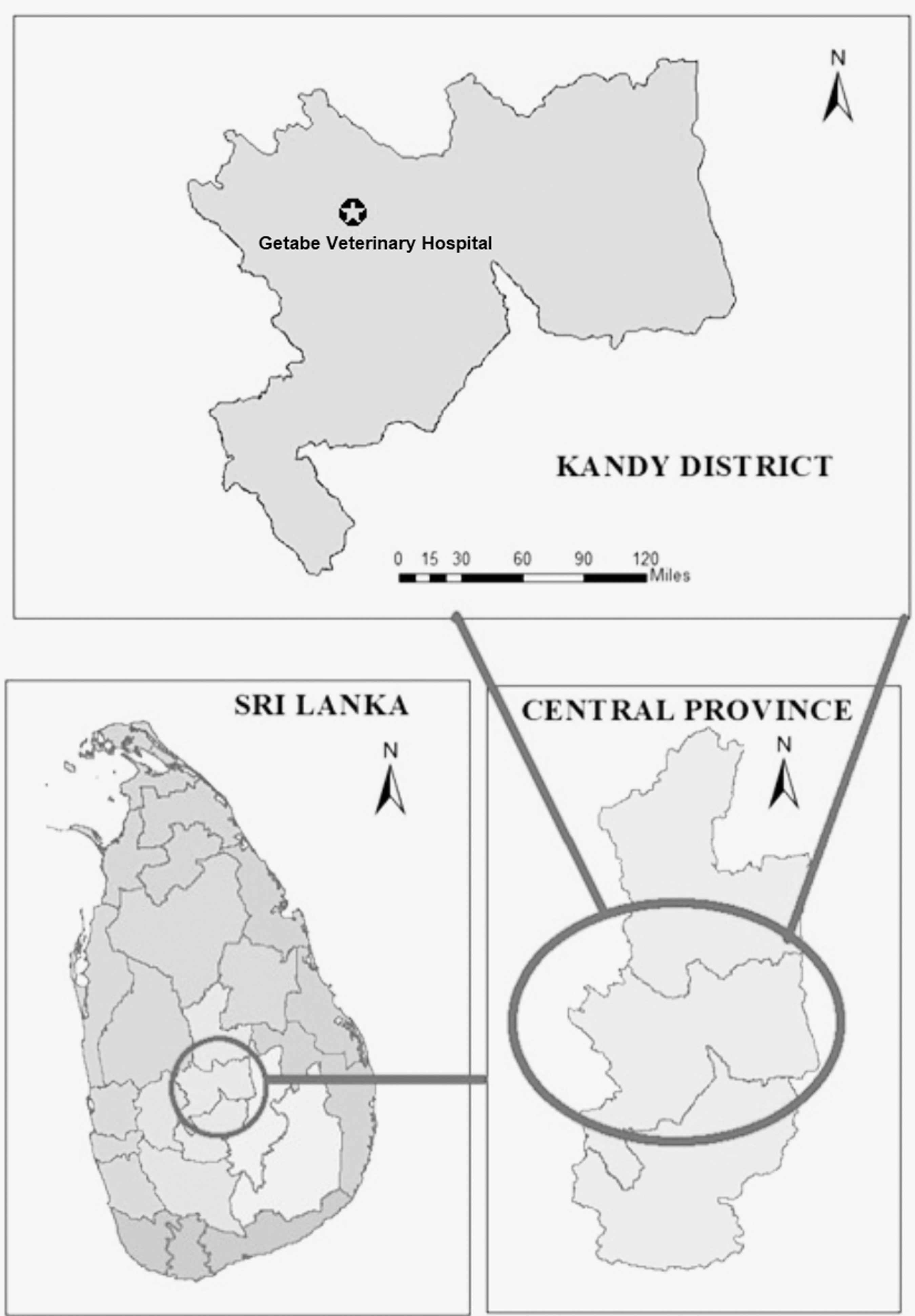
A map of the Kandy District (study area). Dogs that lived in the Kandy District were included in this study.
The animals recruited in this study were classified into vaccinated and unvaccinated groups. Dogs belonging to the vaccinated group underwent complete inoculation of the distemper/hepatitis/leptospirosis (DHL) or distemper/hepatitis/parvovirus infection/leptospirosis (DHPL) vaccines within 1 year, according to the standard vaccination schedule practice before sample collection. Dogs in the unvaccinated group did not receive either DHL or DHPL vaccines before sample collection. In total, 178 apparently healthy dogs were enrolled in this study; blood samples (2 mL) were collected from their cephalic veins and urine samples (10 mL) were obtained by catheterization.
All collected samples were stored at 4°C until they were transported to the University of Peradeniya for processing and further analysis. Blood samples were centrifuged at 3000 × g for 10 min, and the serum was separated and stored at −20°C until the serological test was conducted. Urine samples were centrifuged at 3000 × g for 10 min to remove debris, such as pus cells; 6 mL of the supernatant was further centrifuged at 15,000 × g for 20 min, and the resulting sediments were stored at −20°C until DNA was extracted.
Microscopic agglutination test
To detect the anti-Leptospira antibody, qualitative MAT was conducted using live leptospiral cultures having 13 strains, belonging to the 12 serogroups (Autumnalis, Bataviae, Canicola, Grippotyphosa, Hebdomadis, Icterohemorrhagiae, Javanica, Panama, Pyrogenes, Sejroe, Shermanii, and Tarassovi) according to the World Health Organization guidelines (World Health Organization 2003).
Fifty microliters of the serum sample diluted at 1:50 with phosphate-buffered saline (PBS) was mixed with 50 μL of each culture and incubated at 30°C for 90 min, and the samples with 50% reduction of free cells in the supernatant of the reaction mixture compared with the control (PBS alone) were considered to have a positive result. The positive samples were subjected to quantitative MAT, in which serum samples were serially diluted from 1:100 to 1:6400, and the Leptospira serogroup that tested positive at the highest dilution was considered a positive serogroup. The cutoff titer for the serogroups included in the vaccines was 1:800 and that for serogroups not included in the vaccines was 1:100.
Polymerase chain reaction
DNA was extracted from urine samples using the DNeasy Blood and Tissue Kit (Qiagen, Germany) according to the manufacturer's protocol. Pathogenic Leptospira DNA was detected using a nested PCR targeting flaB, as previously described (Koizumi et al. 2008). For flaB-positive samples, PCR targeting secY was performed as previously described (Gravekamp et al. 1993). PCR amplicons were sequenced by the dideoxynucleotide chain termination method using the BigDye Terminator v 3.1 Cycle Sequencing Kit (Applied Biosystems). The determined sequences were aligned in molecular evolutionary genetics analysis -X software (MEGA-X) (Kumar et al. 2018) using CLUSTALW, and the phylogenetic distance was calculated in MEGA-X using the maximum likelihood method and the Tamura-Nei model (Tamura and Nei 1993).
A phylogenetic tree was created using the flaB sequences obtained in this study (DNA Data Bank of Japan [DDBJ] acc. nos. LC636141–LC636162), those from previously published studies from Kandy, Sri Lanka (Koizumi et al. 2009, Nwafor-Okoli et al. 2012), and those from the public databases. A phylogenetic tree was also created based on the secY sequences obtained in this study (DDBJ acc. nos. LC659313–LC659322) and those from the public databases.
Statistical analysis
Associations of vaccination status with evidence of natural infection and urinary excretion of Leptospira spp. were analyzed using χ 2 test.
Ethical statement
This study was approved by the Animal Ethics Review Committee of the Faculty of Veterinary Medicine and Animal Science, University of Peradeniya, Sri Lanka, under the certificate number VER-2018-003.
Results
The median age of the unvaccinated and vaccinated dogs was 2 years (range: 6 months–15 years) and 2.5 years (range: 6 months–15 years), respectively. The residences of most of the dogs included in this study were within a 30-km radius of the Government Veterinary Hospital, Gatambe, Peradeniya, Sri Lanka (Fig. 1). Although most of the clients allowed their dogs to go outside, we could not obtain information for all of them.
Of the 96 serum samples obtained from unvaccinated dogs, MAT results of 15 (15.6%) of them were positive for the Leptospira spp. and the most probable infecting serogroups were Sejroe (11.4%), Canicola (2.0%), Icterohemorrhagiae (1.0%), and Javanica (1.0%). Of the 82 serum samples obtained from vaccinated dogs, MAT results of 9 (11.0%) dogs revealed positive for Leptospira spp. and reacted only with the Sejroe serogroup (Tables 1 and 2).
Anti-Leptospira Antibodies in Serum Samples from Unvaccinated Dogs
MAT, microscopic agglutination test.
Anti-Leptospira Antibodies in Serum Samples from Vaccinated Dogs
PCR results revealed that 15 of the total 96 (15.6%) urine samples from unvaccinated dogs and 13 of the total 82 (15.9%) urine samples from vaccinated dogs confirmed the urinary shedding of pathogenic Leptospira spp. A phylogenetic tree constructed based on flaB sequences revealed that PCR-positive dogs were infected with L. borgpetersenii, L. interrogans, L. kmetyi, and L. weilii (Fig. 2). Moreover, the secY gene was amplified from 10 of the total 28 flaB-positive samples (Fig. 3).
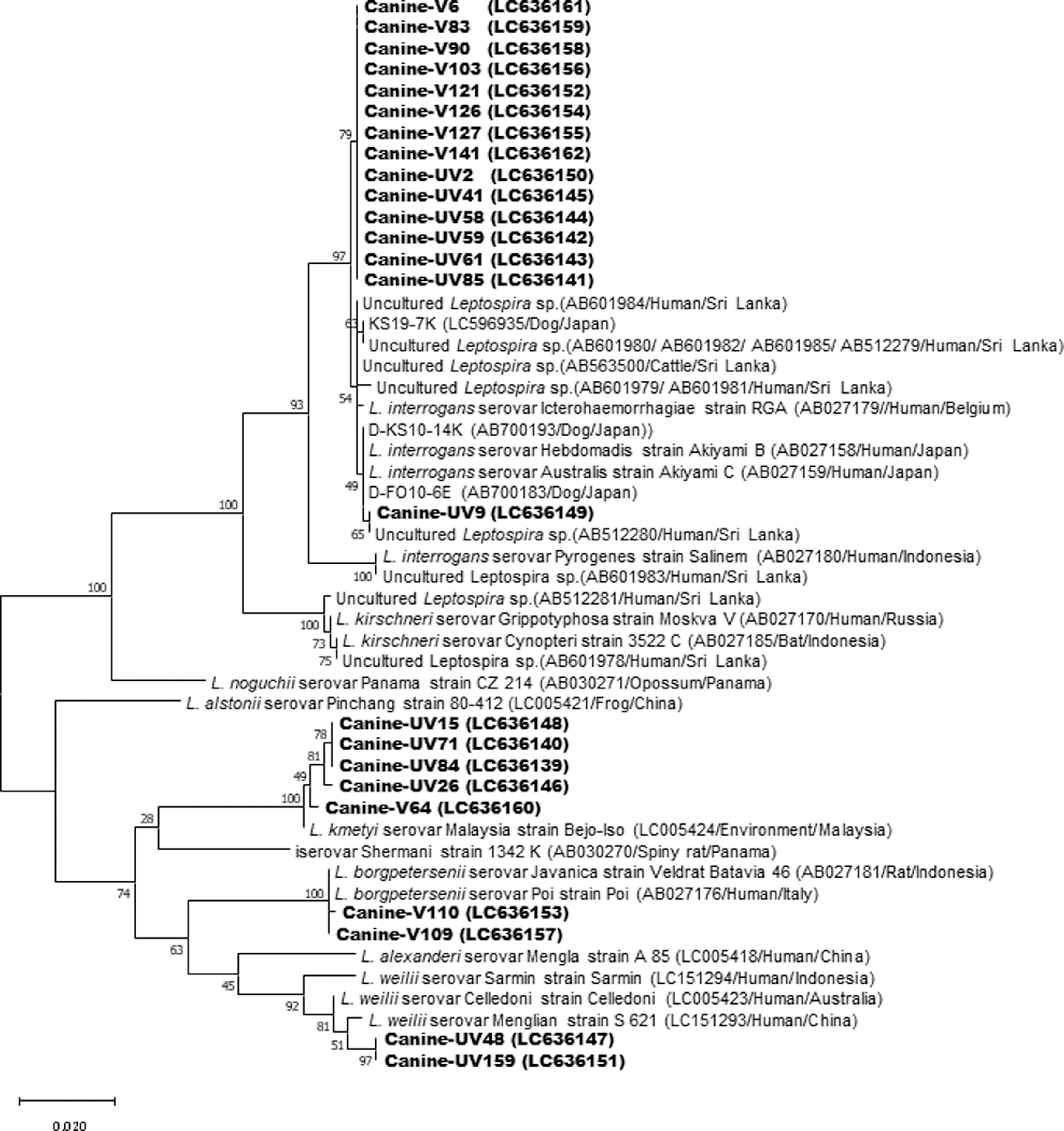
Phylogenetic tree based on flaB sequences of Leptospira spp. Sequences obtained from dogs in this study have been deposited in DDBJ (acc. nos. LC636149–LC636162) and highlighted in bold. Sequences were aligned in MEGA-X using CLUSTALW, and phylogenetic distances were calculated using the maximum likelihood method with 500 bootstrap replicates. Accession number/animal/country from which the strain was isolated indicated in parenthesis. DDBJ, DNA Data Bank of Japan; MEGA-X, molecular evolutionary genetics analysis -X software.

Phylogenetic tree based on secY sequences of Leptospira spp. Sequences obtained from dogs in this study have been deposited in DDBJ (acc. nos. LC659313–LC659322) and highlighted in bold. Sequences were aligned in MEGA-X using CLUSTALW, and phylogenetic distances were calculated using the maximum likelihood method with 500 bootstrap replicates. Accession number/animal/country from which the strain was isolated indicated in parenthesis.
A significant association was not observed between vaccination status and evidence of natural infection (increase in MAT titers against Leptospira serogroups that were not included in the vaccine) (χ 2 = 0.764, p = 0.382) or urinary excretion of Leptospira spp. (χ 2 = 0.002, p = 0.967).
Discussion
This study demonstrated urinary shedding of leptospires in apparently healthy canines in Sri Lanka for the first time, which was detected in ∼16% of dogs, regardless of their vaccination status (Fig. 2). The urinary shedding rate of asymptomatic dogs in this country is comparable with that in the other countries. It has been reported in several countries that the frequency of renal carriage of Leptospira spp. in apparently healthy dogs is 1.5–28.6% based on the molecular methods (Harkin et al. 2003, Rojas et al. 2010, Zakeri et al. 2010, Oliveira et al. 2012, Gay et al. 2014, Llewellyn et al. 2016, Kurilung et al. 2017, Goy-Thollot et al. 2018, Miotto et al. 2018a, Zaidi et al. 2018).
Since chronic carriers intermittently shed leptospires in the urine and their number varies from animal to animal (Harkin et al. 2003, Zaidi et al. 2018), the true prevalence of carriers may be higher than that reported in this study. Moreover, canine urine is acidic, and leptospires can survive for a limited time (Balows et al. 2012). Therefore, to minimize false-negative PCR results, urine samples having maximum volume should be collected over a period of three consecutive days and processed soon after collection (Xu et al. 2014).
This study indicated that the vaccines currently available in Sri Lanka have a limited effect on the prevention of urinary shedding, probably owing to the incompatibility between the vaccine leptospiral serovars and those circulating in the country. Furthermore, none of the commercially available vaccines provides complete protection against renal colonization (André-Fontaine et al., 2003, Klaasen et al. 2003).
Phylogenetic analysis revealed that dogs were chronically infected with four Leptospira species (Figs. 2 and 3). The L. interrogans and L. weilii secY sequences were identical to those of L. interrogans serovar Hardjo from a human patient in Indonesia and L. weilii serovar Coxi from a human patient in Malaysia, respectively (Fig. 3). Detection of L. borgpetersenii, L. interrogans, and L. weilii in dog urine has been reported in several countries, including Algeria, Brazil, Iran, France, Germany, New Caledonia, and Thailand (Zakeri et al. 2010, Gay et al. 2014, Llewellyn et al. 2016, Kurilung et al. 2017, b, Zaidi et al. 2018, Goy-Thollot et al. 2018, Miotto et al. 2018a). L. borgpetersenii, L. interrogans, and L. weilii have also been detected in humans and other animals (Gamage et al. 2011, 2014, Karunanayake et al. 2020, Jayasundara et al. 2021).
Although L. kmetyi is classified as a pathogenic species, it has been commonly detected in environmental samples (Ali et al. 2018). One study conducted in the French West Indies detected L. kmetyi species in the blood samples of two human patients (Bourhy et al. 2013); however, none of the previous studies in Sri Lanka detected L. kmetyi in human or animal samples. Although sequence similarities (and identities) of each Leptospira species were found, further prospective studies are required to verify whether dogs are maintenance hosts of these strains.
The L. interrogans sequence in a canine sample obtained in this study was identical to that previously reported in a human sample obtained from the Kandy District (Koizumi et al. 2009). Further isolation of the L. interrogans strain from dogs and humans and detailed molecular comparisons of the isolates along with an investigation of other potential reservoir animals will reveal the genetic relationship between isolates and provide insights into the maintenance hosts of this strain.
The MAT results revealed that 15.6% of the unvaccinated dogs had antibodies against serogroups Sejroe, Canicola, Icterohemorrhagiae, and Javanica (Table 1), and 11.0% of the vaccinated dogs had antibodies against the serogroup Sejroe; however, none of the samples tested positive for serogroups Canicola and Icterohemorrhagiae, which are contained in the leptospirosis vaccine, used in this country. Previous studies have indicated that leptospirosis vaccines resulted in negative or low antibody titers, but conferred a protective effect against experimental challenges (Klaasen et al. 2003, Miller et al. 2011). Although vaccination protects against infection with the homologous serovars included in the vaccine, dogs can become infected with other serovars (Klaasen et al. 2014).
Serogroup Sejroe was predominantly reactive in both groups of dogs tested in this study. Anti-serogroup antibodies have been detected in both symptomatic and asymptomatic dogs (Scanziani et al. 1995, Miotto et al. 2018b). In addition, serogroup Sejroe strains have been isolated from asymptomatic dogs (Scanziani et al. 1995, Miotto et al. 2018a). Previous studies have shown that antibodies against serogroup Sejroe were detected in samples from cattle and human patients in Kandy (Agampodi et al. 2008, Koizumi et al. 2009, Gamage et al. 2011).
Together, these studies provide a clear picture of environmental contamination with serogroup Sejroe in the Kandy District, for which cattle are generally considered maintenance hosts. Although a small number of households in the Kandy District have small-scale cattle farms, dogs have a higher likelihood of encountering chronically infected cattle because most dogs included in this study were allowed to roam freely.
In conclusion, this study demonstrates that dogs can be naturally infected with various pathogenic Leptospira spp. and shed the bacteria in their urine despite being properly vaccinated against leptospirosis. These facts suggest that dogs may play an important role in environmental contamination by Leptospira spp., which poses a major risk to public health. The isolation and serological typing of Leptospira spp. excreted by dogs are crucial for selecting vaccine candidates for Sri Lankan dogs. Clinically healthy dogs should be screened annually to detect exposure to leptospires and the carrier stage by using molecular methods, and carriers should be treated to control both human and animal leptospirosis in Sri Lanka.
Footnotes
Acknowledgments
We thank all staff members of the Government Veterinary Hospital, laboratory staff members of the Department of Microbiology and Department of Veterinary Public Health and Pharmacology, University of Peradeniya, and staff members of the Microbiology division of the Veterinary Research Institute for their contributions to this study.
Authors' Contributions
Conceptualization: C.G., R.F., N.K., and T.A.; project administration: C.G.; methodology: T.A., P.F., N.K., and C.G.; formal analysis: T.A., N.K., and C.G.; data curation: R.A. and N.S.; validation: N.K.; resources: R.A., M.F., N.S., and C.G.; supervision: R.F., P.F., M.F., N.K., and C.G.; funding acquisition: R.F. and N.K.; writing—original draft preparation: T.A., N.K., and C.G.; writing—review and editing: T.A., R.F., R.A., P.F., M.F., N.S., N.K., and C.G.
Author Disclosure Statement
No conflicting financial interests exist.
Funding Information
This work was supported by the Research Program on Emerging and Re-emerging Infectious Diseases (JP21fk0108139) from the Japan Agency for Medical Research and Development (AMED) (N.K.) and Innovation Commercialization Enhancement grant 6026-LK/8743-LK under the AHEAD project of the Ministry of Higher Education, Technology and Innovation, Sri Lanka (R.F.).