
Abstract
Tick-borne Rickettsia aeschlimannii infection in humans has been described in several countries. This is the first report of R. aeschlimannii in a woman who reported being bitten by ticks in Xingjiang, northwestern China. R. aeschlimannii infection was confirmed by molecular detection in blood and urine of the patient, who presented clinical symptoms of severe edema, partial necrosis, and monocytosis. R. aeschlimannii was also detected in Hyalomma asiaticum ticks around the patient's residence. Infections of spotted fever group Rickettsia species should be included in the differential diagnosis from other tick-borne diseases.
Introduction
Emerging spotted fever rickettsioses, which are transmitted through arthropods and characterized by fever, eschar, headache, malaise, anorexia, nausea, and lymphadenopathy, have become a worldwide health concern (Fang et al. 2015). To date, at least eight spotted fever group (SFG) Rickettsia species, Rickettsia heilongjiangiensis, Rickettsia sibirica, Rickettsia monacensis, Rickettsia raoultii, Candidatus Rickettsia tarasevichiae, Rickettsia xinyangensis, Rickettsia japonica, and Rickettsia sp. XY99, are associated with human disease after tick bites in China (Fang et al. 2015, Liu et al. 2016).
Human infection with Rickettsia aeschlimannii was first reported in Morocco (Beati et al. 1997), and then in Mauritania, Algeria, Tunisia, Libya, Egypt, Greece, and Italy (Demoncheaux et al. 2012). In this study, we report the first case of R. aeschlimannii infection in a woman in Xinjiang Uygur Autonomous Region (XUAR), northwestern China.
Case Description
The patient was a 55-year-old woman who lived in Manas County, XUAR. On September 26, 2019, she noticed a tick embedded on the right lower leg, and removed it herself. A few hours later, the affected area became red, swollen, and painful, with surface blisters slowly appearing afterward. The patient was initially treated with oral amoxicillin (500 mg, thrice daily) for 4 days, but the symptoms persisted. On October 1, she was admitted to the New Lake Regiment Hospital in Mannas County, and a routine examination was conducted (Supplementary Table S1).
Rash, eschar, myalgia, edema, and partial necrosis were found surrounding the tick bite site (Fig. 1). The results of blood cell count and liver enzymes were normal, with the exception of the increased absolute monocyte count (0.65 × 10⁹/L) that accounted for 10.2% among total leukocytes. The patient had no history of familial diseases or infectious diseases. On admission, the patient immediately received debridement due to severe edema on the lower leg. In addition, empirical intravenous cefuroxime sodium (100 mg, thrice daily) and levofloxacin (500 mg, once daily) were administered during October 1–6, however, these treatments did not alleviate her symptoms.
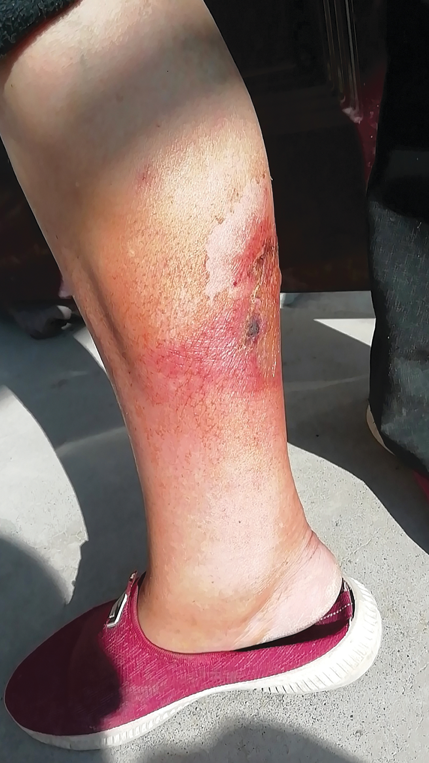
Photo of the tick bite site of the patient at day 5 postonset of illness. Color images are available online.
On October 5, a blood sample was collected to investigate possible pathogens. Both DNA and RNA were extracted, and complementary DNA was synthesized. Known tick-borne pathogens, including Rickettsia spp., Anaplasma spp., Ehrlichia spp., severe fever and thrombocytopenia syndrome virus, Crimean–Congo hemorrhagical fever virus, Tacheng tick virus 1, and Tacheng tick virus 2, were detected using nested PCR or reverse transcription PCR. Rickettsial DNA was detected in both blood and urine through PCR targeting genetic markers of the cell surface antigen 1 (sca1, 553bp) and outer membrane proteins A (ompA, 443 bp), which were further identified as R. aeschlimannii by sequencing.
Subsequently, the patient was treated with doxycycline (100 mg, thrice daily, intravenous injection) during October 7–13, after which the patient's symptoms resolved and she was discharged on October 14. After a 5-month follow-up, the patient recovered with no other complications.
To further investigate the prevalence of rickettsial infection in ticks surrounding the location of the patient's residence, 182 ticks were collected by flagging vegetation, and identified as Hyalomma asiaticum by morphological features and mitochondrial cytochrome c oxidase subunit I gene. PCR showed that 13 of these tick samples (7.14%) were rickettsia positive, in which 1 was confirmed as R. aeschlimannii, 5 were Candidatus Rickettsia barbariae, and the remaining 7 were R. raoulti.
Based on the sca1-ompA concatenated sequences, the results of both BLAST and phylogenic analysis revealed that the rickettsial agent from the patient shared 100% identities to R. aeschlimannii in H. asiaticum sampled at the surrounding of the patient's residence, and was most closely related to R. aeschlimannii strain MC16 (Supplementary Fig. S1).
Nucleotide sequences obtained in the study have been deposited in the GenBank database (ompA: MN794571, MN794572, MW321575, MW321566, MW321578; sca1: MT237577, MT237576, MW321584; and COI: MW498410, MW498406).
Discussion
Ticks infected with R. aeschlimannii have mainly been reported in Africa and Europe (Pretorius and Birtles 2002, Borawski et al. 2019, Getange et al. 2021). In Asia, ticks have been found to be infected with R. aeschlimannii in Turkey, Kazakhstan, China, and Japan (Shpynov et al. 2004, Wei et al. 2015, Keskin et al. 2016, Qiu et al. 2021). However, only a few countries, such as Morocco, Tunisia, Algeria, South Africa, Spain, Turkey, Greece, and Italy, have reported cases of R. aeschlimannii infection (Tosoni et al. 2016).
Clinical signs may include fever, headache, myalgia, vomiting, and eschar or papule at the sites of tick bite. In this study, the patient presented rash, eschar, myalgia, edema, and partial necrosis surrounding the tick bite site. Given obvious edema and partial necrosis on the right lower leg, the patient received debridement. SFG Rickettsia infection has clinical manifestations of fever, facial edema, and fern leaf necrosis of the skin in Sri Lanka (Kumarathunga and Kularatne 2018). In addition, monocytosis has also been described in a patient infected with R. conorii, which was also found in our case report of R. aeschlimannii. Rickettsial agent is usually detected in blood samples.
In this patient, R. aeschlimannii was identified in the urine sample at the early phase of infection. The findings suggest that R. aeschlimannii infection may present with nonspecific clinical syndromes, which is difficult to differentiate from other Rickettsia infections, however, urine testing may prove useful for early diagnosis for these emerging pathogens in endemic areas.
In China, R. aeschlimannii was first reported in Rhipicephalus turanicus in 2015 (Wei et al. 2015). In this study, R. aeschlimannii, R. raoulti, and Candidatus R. barbariae were also detected in H. asiaticum. These results suggest that multiple SFG Ricketsia cocirculate in H. asiaticum as a potential vector in XUAR, northwestern China. To date, at least 19 SFG Rickettsia species have been detected in ticks in China (Liu et al. 2021), with tick-borne diseases highly prevalent in some parts of China, adversely affecting human health (Wu et al. 2013, Wang et al. 2019, 2021, Ma et al. 2021). Testing of SFG Rickettsia should be included as part of all differential diagnoses in symptomatic patients with a recent history of tick bite in this region.
Ethics Approval and Consent to Participate
The research was approved by the First Affiliated Hospital of Shihezi Medical University. Written informed consent was obtained for all research participants.
Footnotes
Author Disclosure Statement
No conflicting financial interests exist.
Funding Information
This work was supported by the National Natural Science Foundation of China (81960379), the Science and Technology Innovation Project in Foshan, Guangdong Province, China (2020001000151) and the Pearl River Talent Plan in Guangdong Province of China (2019CX01N111). Social Sciences Research Projects in Bingtuan (21YB12), High-level Talent Project of Shihezi University (RCZK2018C04), and Non-profit Central Research Institute Fund of Chinese Academy of Medical Sciences (2020-PT330-003).
Supplementary Material
Supplementary Figure S1
Supplementary Table S1
References
Supplementary Material
Please find the following supplemental material available below.
For Open Access articles published under a Creative Commons License, all supplemental material carries the same license as the article it is associated with.
For non-Open Access articles published, all supplemental material carries a non-exclusive license, and permission requests for re-use of supplemental material or any part of supplemental material shall be sent directly to the copyright owner as specified in the copyright notice associated with the article.