




Abstract
Background:
Ehrlichia canis is transmitted by ticks causing Canine monocytic ehrlichiosis, which is considered one of the most critical tickborne pathogens.
Materials and Methods:
This study aimed to identify by PCR technique E. canis in ticks associated with dogs from urban and rural homes in Nuevo Leon, Mexico. The study was conducted at 13 localities in eight municipalities from 2012 to 2021.
Results:
A total of 1873 ticks of three species were captured: Amblyomma tenellum, Dermacentor variabilis, and Rhipicephalus sanguineus s.l. The overall infection rate of E. canis in ticks was 59.12% (149/252). Of the 15 sequences, three haplotypes were identified.
Conclusion:
The urban transmission cycle of canine ehrlichiosis is demonstrated, where the potential vector is the tick R. sanguineus s.l.
Introduction
E
Of the 39 studies performed for the surveillance of Ehrlichia canis in Mexico, 9 are related to the active management of this pathogen in ticks of the Rhipicephalus sanguineus s.l. complex. The prevalence of E. canis in ticks from different regions of Mexico ranges from 9% to 66% (Aragón-López et al., 2021; Escárcega-Ávila et al., 2018; Pat-Nah et al., 2015; Sosa-Gutiérrez et al., 2016a; Sosa-Gutiérrez et al., 2016b). Particularly in the border state of Nuevo Leon, the prevalence varied from 7% to 53% (Dávila-Venegas, 2021; Tamez-González, 2018; Tamez-González, 2015).
Owing to the limited knowledge about the bacteria in ticks, constant surveillance is necessary. Thus, this study aims to identify E. canis in ticks associated with dogs from urban and rural homes in Nuevo Leon, Mexico using molecular biology techniques.
Materials and Methods
The study was carried out at 13 localities of 8 municipalities of Nuevo Leon, Mexico from 2012 to 2021 (Table 1 and Fig. 1). Almost all the municipalities correspond to the metropolitan area of Monterrey, except Montemorelos. The three rural areas were Los Palmitos in Cadereyta Jimenez, San Agustin de Los Arroyos in Montemorelos, and La Boca in Santiago, whereas the other five were considered urban.

Geographic location of the 13 study sites in the state of Nuevo Leon, Mexico. The four gray dots are the sites where Ehrlichia canis–positive ticks were found.
Sampling Data from Each Study Site in Nuevo Leon, Mexico from 2012 to 2021
The study sites belong to the provinces of the Sierra Madre Oriental and the North Gulf Coastal Plain. The altitude of these sites ranges between 287 and 680 meters above sea level. The climate is dry and semi-dry (68%) and warm (20%) sub-humid in the northern Gulf Coastal Plain. The average annual temperature is around 20–25°C, whereas the average yearly rainfall is 650–720 mm, with summer rains in August and September (INEGI, 2022).
The fieldwork was carried out in areas with a history of canine ehrlichiosis or tick infestation. In every home, once the owners of the dogs accepted the inspection to examine the presence of ticks, a search time of 15–20 min was taken for each study site (direct sampling). With the support of the pet owner, we proceeded to locate the ticks. For this, entomological tweezers and gloves were used, and the tick was carefully taken to be removed from the animal. A tick search was conducted (15–20 min per site) in places where the dog was inside the home or in the yard (indirect sampling). After inspection, each collected tick was placed in 8 mL vials with 70% alcohol. Each sample was labeled with the corresponding information. For taxonomic identification, the morphological criteria were used in the keys of Coley (2018) and Walker et al. (2000) as well as the taxonomic keys of Guzmán-Cornejo et al. (2016) and Guzmán-Cornejo et al. (2011).
DNA extraction from ticks was carried out using the phenol-chloroform method of Ferrer et al. (2001) and Halos et al. (2004). Ticks were crushed by the manual process using wooden applicators without cotton. The DNA was quantified in the Epoch Microplate Spectrophotometer (BioTek) by placing 2 μL of the DNA in the sample wells following the specifications indicated by the manufacturer. Finally, the gel was observed in the Axygen® Gel Documentation Systems photo documenter. Pools of 4 and 5 samples were made, taking 5 μL of DNA from each one, with a final volume of 20 and 25 μL in each tube, respectively, forming a total of 50 pools.
The genomic DNA samples of the ticks were amplified by PCR in a 10 μL reaction mixture, with a reaction of 5 μL of 2 × Taq Master Mix (GenScript), 0.5 μL of the mix of primers forward and reverse, 1 μL of sample or control and the volume was completed with 3 μL of H2O. The primers ECC (5′-AGAACGAACGCTGGCGGCAAGCC-3′) and ECB (5′-CGTATTACCGCGGCTGCTGGC- 3′) were used with 478 base pairs, and the amplification was carried out with an already established protocol of Brandão et al. (2019). As a positive control, the reference strain of Ehrlichia canis was used. The amplified fragments were visualized by 1.5% agarose gel electrophoresis in 100 mL of 1 × SB buffer at 90 V for 50 min, using GelRed™ Nucleic Acid Gel Stain and bromophenol blue, under UV irradiation in the MultiDoc-Lt transilluminator. Digital Imaging System (Upland, CA).
The PCR products were sequenced on the ABI Prism 3130 system (Applied Biosystems, Foster City, CA) in Macrogen, Korea. The sequences of the samples were analyzed using BLASTN to compare the available sequences of Ehrlichia canis in GenBank. We performed global alignments using our sequences and those of E. canis recorded in Mexico and deposited in GenBank. We used ClustalW in the MEGA 10.0 software. In addition, we realized a phylogenetic reconstruction based on the maximum likelihood inference method in IQ-TREE. We select the best nucleotide substitution model using the Akaike Information Criterion and the maximum likelihood value (lnL) in the JModeltest v.2.1.7 software. Branch support was estimated using 10,000 nonparametric bootstraps; gaps were excluded from the analysis. We used sequences of Anaplasma phagocytophilum as an outgroup.
Statistical analyses were performed using a chi-square test. Infection rates were compared by study site and positive samples. The Z test of a proportion was used to analyze the significance between females and males, as well as the physiological states of the ticks. A value of p < 0.05 was considered significant. All analyses were conducted using the Epi Info™ program ver. 7.2 from Centers for Disease Control and Prevention, United States (CDC, 2018).
Results
A total 1873 ticks were captured by direct (n = 740) and indirect (n = 1133) sampling. These ticks were identified in three species: Amblyomma tenellum (n = 28), Dermacentor variabilis (n = 23), and Rhipicephalus sanguineus s.l. (n = 1822). The distribution of each species by study area and life stages are given in Table 2. However, the specimens of A. tenellum were captured in a conserved region of Santiago, biting a human. One specimen walked free in a mall of crafts and plants in General Escobedo. The specimens of D. variabilis were captured in a rural area of the ejido Los Palmitos in Cadereyta Jimenez. Almost all R. sanguineus s.l. ticks were captured in the metropolitan area of Monterrey (Table 2). Regarding the spatial distribution of the 13 study sites, in the locality Topo Grande in General Escobedo, a greater number of ticks (n = 639) were captured.
Species of Ticks Captured in 13 Localities of Nuevo Leon, Mexico from 2012 to 2021
L, larvae; N, nymphs; F, female; M, male.
The 13.45% (252 ticks) of the 1873 ticks were analyzed for PCR. The infection rate of E. canis in ticks was 59.12% (149/252). A total of 15 sequences with a size of 478 bp were recovered (GenBank Accession Nos: OQ535742–OQ535756). A total of three haplotypes were identified, haplotype 2 was the most frequent, detected in 10 samples, followed by haplotype 1, recorded in four, and haplotype 3, registered exclusively in one piece. The three recovered haplotypes exhibited a similarity of 98.11–100% with sequences of E. canis recovered from Canis lupus familiaris recently sampled in Cuba (GenBank Accession No: MK507008.1). The phylogenetic analysis grouped our arrangements with E. canis from Mexico and Latin America with a bootstrap value of 100 (Fig. 2).
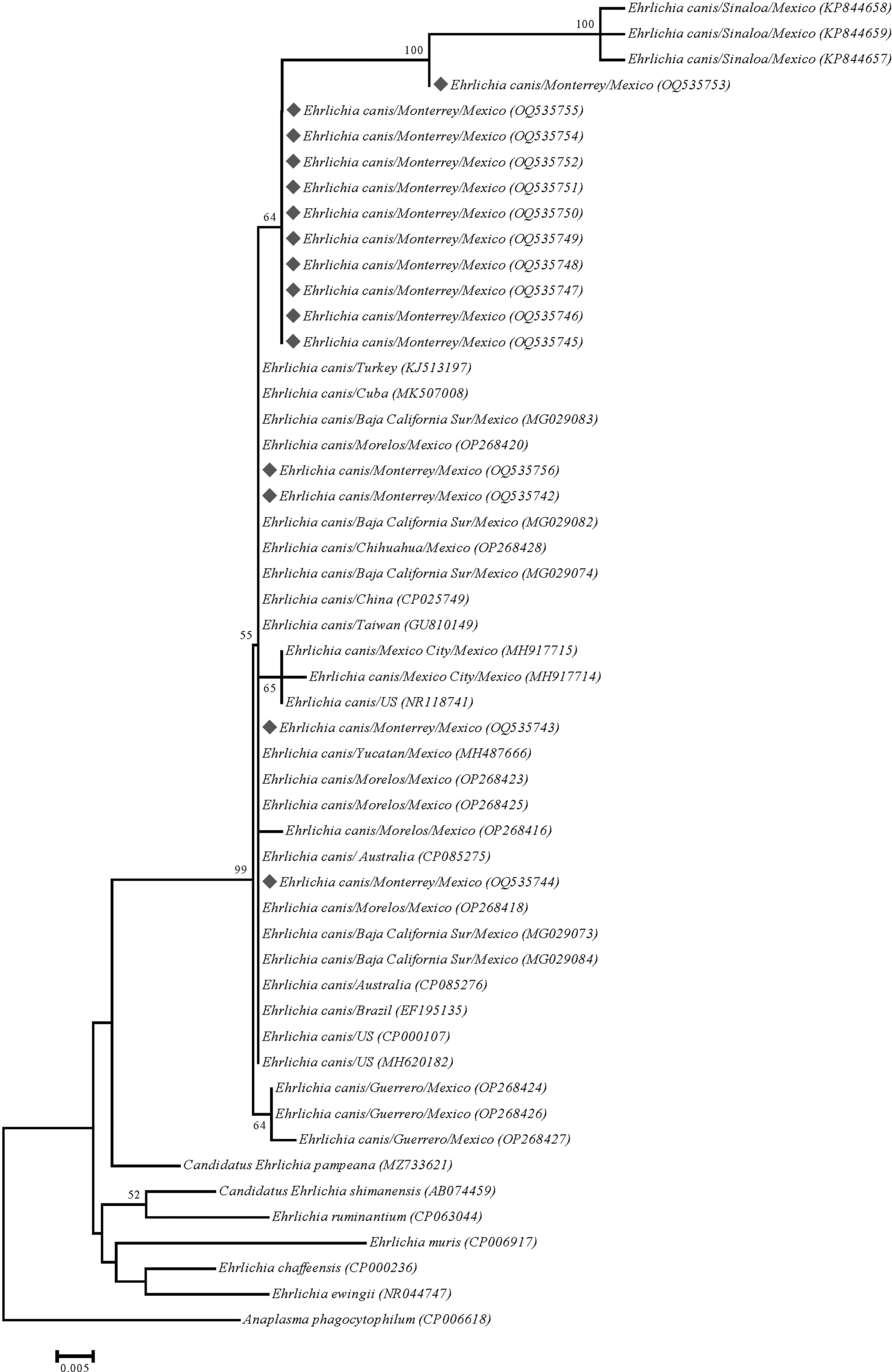
Maximum-likelihood phylogenetic tree generated by using the general time-reversible model using discrete Gamma distribution with 437 bp of the 16S-rRNA gene of several members of the genus Ehrlichia. Sequences in blue indicated the results of this study. Sequences in green represent previous results generated from Mexico deposited on GenBank. Bootstrap values >50 are indicated at the nodes.
Ehrlichia canis infection rates varied by tick species and study sites, as given in Table 3. Of the 122 R. sanguineus s.l. ticks infected that were captured directly from dogs, 57.4% (n = 70) were female, and 42.6% (n = 52) were male [z (1, N = 122) = 3.12, p > 0.001]. The ratio of fed-to-nonfed females was 7.75:2.25 (62/8) [z (1, N = 122) = −17.32, p < 0.001], whereas in males, it was very similar; there were 7.66 times more fed than nonfed males (46/6) [z (1, N = 122) = −14.07, p < 0.001]. The ticks captured from the floor and walls were 25, of which 17 were females (nine fed and eight nonfed) and eight nonfed males.
Ehrlichia canis Infection Rates (%) in Ticks Associated with Dogs by Study Site in Nuevo Leon, Mexico from 2012 to 2021
The localities with the highest infection rates of E. canis in ticks were Adolfo Prieto in Guadalupe (84.21%) and Ex Hacienda el Canada in General Escobedo (71.60%) (Table 2). The relationship between infection rates and study sites was examined with the chi-square test of independence. The relationship between these variables was significant [χ2 (3, N = 401) = 17.59, p < 0.0001]. Thus, infection rates of E. canis in ticks are not uniformly distributed across study sites (Cadereyta Jimenez, General Escobedo, Guadalupe, and Monterrey).
Discussion
In this study, the PCR method identified E. canis in ticks from four localities of Nuevo Leon, Mexico. The amplification of the 16S gene confirmed the presence of this tick-borne pathogen. This bacterium in ticks had already been reported by Tamez-González (2018), Tamez-González (2015), and Dávila-Venegas (2021) in their theses from Nuevo Leon.
The infection rate of E. canis in ticks from this study was higher at 59.92% (154/257) than that of Tamez-González (2015) at 9.17%, and Dávila-Venegas (2021) with 12%, but similar to Tamez-González (2018) who reported 52.73% infection. These infection rates varied depending on the study site and tick species. Tamez-González (2015) found 9.17% E. canis in 32 of 349 R. sanguineus s.l. specimens from 31 of 130 colonies of Monterrey, whereas Tamez-González and Gordillo-Pérez (2017) found an infection rate of 6.92% (18/260) in Monterrey and 12% (6/50) in Montemorelos. Likewise, Tamez-González (2018) detected to E. canis in ticks of R. sanguineus s.l. 222/416 (52.73%) within the metropolitan area of Monterrey because it does not specify in which of the eight municipalities these results came out. In a previous study, Dávila-Venegas (2021) found the presence of E. canis in ticks of R. sanguineus s.l. (48/400; 12%) in seven cities, but with a higher rate in Garcia (19/63; 30%).
These four previous studies obtained samples from Canine Control Centers and Veterinary Clinics, whereas our samples were obtained from homes with tick infestations and a clinical history of canine ehrlichiosis. This is the reason for the high infection rate in this study. The positive tick samples for this species of bacteria from Canine Control Centers, Veterinary Clinics, and homes offer us an overview of the epidemiological situation of canine ehrlichiosis in the metropolitan area of Monterrey, Nuevo Leon, Mexico, particularly in Apodaca, Garcia, General Escobedo, Guadalupe, Santa Catarina, San Nicolas de Los Garza and Monterrey.
There is only one study where serological analysis (SNAP 4Dx® test) revealed E. canis in the blood of dogs (54/391; 13.81%) from the municipality of Monterrey, Nuevo Leon (Salinas-Meléndez et al., 2015). With these works, the urban transmission cycle of canine ehrlichiosis is demonstrated in Monterrey, Nuevo Leon, Mexico, where the potential vector is the tick R. sanguineus s.l.
Previous studies in Mexico have detected multiple E. canis haplotypes in ticks and dogs from six states of the Mexican Republic (Mexico City, Coahuila, Guerrero, Morelos, Sinaloa, and Yucatan). Owing to the variability in the size of the fragments of the E. canis sequences detected in the six states of the republic, the percentage of similarity of the sequences recovered in this work is low, which is why it is possible to assume that the sequences retrieved in our study exhibit a more remarkable similarity with sequences of E. canis from Cuba.
Rhipicephalus sanguineus s.l. is a tick widely distributed in the metropolitan area of Monterrey. It has colonized households in urban areas, but it is also present in rural regions of Nuevo Leon, where it is active throughout the year in both areas. This tick species has been observed to seek refuge inside and around houses, particularly near dog resting places, where they colonize the cracks and crevices of walls and floors. In this study, there was a tick infestation in the yard of the house where the dog lived, a favorable locality for E. canis.
On the contrary, we infer that the tick D. variabilis may play an essential role in the transmission of E. canis in rural areas of Nuevo Leon because we found two specimens (20%) naturally infected with the bacteria in a rural area. However, another infection (60%, 3/5) was also detected within the metropolitan area (Tamez-González, 2018). In addition, D. variabilis was reported in dogs from different urban municipalities (Apodaca, Benito Juarez, General Escobedo, Guadalupe, Monterrey, Pesqueria, and San Nicolas de Los Garza) of Nuevo Leon (Galaviz-Silva et al., 2013). Therefore, its role in transmission in these urban areas is unknown. There is evidence that experimental transstadial transmission of the E. canis is possible in D. variabilis (Johnson et al., 1998). Besides, the medical and veterinary importance of D. variabilis is well documented in the United States (Eisen and Paddock, 2021). The distribution and abundance of these tick species are determined in part by climate, host, and habitat availability (Boorgula et al., 2020).
Conclusions
In this study, we demonstrate the natural infection of E. canis in R. sanguineus s.l. and D. variabilis in four localities of Nuevo Leon, Mexico, at different sampling times. This pathogen requires strategies for its control and prevention, using epidemiological surveillance tools and identifying risk factors. A systematic study is necessary where all landscapes are considered, from the wild, rural to urban, to obtain blood samples from domestic animals and their associated ectoparasites, not only for detecting E. canis, but also other bacteria that affect northern Mexico (e.g., Anaplasma sp., Borrelia burgdorferi s.l., Rickettsia spp.). Therefore, surveillance of these pathogens must be continuous and longitudinal in time and space in Nuevo Leon, bordering the United States.
Footnotes
Acknowledgments
The authors thank the citizens for their participation in the work. Special thanks to veterinarian Gema Esmeralda Guanajuato Puente for her support in the methodology.
Authors' Contributions
J.J.R.-R.: Conceptualization (lead); investigation (lead); writing—original draft (lead); data curation (lead); formal analysis (equal); methodology (equal); visualization (equal); project administration (lead); resources (equal); funding acquisition (equal); supervision (lead); validation (lead). T.L.H.-M.: investigation (supporting); data curation (supporting); methodology (supporting). S.S.-M.: data curation (supporting); methodology (supporting); formal analysis (equal); writing—review and editing (supporting). I.F.-S.: resources (equal); funding acquisition (equal); supervision (equal); validation (equal). R.M.S.-C.: writing–review and editing (supporting); resources (equal); funding acquisition (equal); supervision (equal); validation (equal). J.J.H.-E.: writing—review and editing (supporting); resources (equal); funding acquisition (lead); supervision (lead); validation (equal).
Author Disclosure Statement
Authors declare to have no conflict of interest.
Funding Information
The funding was granted in part by the Autonomous University of Nuevo Leon through the Faculty of Veterinary Medicine and Zootechnics. This research received funding from “Programa de Apoyo a la Investigación Científica y Tecnológica (PAICYT),” Autonomous University of Nuevo Leon, No. 394-CN-2022.