
Abstract
Background:
Francisella tularensis is a Gram-negative bacterium that causes tularemia in both human and animals. Tularemia is a potential serious zoonotic disease that is transmitted by different routes, including tick bites.
Materials and Methods:
This study deals with investigating the prevalence of F. tularensis in the ticks of local animal farms in Kurdistan region since the farmers are normally in close contact with livestock. We used molecular methods for this purpose. A total of 412 tick and 126 blood samples were gathered from goat, sheep, and cow flocks. The existence of F. tularensis 16Sr RNA gene was examined in the samples using nested-PCR technique.
Results:
In the animal blood specimens, no F. tularensis was found. The incidence of F. tularensis was 1.7% (7 out of 412) in the tick samples, representing a very lower possibility of tuleremia infection. Moreover, the two subspecies of F. tularensis novicida and holarctica were identified based on the sequencing of pdpD and RD genes, respectively. The F. tularensis subsp. novicida was isolated from four species of ticks, Hyalomma anatolicum, Rhipicephalus annulatus, Rhipicephalus sanguineus, and Ornithodoros spp., whereas the F. tularensis subsp. holarctica was isolated from Haemaphysalis parva and Hyalomma dromedarii species of ticks.
Conclusion:
Although its prevalence is very low, the isolation of F. tularensis subsp. holarctica from the ticks of farm animals suggests possible transmission of Tularemia through tick bite in Kurdistan region of Iraq. Ref: IR-UU-AEC-3/22.
Introduction
F
This bacterium was isolated from blood, organs, stool, and parasitic ticks of different animals in different countries of the northern hemisphere (Behzadi et al., 2021; Carvalho et al., 2014; Johansson et al., 2004). It can also remain in environment for a long period of time. F. tularensis is also considered as a biological agent because of its high infectiousness and lethality (Appelt and Faber, 2020). Based on their geographical distribution, biochemistry, and virulence, four subspecies of F. tularensis are currently known, tularensis (known as type A), holarctica (known as type B), novicida, and mediasiatica (Harik, 2013). The first two subspecies, tularensis and holarctica, mostly cause human illness (Kingry and Petersen, 2014).
The pathogen was recognized as the causative agent of tularemia since early 1900s (Sjöstedt, 2007). This disease is only common in many countries of the northern hemisphere (Johansson et al., 2004). Tularemia has been reported in both animals and human. It is probably transmitted to humans through different routes, such as touching infected animals, consumption of contaminated water or food, inhalation of aerosols, and ticks bites (Hayes et al., 2005; Petersen and Schriefer, 2005). The clinical manifestations of tularemia are headache, sore throat, joint pain, weakness and fatigue, and fever and chill (Carvalho et al., 2014).
The disease is further characterized as ulceroglandular and glandular tularemia. The former is the most common form that occurs after tick bite and recognized by swelling of the regional lymph nodes and skin ulcer. The latter causes lymphadenopathy without detectable skin ulcer (Petersen et al., 2008).
In addition, ticks are among the medical and veterinary important arthropods in the world. They hurt the hosts through feeding and transmitting different viral, parasitic, and bacterial pathogens, including F. tularensis. Ticks can cause disease and death in livestock, wild animals, and human beings (Anderson and Magnarelli, 2008; Sonenshine, 1993). Besides its role as a biological vector, ticks probably support the survival or growth of F. tularensis in nature for a long period of time (Telford III and Goethert, 2020). Thus, the investigations of tick-borne pathogens are necessary to prevent and control the incidence of the related diseases in animals and human.
The diagnosis of tularemia can not only be done by the clinical examinations. So, the microbiological and serological tests were also used. However, these diagnostic tools have drawbacks in terms of infection risks and long time to obtain the test results (Maurin, 2020). Recently, the molecular techniques are developed to obtain quick and accurate results, using PCR (Bahuaud et al., 2019; Tarnvik and Chu, 2007). PCR-based methods showed more reliability and sensitivity than the serological and histological analyses, when pure culture of F. tularensis and different biological samples were used for the diagnosis of tularemia (Hepburn and Simpson, 2008; Tarnvik and Chu, 2007).
In recent years, tick-borne tularemia cases have been reported in neighboring counties (Behzadi et al., 2021). Also, F. tularensis was recently detected in the blood samples of domestic rabbits in Sulaimaniyah city, Kurdista region, Iraq (Marif et al., 2021). This study aims at the molecular detection of F. tularensis from ticks of livestock in the Kurdistan region, Iraq, using molecular techniques.
Materials and Methods
Study area and sample collection
The study was conducted in Sulaymaniyah governorate (35°33′40″N and 45°26′14″E, and elevation is ∼830 meters above sea level) from March 2021 to July 2022. A total of 412 tick specimens were collected from the body surface of the host animals (186 sheep, 72 goat, and 154 cows), also from cracks and crannies in the sleeping places of the animals in four districts: Chamchamal in the west, Dokan in the north, Penjwin in the east, and Darbandekhan in the south of the Sulaymaniyah Province of Iraq (Fig. 1).
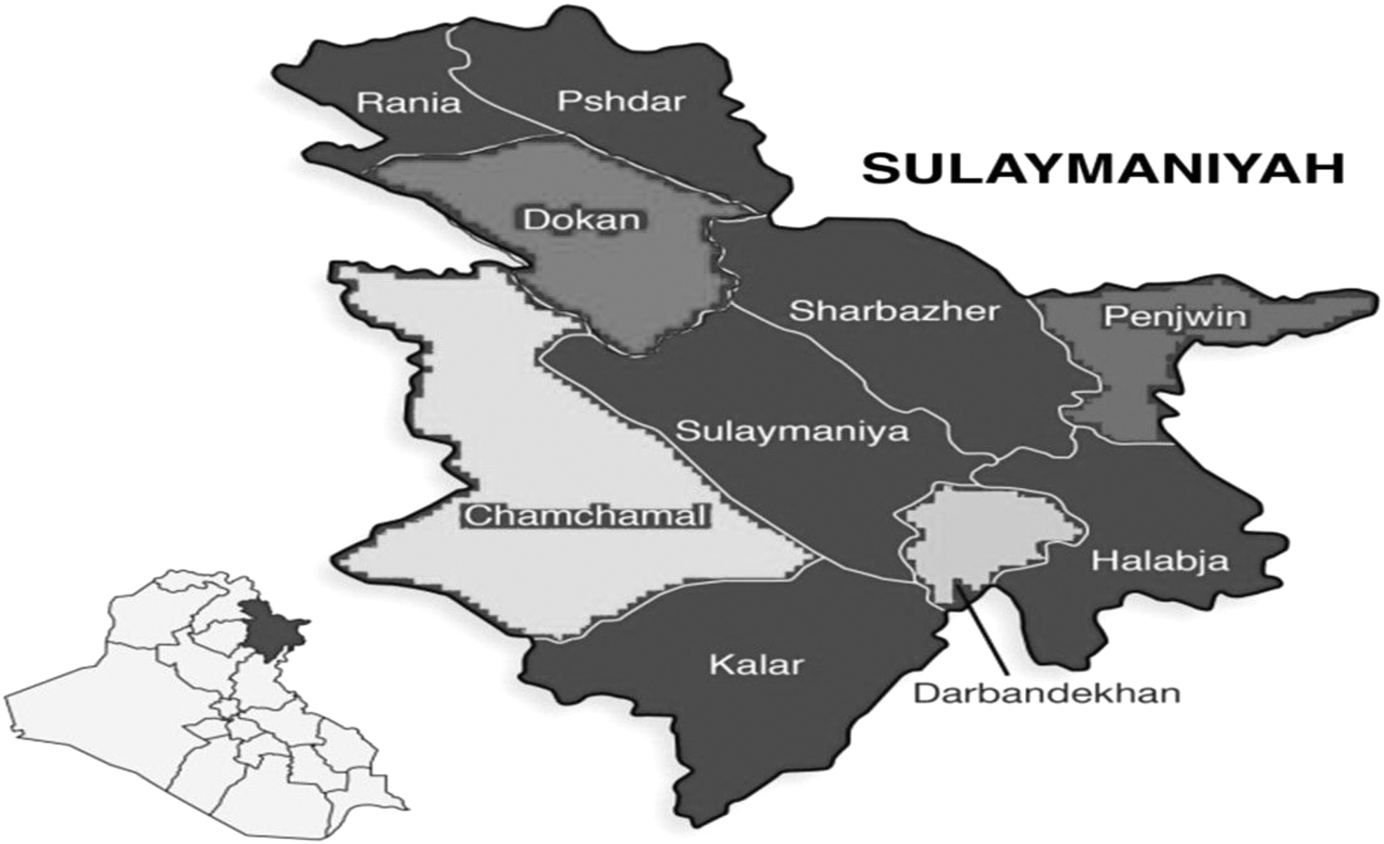
Map of the study locations in Sulaymaniyah province, Kurdistan region, Iraq.
Besides, 126 blood samples were randomly taken from 56 sheep, 38 goats, and 42 cows. Clinically, the animals were healthy, hence, no acaricide treatment was conducted. The ruminants' blood samples were added into tubes comprising ethylenediaminetetraacetic acid (EDTA) and then kept at 4–8°C until transferring to the laboratory. The species and genus of the gathered ticks were assessed and recognized by microscopic examination based on their morphology (Barbieri et al., 2013).
DNA extraction
First, using sterile distilled water, the preserved tick samples in 70% ethanol were first rinsed for 10 min. Then they were dried on sterile filter paper and suspended in phosphate-buffered saline (200 μL) (pH 7.4) in sterile 1.5 mL microcentrifuge tubes. A sterile scalpel was used to chop the ticks into small pieces, which were then homogenized using sterile micropestle. The total genomic DNA was extracted and eluted in 50 μL of tris EDTA buffer, through the (AddPrep Genomic DNA Extraction; AddBio, Korea) tissue kit based on the instructions of the manufacturer.
Furthermore, genomic DNA was extracted from animal blood samples utilizing a commercial kit QIAamp Blood/Tissue Kit (Qiagen, Hilden, Germany) based on the protocol of the manufacturer. All DNA specimens were kept at −20°C until using the samples for PCR analysis.
Molecular detection of F. tularensis
F. tularensis 16S rRNA gene was amplified using primer sets designed from Amplifx (Version 1.7.0) software by nested PCR as given in Table 1. The PCR was carried out in 25 μL comprising 4 μL of template DNA, 1 μL of each primer with 12.5 μL of master mix, and the total volume was made up using sterile distilled water. In addition to minimize nonspecific amplification, a so-called touchdown PCR program was used (Don et al., 1991).
Selected Primer to Amplify a Fragment of 16S rRNA Gene for the Identification of Francisella tularensis
The touchdown and 16S rRNA-PCR thermal program (Table 1) were defined in the thermal cycler (Quanta Biotech, England). For the nested PCR, the normal PCR product (2.5 μL) was utilized as template and PCR products were electrophoresed on a 2% agarose gel comprising safe stain (Labnet, ENDURO, USA). Then they were visualized through Genius Gel Documentation (Syngene Bio-Imaging, United Kingdom).
Molecular detection of F. tularensis subspecies
The existence of RD and pdpD genes was also assessed for identification of F. tularensis subspecies through PCR subtyping assays based on WHO guidelines. A fragment of pdpD gene was amplified using a pair of primers in 280 bp in size: Forward primer: 5′-TGGGTTATTCAATGGCTCAG-3′ Reverse primer: 5′-TCTTGCACAGCTCCAAGAGT-3′ that was used to identify F. tularensis subsp. novicida. Moreover, another pair of primers was applied to identify F. tularensis subsp. Holarctica: Forward primer, 5′-TTTATATAGGTAAATGTTTTACCTGTACCA-3′ and Reverse primer, 5′-GCCGAGTTTGATGCTGAAAA-3′ fragment of RD gene with the size of 1150 bp.
The conditions for RD gene were performed: initial denaturation at 95°C for 15 min, and then 36 cycles of 95°C, 57°C, and 72°C for 30 s, 45, and 60 s, respectively. Then, 72°C for 10 min. For pdpD gene: initial denaturation at 95°C for 15 min, and then 40 cycles of 95°C, 54°C, and 72°C for 30 s, 30 s, and 45 s, respectively, then, 72°C for 10 min (Rahravani et al., 2022). To confirm positive F. tularensis, sequencing of pdpD, RD, and 16Sr RNA amplicons was performed using the Sanger sequencing method. Phylogenetic analyses were done by Mega (version 11) software.
Nucleotide diversity and phylogenetic tree construction
Sequences were uploaded to the National Center for Biotechnology Information (NCBI) to seek the most analogous reference sequences; furthermore, the COI (Country of Origin Information) positions were identified using NCBI's BLAST. For phylogenetic analysis, all Francisella COI sequences accessible in the Gene Bank were utilized. By using the Clustal alignment program, the alignment was manually adjusted to eliminate any related errors before being released as MEGA and FASTA files (Kumar et al., 2018).
All of the acquired nucleotide sequences were entered into the Gene Bank and given accession numbers. After that, the phylogenetic association was investigated and developed using the Molecular Evolutionary Genetics Analysis software version 10 by the maximum-likelihood method. One thousand bootstraps were used to test the reliability of an inferred tree. BioEdit version 7.0.1 and BLASTN software were used to analyze DNA sequence polymorphism to assess nucleotide diversity (Kumar et al., 2018).
Statistical analysis
To conduct statistical analysis of the data, SPSS (version 20) software (IBM, Armonk, NY, USA) was utilized. The variables were compared using chi-square test during the analysis (p ≤ 0.05).
Results
Prevalence of F. tularensis
The detection of F. tularensis was done based on the PCR of 16S RNA gene. None of the animal blood samples contained F. tularensis. However, out of 412 tick specimens, 7 were positive for F. tularensis (Fig. 2 and Table 2). The molecular prevalence was 0% and 1.7% in the animal blood and ticks, respectively. F. tularensis was almost equally isolated from three genera of ixodidae (hard) ticks and one genus of Argasidae (soft) ticks.
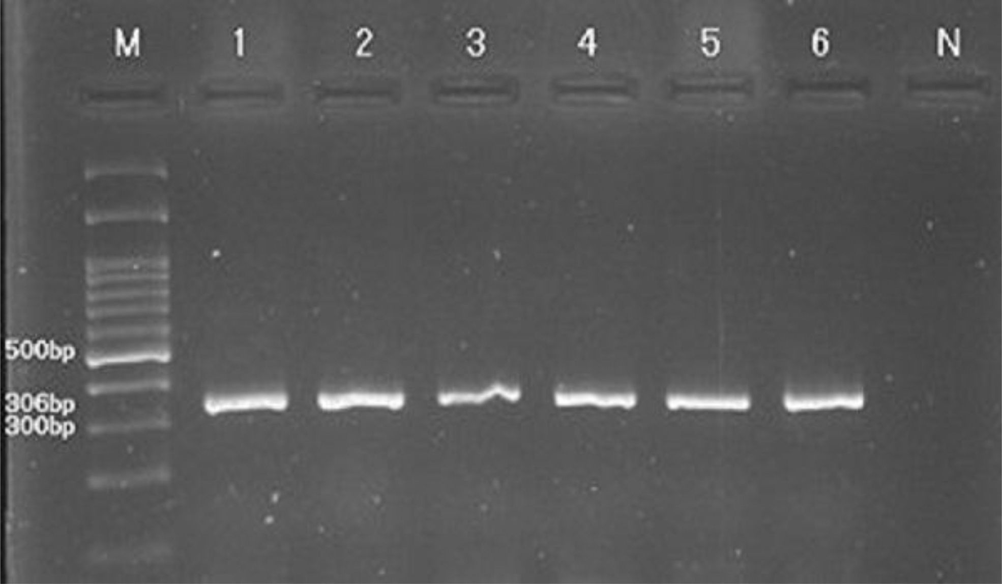
The amplicons of 16Sr RNA gene (306 bp) of Francisella tularensis. Lanes 1–6: positive specimens, and lane N: negative specimen.
Prevalence of Francisella tularensis Based on Ticks Species, Livestock Species, and Geographical Location
CI, confidence interval.
The species of ticks that harbored F. tularensis were Hyalomma anatolicum, Hyalomma dromedarii, Haemaphysalis parva, Rhipicephalus annulatus, and Rhipicephalus sanguineus. In addition, F. tularensis was isolated from the soft ticks of Ornithodoros spp. In contrast, Rhipicephalus turanicus, Hyalomma asiaticum, and Dermacentor marginatus were negative for F. tularensis (Table 2).
The prevalence of F. tularensis according to the livestock species was 2.15% (cow), 1.29% (sheep), and 1.38% (goat) (Table 2). Meanwhile, the prevalence of F. tularensis in different districts of Sulaymaniyah province was 2.18% in Chamchamal, 1.47% in Dokan, 1.26% in Penjwin, and 1.56% in Darbandekhan (Table 2).
Subspecies of F. tularensis
According to the results of pdpD and RD gene sequences and phylogenetic analyses (Fig. 3), novicida and holarctica subspecies of F. tularensis were identified and their sequences were uploaded to the Genbank database with the acc. nos. OP555286 and OP622846, respectively (Figs. 3 and 4). The F. tularensis subsp. novicida was isolated from the ticks of H. anatolicum, R. annulatus, and Ornithodoros spp. that parasitized cows, sheep, and goats. In contrast, the F. tularensis subsp. holarctica was found in H. parva and H. dromedarii ticks.
Francisella tularensis subsp. novicida phylogeny based on the sequence of pdpD gene. The phylogenetic tree is constructed by MEGA 11 software.

Francisella tularensis subsp. holarctica phylogeny produced by the neighbor-joining method based on sequence of RD gene.
Discussion
The ticks are vectors of microbial pathogens that can cause infections in both human and animals (Gehringer et al., 2013). F. tularensis is a tick-borne and zoonotic bacterium causing tularemia disease in human. In this study, the incidence of F. tularensis in farm animals (sheep, cows, and goats) was assessed. It was indicated that the incidence of F. tularensis associated with ticks is 1.7% in the Kurdistan region in Iraq, which is very lower than the incidence (16%) of F. tularensis in domestic rabbits in Sulaimaniyah city, Kurdista region, Iraq (Marif et al., 2021).
Consistently, a recent study detected a very low prevalence (0.82%) of F. tularensis subsp. holarctica in D. marginatus ticks in Iran (Rahravani et al., 2022). In our study, the highest positive rate of F. tularensis was found in Ornithodoros spp. (2.46%), Rhipicephalus spp. (1.85%), followed by H. parva (1.69%) and Hyalomma spp. (1.63%). No F. tularensis was isolated in Dermacentor spp. Moreover, the F. tularensis subsp. holarctica was isolated from H. parva and H. dromedarii ticks. The positive rate was significantly different among different tick species (χ 2 = 33:04, p < 0.05).
Similarly, F. tularensis was reported in Rhipicephalus, Hyalomma, and Haemaphysalis genuses of ticks in Bulgaria (Ivanov et al., 2011), Japan (Suzuki et al., 2016), Iran (Rahravani et al., 2022) and Ethiopia (Szigeti et al., 2014). In contrast, F. tularensis was not detected in >1000 samples of Haemaphysalis ticks in California, USA (Duzlu et al., 2016). Also, 1477 adult hard ticks of Rhipicephalus, Haemaphysalis, Hyalomma, Dermacentor, and Ixodes genuses were free of F. tularensis in Turkey (Duzlu et al., 2016). Accordingly, the incidence of F. tularensis in ticks is very lower and different based on the tick species. Therefore, the likelihood of Tularemia is very low in the Kurdistan region, although the discovered F. tularensis subsp. Holarctica may still result in human illness (Kingry and Petersen, 2014).
Conclusion
This is the first report of isolation and genetic characterization of F. tularensis from ticks of livestock (cow, sheep, and goat) in the Kurdistan region, Iraq. F. tularensis was detected in a very low prevalence of 1.7%. F. tularensis subsp. holarctica and F. tularensis subsp. novicida were isolated from both hard and soft ticks. These results recommend careful treatment with the animals that are parasitized with ticks, hoping to prevent the incidence of Tularemia.
Footnotes
Acknowledgments
We thank all members of the molecular laboratory of the Urmia University, Charmo University, Lorestan University, and Zabol University. The authors thank Dr Shorsh Hamaxwrshid and Ismail Rafaat from Vet Plus Veterinary Clinic, Chamchamal, Iraq, for their support throughout this study. Also we thank the farmers who participated in this study.
Authors' Contributions
R.R.M., A.E., P.K., and S.S. conceptualized the study. K.R.S., and R.R.M. collected, analyzed, interpreted the data, and conducted the statistical analysis. P.K., K.R.S., and S.S. wrote the first draft of the article. All authors revised the article and approved the final version. P.K., R.R.M., and A.J. had full access to all of the data in the study and take responsibility for the integrity of the data and the accuracy of the data analysis.
Author Disclosure Statement
No competing financial interests exist.
Funding Information
No funding was received for this article.