
Abstract
Background:
Theileria spp. are responsible for ovine and caprine theileriosis, leading to significant morbidity and mortality in small ruminants. The present study aims to investigate Theileria spp. infections in small ruminants from Southern Punjab in Pakistan, and genetic characterize revealed Theileria spp. isolates.
Methods:
A total of 93 sheep and 107 goats were sampled between May and August 2022. Blood smears were examined microscopically, and PCR amplification targeting the 18S rRNA gene was performed to detect Theileria spp. Additionally, specific PCR assays targeting 18S rRNA and ms1 partial sequences were used to identify Theileria ovis and T. lestoquardi, respectively.
Results:
The prevalence of Theileria spp. was significantly higher using PCR (13.5%) compared to microscopic screening (5%). Sheep showed a higher prevalence rate (19.4%) compared to goats (8.4%) (p = 0.024). Young sheep aged ≤ 1 year were more commonly infected with Theileria spp. (41%) compared to older sheep (p = 0.006). The prevalence of Theileria spp. was higher in sheep-only herds (37.3%) compared to goat-only herds (18%) or mixed-species herds (8.1%) (p = 0.015). The prevalence rates of T. ovis and T. lestoquardi were 9% and 2.5%, respectively, with four animals (2 goats and 2 sheep) showing co-infection. Phylogenetic analysis revealed that our T. ovis 18S rRNA sequence clustered with previously reported sequences from sheep in Turkey, China, Spain, and goats in Tanzania. The obtained T. lestoquardi ms1 partial sequence formed a distinct cluster from other T. lestoquardi isolates in Pakistan and neighboring countries.
Conclusion:
Theileria spp. co-circulation in Pakistani small ruminants, particularly the presence of T. ovis and T. lestoquardi, highlights the need for attention from animal health decision-makers due to their financial and health impacts.
Introduction
Sheep and goats are important livestock species that rose in developing countries to improve the economic conditions of rural dwellers in Pakistan (Ahmad and Tasawar, 2016). A total of 1,614 million sheep and 475 million goats are reared worldwide, of which 65% of sheep and 95% of goats are raised in developing countries. Asia, Africa, and Latin America are the zones that contain 53%, 33%, and 14%, respectively of the total population of small ruminants, worldwide (Haenlein, 2001; Khan et al., 2022).
Small ruminants in the Asian farming system maintain a valuable economic role, as most livestock owners manage their income from surplus animals and by-products (Irshad et al., 2010). In 2014–2015, in Pakistan, the sheep population was 29.4 million, whereas the goat population was 68.4 million. In 2014–2015, ewe milk production was 38 tons, whereas goat milk production was 845 tons. Small ruminants can greatly adapt to extreme climatic situations (Irshad et al., 2010).
Pakistan, next to China and India, is the main goat-producing country in the world. The major occlusion of livestock manufacturing in Pakistan is attributed to many factors, including parasite infestation (both ectoparasites and endoparasites) (Sajid et al., 2008; Khan et al., 2022).
Ticks and tick-borne diseases (TTBDs) are a major constraint to livestock expansion in Pakistan (Ahmed et al., 2007). TTBDs cause an annual loss of US$7000 million worldwide (Ahmed et al., 2007), and the cost of treating tick-borne diseases in cattle ranges between US$13.9 and US$18.7 billion per year (De Castro et al., 1997). Mcleod and Kristjanson (1999) reported that TTBDs severely damage the livestock industry by reducing 14% lactation in infected animals. As a result, ticks act as not only impending vectors of tick-borne diseases but also reservoirs of contagious agents of certain diseases in humans and other animals (Jongejan and Uilenberg, 1994).
Ovine and caprine theileriosis are caused by intracellular parasitic protozoa of the genus Theileria, transmitted by ixodid ticks (Razmi et al., 2003) and result in severe morbidity and mortality in infected animals, depriving the rural inhabitants of their livelihood (Li et al., 2014). Theileriosis is caused by six Theileria species (Alanazi et al., 2019), among which T. ovis, T. recondite, and T. separata are deliberately mild, whereas T. lestoquardi, T. luwenshuni, and T. uilenbergi are extremely pathogenic for small ruminants (Razmi et al., 2003; Yin et al., 2004; Aktaş et al., 2005; Tanveer et al., 2022).
Malignant ovine theileriosis caused by T. lestoquardi infection has been reported by many authors around the world. It is widespread in Eastern Europe, the Middle East, North Africa, Iran, Iraq, and Sudan (Soulsby, 1982). Theileria lestoquardi induces a lymphoproliferative disease characterized by symptoms such as fever, anemia, emaciation, reduced activity, abortion, and, ultimately, fatality (Jianxun and Hong, 1997). Pathological effects, including lymphadenopathy, rapid weight loss, and jaundice, have been observed in sheep and goats (Rehman et al., 2010). Moreover, the growth of infected animals is further reduced due to inefficient feed conversion, which negatively affects weight gain and milk production and leads to considerable economic losses in terms of low productivity and mortality (Irshad et al., 2010; Tanveer et al., 2022).
Theileria detection can be achieved by either microscopic screening, which is rapid and inexpensive but not infallible to species demarcation due to the morphological resemblance of Theileria piroplasms, or polymerase chain reaction (PCR) amplification, which is sensitive and specific but precious (Riaz et al., 2019; Riaz et al., 2023). Combining the two diagnostic techniques can represent a practical gadget to detect animals afflicted with theileriosis.
In this study, we present a novel epidemiological study conducted on sheep and goats to assess the prevalence of Theileria piroplasms in Bund Bosan, Multan district, Southern Punjab, Pakistan. The investigation also aims to identify potential risk factors associated with the spread of Theileria spp. infections in sheep and goats. Additionally, we explore the phylogeny of the revealed isolates based on ms1 and 18S rRNA markers, adding a distinctive contribution to the understanding of these infectious agents in the region.
Materials and Methods
Study area
This survey was conducted between May and August 2022 in Band Bosan town of Multan district, Southern Punjab, Pakistan. The district of Multan is located between 29ʹ-22ʹ north latitude and 71ʹ-4ʹ east longitude with an extreme temperature of 49°C in summer and 1°C in winter and an average rainfall of 127 mm (Fig. 1). Bosan town is located on the northern side of Multan district, containing a total area of 500 km2 comprising 24 union councils. Sheep farming and goat farming are the main livelihoods of this town. Most small ruminants (sheep and goats) are kept in mixed herds containing 30–100 animals per herd.

Maps of Pakistan showing Punjab province
Blood sampling
Blood samples were taken from 200 healthy small ruminants (93 sheep and 107 goats) from arbitrarily selected herds in eight Bund Bosan town councils, Multan district, Pakistan. Blood samples were taken from the jugular vein and stored instantaneously in 5-mL Eppendorf tubes containing 0.5 M EDTA (a few drops) as a preservative for DNA extraction. Data on animal characteristics, including species, age, gender, and presence of ticks, and herd characteristics, comprising location, size, and herd composition, were collected during sampling to determine the potential risk factors for spreading Theileria infection. All experiments were approved by the Ethics Committee of the Institute of Pure and Applied Biology at Bahauddin Zakariya, University Multan, Pakistan.
Microscopic examination of thin blood smears
Blood smears (thin and thick) were prepared and air-dried and then immersed in absolute methanol for fixation. Blood smears were stained with 5% Giemsa (pH = 7.2) for 30 min in the laboratory of the Institute of Pure and Applied Biology (IP & AB), Bahhudin Zakaryia University, Multan, and then analyzed by light microscopy for the detection of Theileria spp. in erythrocytes. Morphological characteristics of Theileria piroplasms were used for identification, as described by Soulsby (1982) and Urquhart et al. (1996). The piroplasms were polymorphic and, morphologically, most occurred as rods, needle-like, pear-shaped, or spherical forms.
DNA extraction
DNA was extracted following the inorganic method previously described by Shaikh et al. (2005). The quality of the extracted DNA was checked on a 0.8% agarose gel under an ultraviolet illuminator. The extracted DNA was stored at −20°C for further PCR amplification.
Molecular detection by PCR
Three sets of primers were used in the present study to diagnose theileriosis based on the 18S rRNA gene. The first set of primers was forward 5ʹ-AGTTTCTGACCTATCAG-3ʹ and reverse 5ʹ-TTGCCTTAAACTTCCTTG-3ʹ, formerly used by Allsopp et al. (1993) for the amplification of an 18S rRNA gene specific for the genus Theileria. The reaction volume included 5 µL of template DNA, 5 µL of 10× PCR buffer (100 mM Tris–HCl [pH 9], 500 mM KCl, 1% Triton X-100), 10 pm of primers, 250 M of each of the four DNA bases, and 2 U Taq DNA polymerase (Vivantis, UK) with a total reaction volume of 50 µL. The thermoprofile contained 94°C for 3 min for the initial denaturation of DNA, followed by 35 cycles at 94°C for 1 min for denaturation, 60°C for 1 min for annealing, and 72°C for 1 min for the extension. The reaction ended with a final extension step of 72°C for 7 min.
The second set of primers was forward 5ʹ-GTGCCGCAAGTGAGTCA-3ʹ and reverse 5ʹ-GGACTGATGAGAAGACGATGAG-3ʹ, used for the specific detection of T. lestoquardi based on the amplification of a partial sequence (785 bp) of ms1 gene coding for the 30-kDa merozoite surface antigen (Kirvar et al., 1998). The third set of primers was forward 5ʹ-TCGAGACCTTCGGGT-3ʹ and reverse 5ʹ-TCCGGACATTGTAAAACAAA-3ʹ, used for the specific identification of T. ovis based on the amplification of 520 bp of 18S rRNA gene (Altay et al., 2005). The PCR for these two Theileria spp. was performed in a total reaction volume of 25 µL comprising 3 µL of template DNA, 3 µL of 10× PCR buffer (100 mM Tris–HCl [pH 9], 500 mM KCl, 1% Triton X-100), 10 pm of each primer, 250 M each of the four dNTP, and 2 U Taq DNA polymerase (Vivantis UK).
Cycling conditions for T. lestoquardi consisted of 35 cycles, 94°C for 3 min for initial denaturation of DNA; each cycle involved denaturation at 94°C for 1 min, annealing at 56°C for 1 min, and extension step at 72°C for 1 min with a final extension step of 72°C for 7 min. Cycling conditions for T. ovis were 3 min at 96°C, followed by 5 cycles, 30 s at 94°C, 30 s at 56°C, and 1 min at 72°C. These five cycles were followed by 30 cycles. Each cycle consisted of 30 s at 94°C, 30 s at 54°C, and 1 min at 72°C with a final extension step of 7 min at 72°C. Positive DNA samples of T. lestoquardi and T. ovis were provided by Ulrike Seitzer (VIIRC Center, Borstel, Germany).
The PCR products produced during the amplification were separated on a 1.5% agarose gel. After gel electrophoresis, small ruminant samples showing amplified DNA fragments of 1098, 520, and 785 bp were considered positive for Theileria spp., T. ovis, and T. lestoquardi, respectively.
Statistical analysis
For statistical analysis, three age classes were made, namely, ≤1 year, >1 year and <3 years, and ≥3 years. On the basis of size, the herds were classified into three categories, that is, 1–30, 31–60, and >60 animals, and three groups of herds were made on the basis of composition, namely, only sheep, only goats, or mixed herds with both sheep and goats. Fisher’s exact test was used to assess the correlation between theileriosis and potential risk factors, that is, animal species, gender, breed, and presence of ticks on animals. Pearson’s chi-square analysis was used to determine the association between theileriosis and different age-groups and herd composition of studied animals. For statistical analysis, MiniTab, USA (Version 16), was used, and the significance level was set at p < 0.05 (two-tailed). Data were expressed as mean values ± standard error of the mean.
Results
Microscopic and molecular survey of Theileria species
The detection for theileriosis was significantly higher by PCR (13.5%, n = 27) compared with microscopic screening for blood smears (5%, n = 10) (Table 1, p = 0.003). By using PCR, theileriosis was found to be more prevalent in small ruminants from Haji Pur (23.3%) than in other sampling sites, but the difference was not significant (Table 1, p = 0.595). Theileriosis was more prevalent in sheep (19.4%) than in goats (8.4%) (p = 0.024, Table 2). Sheep infested with ticks had a higher incidence of theileriosis (37.3%) compared with those free of ticks (15.4%) (p = 0.041, Table 3). In addition, sheep of age ≤1 year had a higher incidence of theileriosis (41%) compared with older ones (17.5%) (p = 0.006, Table 3). Moreover, theileriosis is more prevalent in herds of sheep only (37.3%) than those formed by goats (18%) or by both species (8.1%) (p = 0.015, Table 4).
Microscopic and Molecular Prevalences of Theileria Spp. According to Different Sampling Sites
The sampling sites are eight Bund Bosan town union councils from Multan district, Pakistan.
CI, 95% confidence interval.
Statistically significant, p < 0.05.
Prevalence Infection Rates of Theileria Spp., Theileria ovis, and Theileria lestoquardi Overall and According to Different Animal Species
CI, 95% confidence interval.
p-Value was calculated between infection rates of Theileria ovis and Theileria lestoquardi overall and according to different animal species.
Statistically significant, p < 0.05.
Prevalence of Theileria Spp. According to Different Risk Factors Related Studied Sheep and Goats from Band Bosan Town of Multan District, Southern Punjab, Pakistan
CI, 95% confidence interval.
Statistically significant, p < 0.05.
Prevalence of Theileria Spp. According to Size and Composition of Herds of Studied Sheep and/or Goats from Band Bosan Town of Multan District, Southern Punjab, Pakistan
CI, 95% confidence interval.
Statistically significant, p < 0.05.
Specific detection of T. ovis and T. lestoquardi
The overall prevalence rates of T. ovis and T. lestoquardi were 9% and 2.5%, respectively, with a coinfection rate of 2% (Table 2). In particular, in sheep, the prevalence rates of T. ovis and T. lestoquardi were 15.1% and 2.2%, respectively, with a coinfection rate of 2.2%. The difference between the infection rates is statistically significant (p = 0.001, Table 2). In goats, prevalence rates of T. ovis and T. lestoquardi were 3.7 and 2.8%, respectively, with a coinfection rate of 1.9%. The difference between the infection rates is not significant (p = 0.692, Table 2). In addition, the difference in T. ovis infection estimated in sheep and goats was statistically significant (p = 0.005), whereas for T. lestoquardi, this difference was not significant (p = 0.768, Table 2).
Phylogenetic analysis of T. lestoquardi and T. ovis isolates
The specific infection of T. ovis was validated by sequencing 322 bp of the 18S rRNA gene from one randomly selected positive sheep sample. To1 sequence (GenBank acc. no. OP735589) obtained after sequencing was 100% homologous on comparisons with T. ovis partial sequences published in the GenBank. Phylogenetic analysis based on the alignment of our sequence obtained in this study with 18 sequences of T. ovis and other Theileria species obtained from GenBank was performed (Fig. 2). Our sequence was clustered with other T. ovis sequences in the monophyletic cluster (Fig. 2).

Phylogenetic tree based on a partial sequence (322 bp) of the 18S rRNA gene of Theileria ovis isolated in this study and those of T. ovis and other Theileria species available in GenBank from other countries around the world. Numbers in nodes represent the percentage of 1000 bootstrap iterations supporting the nodes (only percentages greater than 50% are shown). The host, strain, isolate or clone identification, country of origin, and GenBank accession number are indicated in the tree for each sequence. Our sequence newly obtained in this study is highlighted in bold and marked with asterisks. One T. mutans 18S rRNA partial sequence was added as an outgroup.
The specific infection of T. lestoquardi was confirmed by sequencing 420 bp of the ms1 gene from one randomly selected positive sheep sample. Tl1 sequence (GenBank acc. no. OP750049) revealed in this study was 100% to 98.33% homologous on comparisons with T. lestoquardi partial sequences published in the GenBank. A phylogenetic tree based on the alignment of our sequence obtained in this study with 19 sequences of T. lestoquardi and other Theileria species obtained from GenBank was constructed (Fig. 3). Our sequence was classified in T. lestoquardi cluster close to those isolated from small ruminants in Iran and Sudan (Fig. 3).
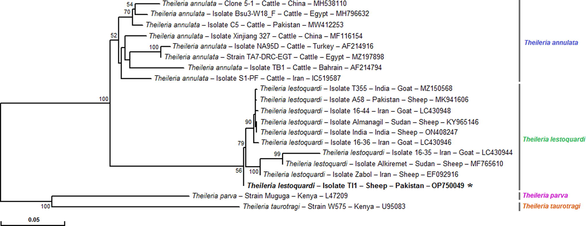
Representative neighbor-joining tree based on multiple sequence alignments of the partial ms1 nucleotide sequences (420 pb) of our T. lestoquardi isolate with those of T. lestoquardi and other Theileria spp. isolates and strains published in GenBank. Partial sequence from this study is represented in bold and marked with asterisks. Numbers associated with nodes represent the percentage of 1000 bootstrap iterations supporting the nodes (only percentages greater than 50% were represented). The host, strain or isolate name, the country of origin, and the GenBank accession number are indicated. Two ms1 partial sequences of T. parva and T. taurotragi were added as outgroups.
Discussion
Ovine and caprine theileriosis cases have been detected by clinical symptoms and microscopic examination in previous studies in Pakistan (Rehman et al., 2010; Irshad et al., 2010; Naz et al., 2012). The microscopic examination method and the presence of clinical symptoms are reliable only in critical cases, whereas it is deficient in carrier animals due to morphological similarities between piroplasms (Inci et al., 2010; Zakkyeh et al., 2012; Riaz et al., 2023).
For epidemiological studies, an accurate diagnosis of the parasite species is essential, which is possible using molecular detection methods compared with other conventional methods used for ovine and caprine theileriosis, such as microscopic and serologic techniques (Altay et al., 2005; Riaz et al., 2023). In particular, PCR is more applicable and useful to understand disease behavior in studied small ruminant populations, as these types of investigation comprise different aspects such as genetic diversity of populations, possible vulnerability to infections according to biotic factors related to the animal, the environmental conditions, the distribution of parasitic infections, and the risk of probable mutations. Also, for the amplification of parasite DNA, only a small amount of DNA is needed rather than a greater DNA quantity, which is not always available during the entire life cycle of the parasite in hosts (Gasser, 2006; Nehra et al., 2022).
The current investigation used PCR amplification and blood smear screening methods to determine theileriosis in Bund Bosan, Multan district, Pakistan. Of the 200 blood samples examined, 27 (13.5%) and 10 (5%) were found positive for Theileria spp. using PCR and microscopic examination, respectively. However, several previous reports reported higher prevalence of theileriosis in sheep and goats than our study, estimated at 19.35% and 54.03% in Turkey (Altay et al., 2005), 21% and 56% in Iran (Heidarpour et al., 2009), 22.27% and 60% in the eastern half of Iran (Heidarpour et al., 2010), 46% and 76% in Iran (Yaghfoori et al., 2013), and 69.7% and 89% in Iran (Jalali et al., 2014) by using, respectively, microscopic examination and PCR amplification. These disparities in infection rates could be due, in part, to differences in geoclimatic conditions such as variations in annual temperature and rainfall, differences in screening techniques, number of tested animals, and sampling period and procedure.
Various studies in the literature have documented the prevalence rates of T. ovis and T. lestoquardi in sheep and goats from Pakistan using molecular tools. For instance, Irfan et al. (2023) conducted a study in the Muzaffar Garh district of Punjab province, collecting 1084 blood samples from apparently healthy goats, and found that 1.11% of the tested animals were infected with T. ovis. Arif et al. (2023) reported a 3% infection rate of T. ovis in small ruminants from the Fort Munro region of Punjab province. Abid et al. (2021) discovered a 10.6% prevalence of T. ovis in sheep from Layyah district in Punjab province. Tanveer et al. (2022) revealed that 6.1% and 1.2% of analyzed sheep blood samples from Rajanpur district in Punjab were positive for T. ovis and T. lestoquardi, respectively, using PCR assays. Durrani et al. (2012) recorded a relatively low prevalence (6%) of T. ovis when analyzing small ruminant blood samples from Punjab and Khyber Pakhtunkhwa provinces in 2012. They observed higher T. ovis prevalence in small ruminants from Khyber Pakhtunkhwa compared with Punjab, attributing this variation to distinct climatic conditions and farming techniques in the two sampling sites. Finally, Saeed et al. (2015), and Fatima et al. (2015) estimated the prevalence of T. lestoquardi at 3% in small ruminants from Khyber Pakhtunkhwa and Punjab provinces.
In the present study, theileriosis was significantly higher in sheep (19.4%) than in goats (8.4%) (p = 0.024). Previous studies by Irshad et al. (2010), Naz et al. (2012), Iqbal et al. (2013), and Niaz et al. (2021) reported a higher prevalence in sheep compared with goats in Pakistan, with estimated rates ranging from 7.36% to 3.8%, 13.9% to 8.2%, 43.7% to 37.5%, and 14.5% to 8.8%, respectively, in both species. Similar higher infection rates in sheep compared with goats have also been reported in other countries, such as those in China (27.63–13.12%) by Guo et al. (2002) and Turkey (28.90–4.10%) by Altay et al. (2012). These findings suggest that sheep may be more susceptible to theileriosis. Indeed, the higher infection in sheep could probably be due to the difference in the nature of the skin, given that the skin of goats is thick and more resistant to the attachment to ticks than the skin of sheep. Also, the higher infection could be due to the presence of sheep wool, in which ticks could easily hide, thus causing theileriosis after their attachment and transmission of Theileria pathogens. In contrast, goats have a lower risk of tick infestation, as they are able to reside in isolated and steep areas. These results are consistent with previous studies in Pakistan and other countries that also reported a higher prevalence of Theileria infections in sheep compared with goats (Irshad et al., 2010; Naz et al., 2012; Iqbal et al., 2013; Guo et al., 2002; Altay et al., 2012; Kose et al., 2022).
Notably, T. ovis and T. lestoquardi are suspected of causing ovine and caprine theileriosis in Pakistan (Durrani et al., 2011). In the present study, the prevalence of T. ovis was found to be higher (9%) than that of T. lestoquardi (2.5%, p < 0.05), whereas the mixed infection of the two Theileria species was lower (2%) in small ruminants. Our results are in agreement with those of Rehman et al. (2010) and Durrani et al. (2012), who reported a higher prevalence of T. ovis compared with T. lestoquardi in Pakistan. However, our results contradict those of Heidarpour et al. (2009) and Heidarpour et al. (2010) in Iran, who reported higher prevalence rates of T. lestoquardi infection (87.5% and 55.3%, respectively) compared with those estimated for T. ovis (12.5% and 44.7%, respectively). The difference in prevalence rates between different Theileria species could be caused, in part, by the differential infestation with various tick species, vectors of each Theileria spp., the differential genetic resistance against each Theileria species, and the difference in host immunity levels according to each Theileria species.
In our study, we identified age and the presence of ticks as potential risk factors for Theileria spp. infection in small ruminants. However, there was no significant difference observed between the genders of both animal species (p > 0.05). Our results indicated that for sheep, these two factors were significant concerning theileriosis (p < 0.05). Conversely, for goats, no significant association was found between Theileria spp. infection rates and different age-groups (p > 0.05). Our findings are consistent with prior research, such as the studies conducted by Zangana and Naqid (2011) in Iraq and by Naz et al. (2012) in Pakistan, which reported higher prevalence rates of theileriosis in older animals. This increase in infection rate in older small ruminants is believed to be partially explained by the declining immunity that occurs with age, as suggested by Hall et al. (2001) and Roberts et al. (2001). However, our results diverge from the findings of Iqbal et al. (2013) and Niaz et al. (2021), who reported higher infection rates in small ruminants <1 year old in Pakistan, and those of Guo et al. (2002), who found a higher incidence of theileriosis in young small ruminants in the Ganan region of China.
Although the statistical analysis indicated a nonsignificant association between theileriosis and the gender of small ruminants (p > 0.05), it is noteworthy that the overall prevalence of ovine theileriosis was higher in males than in females. This pattern aligns with the findings of Iqbal et al. (2013), who reported a higher rate of infection in males in Pakistan. However, it contradicts the results of Gebrekidan et al. (2014) and Niaz et al. (2021), who reported a higher prevalence of Theileria spp. in females in Ethiopia and Pakistan, respectively, although the gender difference was not significant. Additionally, Naz et al. (2012) also reported a higher prevalence of theileriosis in females compared with males in Lahore, Pakistan. These study results may be partially explained by the decrease in immunity and lower resistance to ticks during pregnancy, potentially increasing the risk of Theileria spp. infection in females compared with males (Khan et al., 2022).
The disparity in Theileria spp. prevalence based on the presence or absence of ticks in the analyzed small ruminants was statistically significant (p < 0.05). Specifically, 19.2% of small ruminants testing positive for Theileria spp. were found to be infested with ticks, whereas a mere 2.9% of the animals positive for Theileria spp. were tick free. These results are consistent with previous research conducted by Altay et al. (2007), Iqbal et al. (2013), and Niaz et al. (2021) and corroborate the findings of Ananda et al. (2009), who observed a systematic rise in the prevalence of hemoprotozoan diseases after the peak of tick infestation in domestic ruminants. The elevated prevalence of ovine theileriosis in small ruminants infested with ticks may be attributed to the fact that the prevalence of hemoprotozoa is contingent upon tick activity (Yeruham et al., 1995) and the intensity of tick infestation, which is influenced by climatic conditions (Zaeemi et al., 2011; Khan et al., 2022).
Furthermore, the prevalence of theileriosis was significantly influenced by herd composition (p < 0.05). Herds exclusively comprising sheep exhibited a higher incidence of Theileria spp. infection compared with herds consisting solely of goats or those including both sheep and goats. However, these findings conflict with those of Saeed et al. (2015), who reported a higher infection rate with Theileria spp. in mixed herds in their study in Khyber Pakhtoon Khaw, Pakistan. Additionally, herd size was identified as another critical risk factor contributing to the spread of ovine theileriosis.
In our current study, a higher incidence of Theileria species infection was observed in herds with a size ranging from 1 to 30 animals compared with larger herds. The overall association between herd size and Theileria species infection was significant (p < 0.05). These results contradict the findings of Durrani et al. (2012), who reported that T. ovis infection was not affected by herd size in small ruminants from Punjab and Khyber Pukhtoon Khwa provinces in Pakistan.
Information regarding the diversity of T. ovis isolates circulating in small ruminants in Pakistan, as well as the phylogenetic relationships between Pakistani isolates and those from neighboring countries, remains quite limited. In our study, we obtained partial sequences of the 18S rRNA gene from a T. ovis isolate infecting a sheep. This sequence exhibited perfect homology with partial 18S rRNA sequences from various T. ovis isolates previously documented in sheep from Turkey (Schnittger et al., 2004; Altay et al., 2005), China (Li et al., 2014), and Spain (Nagore et al., 2004) and in goats from Tanzania (Schnittger et al., 2004) (Fig. 2).
However, when we conducted a phylogenetic analysis based on the partial ms1 sequence of T. lestoquardi found in our study and those available in GenBank, we observed a notable genetic diversity. The selected sequences were categorized into three distinct clusters. Notably, our T. lestoquardi ms1 sequence was uniquely classified within the third cluster (Fig. 3), which was relatively distant from sequences isolated from other small ruminants in Pakistan (Nasreen et al., 2020), Iran (Hakimi et al., 2019), India (Nangru et al., 2022), and Sudan (Hassan et al., 2019).
Conclusion
In conclusion, our study revealed a higher prevalence of T. ovis compared with T. lestoquardi in blood samples from small ruminants in Southern Punjab, Pakistan. However, our study focused solely on blood samples, and further investigations involving additional sample types, such as ticks and flies, could provide a more comprehensive understanding of Theileria spp. transmission. In addition, other studies are also needed to search potential coinfections with other tick-borne pathogens in small ruminants, determine the reservoirs of Theileria species in Pakistan, and improve our knowledge of their financial and health impacts.
Footnotes
Acknowledgments
The authors thank all veterinarians for helping in the sample collection.
Authors’ Contributions
Conceptualization: M.R.; methodology: M.R.; software: M.R., A.K., and M.B.S.; validation: M.R., A.K., and M.B.S.; formal analysis: C.-C.C., M.R., A.K., and M.B.S.; investigation: M.R. and S.-C.C.; resources: M.R.; data curation: S.-C.C. and M.B.S.; writing—original draft preparation: S.-C.C., M.R., and M.B.S.; writing—review and editing: M.R., Z.T., N.N., J.F., M.S., R.C., and A.D.A.; visualization: S.-C.C., I.A., A.D.A., and M.B.S.; supervision: A.K., C.-C.C., and M.B.S.; project administration: C.-C.C. and M.R.; funding acquisition: C.-C.C. and M.R. All authors have read and agreed to the published version of the article.
Institutional Review Board Statement
Ethics Research Committee of the Institute of Pure and Applied Biology, Bahauddin Zakariya University, Multan, Pakistan, approved all the experimental procedures and protocols applied in this study.
Informed Consent Statement
Informed consent was obtained from livestock owners before including their animals in this study.
Data Availability Statement
Not applicable.
Author Disclosure Statement
No conflicting financial interests exist.
Funding Information
This research received no external funding and was supported by the Higher Education Commission, Islamabad, Pakistan, which provided the grant for the completion of this research project under Indigenous 5000 PhD scholarship Batch: IV (PIN: 074-3437-Bm4-216).