
Abstract
Objective:
To investigate the epidemic factors of hemorrhagic fever with renal syndrome (HFRS) and compare the S and M gene sequences of hantavirus (HV) between rodents and the infected cases.
Methods:
Detailed epidemiological investigations were conducted on the cases’ working and living areas. Captured rodents were classified by night trapping method, and their lungs and blood were collected for virus carriage detection after aseptic dissection. Viral S and M fragments of HV RNA were amplified and sequenced from positive samples of cases and mice, and their homology was analyzed.
Results:
After reconstruction, the geographic and living environment changed significantly, altering rodent behaviors. The industrial park, characterized by high population density, poor living conditions, and frequent contact of rodent (feces) and humans, had a high rodent density and HV virus infection ratio. Four workers infected with HV were positive for anti-HV immunoglobulin G (IgG) and IgM. Among the positive samples, HV RNA was detected in all two cases, and four Rattus norvegicus specimens were Seoul type HV S3 subtype. The virus had the closest relationship with Rod/2012/QHD/4/Gc (Hebei, China) and RuianRn180 (Zhejiang, China), with the 100% homology of M gene segment. The homology of viral S gene segment exhibited the closest relationship with the Jiangxi isolated JiangxiXinjianRn-09–2011, ranging from 99.6% to 99.8%.
Conclusion:
The HV sequencing showed a strong epidemiological relationship between the cases and host rodents. Improving living environmental health conditions, administering HFRS vaccine, and reducing rodent density and human–rodent contact can mitigate the risk of HFRS.
Introduction
Hemorrhagic fever with renal syndrome (HFRS) is an infectious disease caused by viruses in the hantavirus (HV) genus, which are transmitted by rodents. The disease is characterized by symptoms such as fever, bleeding, headache, abdominal pain, and acute kidney injury (Li et al., 2014; Zhang et al., 2010). HV, Seoul virus (SEOV), Puumala Virus (PUUV), and Dobrava-Belgrade virus (DOBV) are the main causative agents of HFRS (Clement et al., 2003; Maes et al., 2004). Rodents are the primary natural hosts of HV, and the asymptomatic infected animals maintain a long coevolved relationship with the virus. Humans typically contract HFRS by inhaling aerosolized rodent feces (e.g., saliva, urine, or feces) or by being bitten by rodents or through blood transfusions (Schmaljohn and Hjelle, 1997). China has the highest incidence of HFRS, accounting for about 90% of global cases in recent decades. Although the incidence of HFRS has fluctuated in recent decades, it remains one of the top nine infectious diseases in mainland China (Zou et al., 2016). Zhoushan has been a constant epidemic area of HFRS, with frequent cases reported before 2000. However, with the promotion of rodent control and environment improvement, the rat density has significantly decreased, and the incidence of HFRS has been controlled as well. No local infections have been reported between 2005 and 2019. Unfortunately, four cases were identified consecutively in the same industrial park from December 2019 to June 2020. We conducted this investigation to analyze the epidemic factors of HFRS and provide useful methods to prevent its occurrence.
Materials and Methods
Basic information collection
The epidemic factors include the basic information of the local population, the living environment of both the patients and the healthy residents, the natural geographical landscape, and the intensity and situation of the previous outbreak.
Case investigation
The detailed case investigation was carried out according to the case questionnaire provided in the national HFRS surveillance program (Luo et al., 2022), and blood samples were collected for detection.
Host animals’ investigation
Host animals’ survey was conducted according to the method in the national surveillance program for HFRS in the outbreak area. The specimens such as lungs and blood were collected from the captured host animals after classification and identification. The specimens were identified according to a previous report (Zhou et al., 2022).
Sample examination
Antibody detection
Serum samples were tested for HV-specific immunoglobulin M (IgM) and IgG antibodies according to the instructions of HV antibody detection kit (colloidal gold method), which was purchased from Xiamen Bosheng Biotechnology Co., Ltd.
HV fluorescence quantitative reverse transcription-polymerase chain reaction detection
The virus RNA was extracted from the tissues according to the RNA extraction kit to collect total RNA, and the genotypes of HV I and II were determined by fluorescence PCR. The viral gene was amplified according to the primer sequences recommended in the surveillance protocol. The gene loci and primer information are presented in Table 1. The reaction system and conditions of real-time quantitative reverse transcription-polymerase chain reaction (RT-PCR) were carried out according to the instructions of the kit. RNA extraction kit was purchased from Qiagen Company, and HV Type I and II Nucleic Acid Detection Kit (fluorescence PCR) was purchased from Shanghai Zhijiang Technology Co., Ltd.
Primer Sequences of Hantavirus Amplified by Nested PCR
PCR, polymerase chain reaction.
Viral M and S gene amplification and sequencing
The M gene (MK360788.1) and the S gene (MK360780.1) were amplified by nested PCR, and the gene loci and primers are also listed in Table 1. For amplification, the first cycle of sleeve amplification was 50°C (30 min), 95°C (3 min), 95°C (30 s), 55°C (30 s), and 72°C (1 min) for 30 cycles, and the second cycle was 95°C (30 s), 55°C (30 s), and 72°C (1 min) for 30 cycles. After electrophoresis for 50 min, the specific band concentration was determined on the Imager instrument to meet the requirement of sequencing. One-step RT-PCR kit (RR055A) and PCR kit (RR902A) were used for outer and inner amplification, respectively, and all the kits were purchased form Bao Bio-engineering (Dalian) Co., Ltd. (Dalian, China).
Genetic evolution analysis
After removing the primer sequences, the viral sequences were spliced, compared, and analyzed by DNAStar software. The reference sequences of human and rodent M and S genes were downloaded from the GenBank of National Center for Biotechnology Information. The sequence homology comparison and the M and S genes’ phylogenetic tree construction were carried out by MEGAX software (version 10.1.8). The construction strategy was choosing the smallest BIC Tamura 3-parameter model, and the maximum likelihood method was used for data analysis.
Results
Characteristics of the epidemic area
The epidemic outbreak occurred in an island industrial park that was originally a township with a permanent population of around 500 and a land area of about 7 km2. In 2015, the entire relocation was relocated, and adjacent small islands were reclaimed to form a larger island spanning 23 km2, leading to significant changes in the geographic landscape. The industrial park housed nearly 60,000 workers, none of whom had been vaccinated against HFRS. The living quarters consisted mainly of simple board-based house with high population density, dusty conditions, and poor hygienestandards. Rodents and their feces were easily found in both residential and working areas.
Cases clinical characters
All four cases were mild, with atypical early clinical symptoms and relatively indistinct disease stages. Table 2 provides an overview of the main clinical signs and symptoms observed in these cases. Routine symptomatic treatment and anti-infection treatment were administered to all patients, whereas antiviral treatment was not given. All patients successfully recovered after 1 week of treatment.
Clinical Symptoms and Signs of HFRS for Patients
F, female; HFRS, hemorrhagic fever with renal syndrome; M, male; N, no; Y, yes.
Cases’ epidemic characters
The first case was reported in December 2019, followed by one case per month from April to June 2020. Among the four cases, two occurred in campsite 15, one in campsite 19, and one in campsite 2 (Fig. 1). All cases were male, aged between 30 and 56 years old, two cases were supervisors, one case was a technician, and one case was a construction worker. The duration from symptom onset to clinical presentation ranged from 2 to 8 days, and the time from presentation to diagnosis varied from 0 to 5 days. The period from symptom onset to recovery ranged from 7 to 15 days (Table 3). In addition, there was a positive correlation between the interval from symptom onset to presentation and the duration of the illness (R = 1.29, p < 0.05).
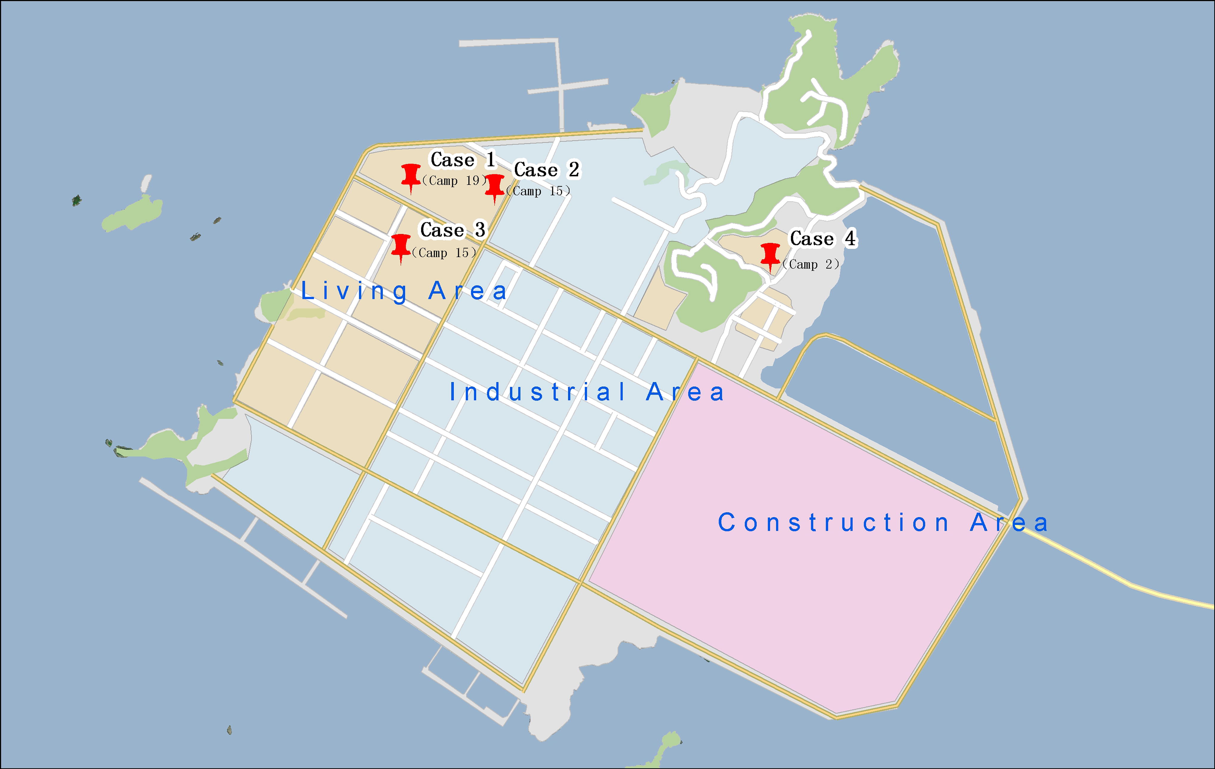
Distribution map of cases. The industrial area, living area, and construction area were all indicated on the map, and the red pins indicated the cases’ locations in this outbreak.
Partial Epidemiological Characteristics of Cases
Host animals’ survey results
Forty rat cages were set up in campsites 2, 15, and 19, and 8, 18, and 12 rodents were accordingly captured, respectively. All of the captured rodents were Rattus norvegicus. The rodent density was 20%, 45%, and 30% for these areas, respectively, with an overall rodent density of 31.67%. HV RNA was detected positive by quantitative RT-PCR of one rodent from campsite 2, as well as two each from campsite 15 and campsite 19 (Table 4).
Survey of the Population Density of Rat and HV Detection Results
HV, hantavirus.
Samples’ detection results
All cases were tested positive for both anti-HV IgG and IgM. HV RNA was detected positive in the serum samples of cases 2 and 3, whereas cases 1 and 4 showed no presence of HV RNA. HV RNA was detected in 4 of 38 R. norvegicus, indicating a viral infection rate of 10.53% among the rat population.
S and M gene sequencing and phylogenetic analysis
The M and S gene segments of HV RNA were amplified and sequenced from two cases and four R. norvegicus specimens. The sequences were uploaded to GenBank under accession number MT822296-MT822307. The phylogenetic analysis showed that the M gene fragment exhibited 100% homologous and belonged to SEOV S3 subtype, which was evolutionary distinct from Hantaan virus (HTNV). Specifically, the M gene was found to be most closely related to Rod/2012/QHD/4/GC isolated from Hebei and RuianRN180 in Zhejiang province (Fig. 2). The S gene was identified on the same evolutionary branch with 99.6 − 99.8% homology and was most closely related to the Jiangxi isolated JiangxiXinjianRn-09-2011 while having the most distant relationship with the French isolated REPLONGES (Fig. 3). The length of M gene is 417 bp, and the length of S gene was 560 bp.

Phylogenetic analysis of partial M gene of SEOV. The viral sequences were analyzed by DNAStar software, and the phylogenetic analysis of nucleotide sequences was carried out with the maximum likelihood method. SEOV, Seoul virus.
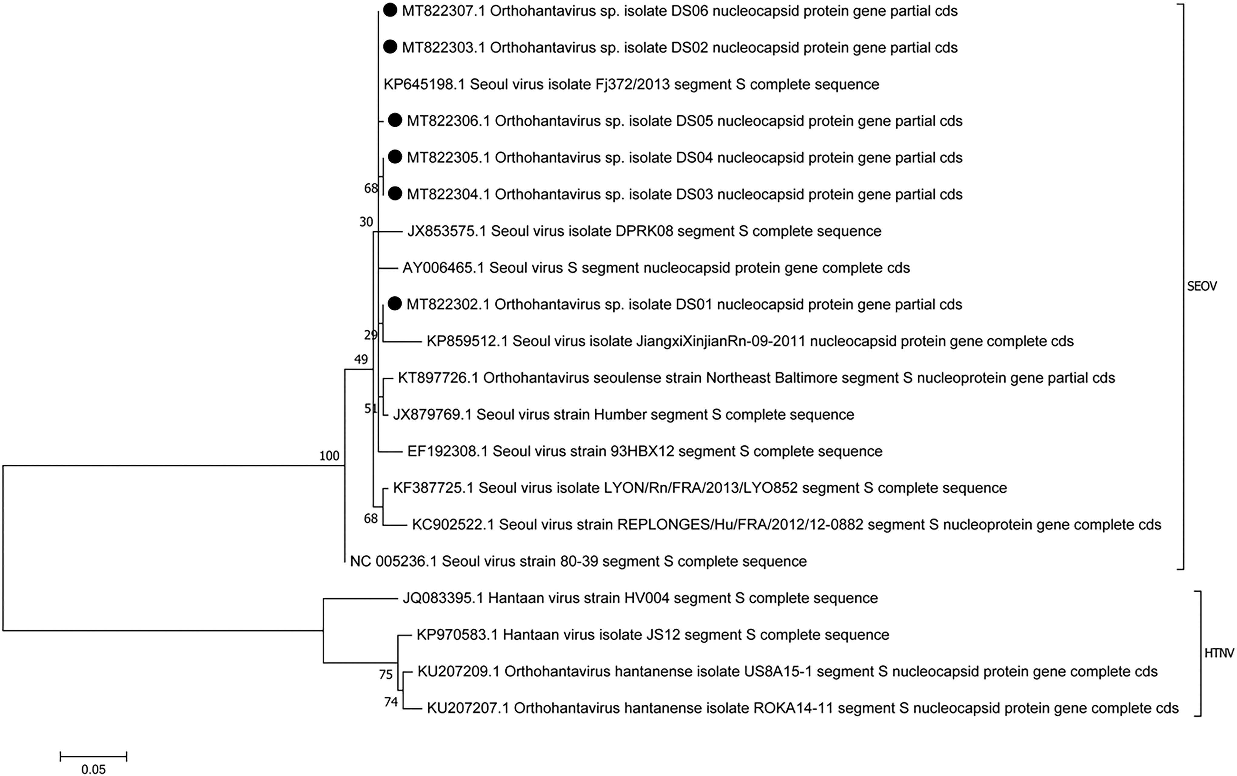
Phylogenetic analysis of partial S gene of SEOV. The viral sequences were analyzed by DNAStar software, and the phylogenetic analysis of nucleotide sequences was carried out with the maximum likelihood method.
Discussion
Rodent habitat disturbance can lead to increased rodent activity, raising the likelihood of contact with individuals working or residing in the same area (Ma et al., 2014). Following the reconstruction of the industrial park, significant changes occurred in the geographical environment and the living habitat of rodents. This transformation resulted in a substantial increase in population density, maintaining high levels over an extended period, along with a heightened viral carrying rate. The primary determinant influencing the prevalence of HFRS in humans is the periodicity and abundance of rodents, particularly in relation to the development of epidemics within their expanding populations. Prolonged cohabitation with a large number of rodents, coupled with exposure to rodents and their excreta, escalates the risk of HFRS infection (Tkachenko et al., 1999).
HVs exhibit remarkable stability in the air, surviving for over 10 days at room temperature and even longer periods exceeding 18 days at 4°C or −20°C (Hardestam et al., 2007, Kallio et al., 2006). Our investigation revealed that the virus is primarily transmitted to humans through the inhalation of virus-contaminated aerosols from rodent excreta and secretions, as well as contaminated food, rather than through rodent bites, consistent with findings from a previous study (Pedrosa and Cardoso, 2011). The industrial park’s environment is characterized by poor conditions and high levels of dust, with workers often neglecting to wear masks. Inhalation of virus-laden aerosols from contaminated excreta and secretions is likely a significant mode of transmission. Furthermore, the substandard food and lodging arrangements for workers contribute to the problem, as rats and their excreta are frequently encountered in canteens and dormitories. Insufficient measures are in place to prevent rodents from accessing raw food materials, finished products, and tableware, further exacerbating the risk of infection.
All rodents captured during our investigation were identified as R. norvegicus, with the viruses detected in both the rodents and the patients samples being SEOV. This finding aligns with the established knowledge that HV typically originates from Apodemusagrarius, whereas SEOV is associated with R. norvegicus (Zhang et al., 2014). Symptoms of HFRS caused by HV are more pronounced and carry a higher mortality rate than those caused by SEOV. Conversely, HFRS symptoms resulting from SEOV infection are milder, with atypical clinical manifestations, lower mortality rates, and better prognoses. In this outbreak, all cases were attributed to SEOV. The duration between disease onset and the initiation of effective treatment positively correlates with the disease course; early diagnosis and intervention can reduce the disease duration and enhance prognosis.
Gene sequencing and comparison of HV virus strains from the infected rodents and human cases revealed a high level of homology, suggesting a strong epidemiological relationship with R. norvegicus. The outbreak of HFRS was closely related to the change in geographical environment, elevated rodent density, high prevalence of virus-carrying rodents, large population density, poor working and living conditions, unhealthy habits, and the absence of HFRS vaccination among workers.
Vaccination and rodent extermination are the primary methods used to control HFRS (Ma et al., 2012). Expanding vaccine immunization programs, as well as promoting earlier diagnosis and effective treatment, can help reduce the morbidity and mortality of HFRS in endemic areas (Ma et al., 2014). Currently, the most effective strategy for preventing HV infections is to limit human–rodent contact and avoid respiratory inhalation of fecal-contaminated air. Furthermore, increasing public awareness and knowledge of pathogen sources and transmission routes (including how to avoid contact with HV), improving housing conditions, maintaining good hygiene, and reducing human migration from rural to urban areas can all contribute to a decline in HFRS cases (Zhang et al., 2010).
Footnotes
Availability of Data and Materials
All data are available upon reasonable request from the corresponding author.
Authors’ Contributions
Qilong Tan and Jiwei Shu carried out the epidemic survey, Lin Ye, Sen Zhang, and Zhiping Wang helped with survey preparation. Tongjie Zhang was responsible for the gene classification work and Shibo Li helped with grammar checking. Zhilei Mao wrote the original manuscript and was responsible for the data quality control.
Author Disclosure Statement
The authors declare no competing interests.
Funding Information
The study was supported by the Top Talent of Changzhou “The 14th Five-Year Plan” High-Level Health Talents Training Project (2022CZBJ088), Natural Science Foundation of Zhejiang Province (LY21H100002), Project Foundation of Zhoushan Science and Technology Bureau (2021C31089), and Project Foundation of Zhoushan Science and Technology Bureau (2024C31025).