




Abstract
Introduction:
COVID-19 is an infectious disease caused by SARS-CoV-2 that has become a serious threat to public health owing to its rapid spread from aerosols from infected people. Despite being considered a strictly human disease, there are reports in the literature about animals with confirmed presence of the virus.
Aim:
Owing to the scarcity of scientific literature on the potential for infection of animals and their importance for One Health, the objective of this work was to research SARS-CoV-2 RNA in felines (Felis silvestris catus) and dogs (Canis lupus familiaris) domiciled.
Materials and Methods:
Oropharyngeal swabs were collected from domestic dogs and cats belonging to patients diagnosed with COVID-19 from August to October 2021 and residents of the northwest and west regions of Paraná, Brazil.
Results:
Of the 34 samples collected, 14 were from dogs and 20 from cats. Three of these samples tested positive in real-time PCR, and two of them were also positive in the immunochromatographic test. After testing positive in real-time PCR, the samples underwent genetic sequencing using the Illumina COVIDSeq test. Of the 34 samples collected, three (9%), all of them female and from the feline species, tested positive in real-time PCR, with two of these (67%) also testing positive in the immunochromatographic test. Regarding sequencing, it was possible to sequence the three samples aligned with the AY.101 lineage, corresponding to the Delta variant.
Conclusion:
The occurrence of SARS-CoV-2 infection in dogs and cats is seen as an unintended event with significant implications for public health, including its potential transmission to other animal species. Further research is required to enhance our understanding of how this disease spreads among these animals and its broader impact on One Health initiatives.
Introduction
First reported in 2019, SARS-CoV-2, a single-stranded RNA virus with an envelope responsible for the death of 6.61 million people by November 2022, triggered one of the major pandemic scenarios in recent times (Limongi and Oliveira, 2020; Lopes et al., 2020; Singhal, 2020; Velavan and Meyer, 2020). Its zoonotic potential in domestic dogs (Canis lupus familiaris) and cats (Felis silvestris catus) has significant implications for public health and One Health strategies.
As highlighted by Han et al. (2022) and Our World in Data (2022), the COVID-19 pandemic, caused by SARS-CoV-2, has resulted in a substantial global burden with millions of deaths reported by November 2022. Although the primary focus has been on human health impacts, it is crucial to consider the potential transmission of the virus to domestic animals.
Research by Delai et al. (2020), Lopes et al. (2020), Ristow et al. (2020), and Manzini et al. (2021) indicates that dogs and cats can be exposed to SARS-CoV-2, especially in households where humans are infected or during quarantine periods. Although the transmission from animals to humans is considered unlikely, the prolonged and close contact between infected individuals and their pets raises concerns about possible cross-species infections.
Recent studies, such as Leroy et al. (2020) and Bienzle et al. (2022), have detected SARS-CoV-2 RNA and antibodies in asymptomatic domestic dogs and cats, suggesting potential viral shedding and infection in these species. However, the zoonotic risk and clinical manifestations in animals remain areas of ongoing investigation, underscoring the need for further research.
Given the scarcity of scientific literature on the zoonotic potential of SARS-CoV-2 in canine and feline species and their importance for One Health, the objective of this work was to investigate the presence of SARS-CoV-2 RNA in domiciled felines (Felis silvestris catus) and dogs (Canis lupus familiaris) from the state of Paraná, Brazil. We identified three felines that resulted positive to SARS-CoV-2 immunochromatographic and/or real-time PCR tests, with all of them being infected by lineage AY.101 of the Delta variant of concern. We believe that these findings warrant attention from public health authorities and veterinary professionals to implement appropriate measures for surveillance, prevention, and control of zoonotic diseases.
Materials and Methods
Ethics committee
This study was approved by the Research Ethics Committee Involving Animal Experimentation at UNIPAR under protocol 1002/2020.
Sample and location
A sterile rayon-tipped swab (Neolab®) was used to collect oropharyngeal swabs from dogs and cats belonging to patients diagnosed with SARS-CoV-2 (COVID-19) between August and October 2021 in the northwest and west regions of the state of Paraná, Brazil. These animals, 14 dogs and 20 cats, belonged to 11 patients. For all animals analyzed in the study, their guardians authorized the collection of biological samples by signing a Term of Consent and Authorization provided by the researchers.
Of these animals, 23 were found in a municipality situated in the northwest region (Umuarama city), whereas 11 were in a municipality in the western region (Cascavel city), both situated in the state of Paraná and near the Paraguayan border (Fig. 1).
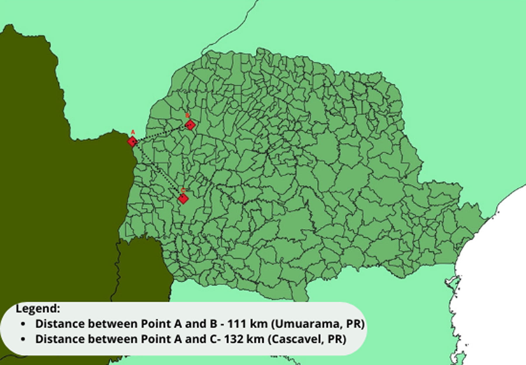
Map of the state of Paraná, Brazil, containing the location of the collection areas for biological samples of dogs and cats and their distance to the Paraguayan border. Legend: A—Paraguayan border; B—City of Umuarama; C—City of Cascavel.
Veterinarians examined the animals during collection to check for any clinical signs or physiological alterations. It’s important to note that the collection of biological material occurred after the quarantine period (∼20 days postdiagnosis confirmation) of the animal’s respective owner, ensuring the physical and sanitary integrity of the researchers involved.
The biological samples were placed in sterile bottles containing 1% phosphate-buffered saline (PBS) transport medium and promptly refrigerated before being sent to the Laboratory of Molecular Biology within the Graduate Program in Animal Science with a Focus on Bioactive Products at UNIPAR. There, they were stored frozen at −80°C. Once all samples were collected, they were forwarded to the Tropical Medicine Center of the Itaiguapy Health Foundation in Foz do Iguaçu, Paraná, and to the Bioinformatics Laboratory at the National Scientific Computing Laboratory in Petrópolis, Rio de Janeiro, for diagnosis and sequencing, respectively.
Serological and molecular diagnostics
Serological test—rapid immunochromatographic test
For the analysis of positive samples in the real-time PCR research, a rapid immunochromatographic test (COVISTIX-Sorrento Therapeutics®) was used, which uses a platinum nanocatalyst specifically directed to the nucleocapsid (N) or matrix antigens (M) of the virus. Thus, the nasal swabs confirmed through molecular biology were submitted to a tube containing a lysis buffer; after 2 min, four drops were discarded in the center of the cassette sample well; and after 15 min, the tests were read. Samples that showed two clear lines, one in the Test band (T) and the other in the Control band (C), were considered positive. Samples that showed only one line evident only in the Control (C) lane were considered negative.
Nucleic acid extraction
For the extraction of genetic material, the Zymo Quick DNA/RNA magBEad kit (Zymo Research®) was used, which uses electromagnetic beads technology during the sample extraction and purification process, according to the manufacturer’s specifications. After this procedure, the samples were submitted for amplification through real-time PCR.
Real-time PCR
The amplification of the genetic material was performed in the QuantStudioTM 7 Flex Real-Time PCR equipment (ThermoFisher®) using the Biomol One Step/COVID-19 Kit (IBMP®), which is a molecular test for detecting the presence or absence of SARS-CoV-2 nucleic acids in nasopharyngeal and/or oropharyngeal swab samples using the real-time polymerase chain reaction with reverse transcription (RT-PCR) technique, allowing the detection of specific sequences in an RNA sample extracted from measurements of fluorescence intensity during the reaction. In this case, two targets were investigated: ORF1ab (ORF) conserved region and nucleocapsid N protein region. For this, the cycling conditions proposed by the kit manufacturer were used.
Samples that showed characteristic amplification for only the ORF target or both ORF and N targets up to cycle threshold (Ct) 40 and amplification only for the N target with Ct less than 37 were considered positive. Samples that did not show amplification of any of the genes and those that only showed amplification of the N target above Ct 37 were considered negative. To carry out the diagnosis, positive and negative controls provided by the kit manufacturer were used, following their preparation and interpretation guidelines.
Sequencing methodology
Libraries were constructed with Illumina COVIDSeq Test (Illumina), according to the manufacturer’s protocol without any modifications. The TapeStation System (Agilent) was used for the libraries’ quality control. Sequencing was conducted using a NextSeq 500/550 Mid Output Kit v2.5 (300 Cycles) in a NextSeq 500 equipment (Illumina) set to generate 2 × 149 bp reads.
Sequence analysis, consensus building, and variant calling were performed with DRAGEN COVID Lineage v3.5.9. The assembled genomes were classified according to the Pango Lineage classification using the PangoLEARN model database (v4.0.6) and SNPs annotated with snpEff/SnpSift (v.5.0e). All genomes were then submitted to the Global initiative on sharing all influenza data (GISAID) database and made publicly available under the identifier EPI_SET_221206ps.
To further confirm the lineage classification of genomes, we have selected from the GISAID database the oldest sequence from Paraná of each SARS-CoV-2 lineage present in the state up to the sampling period. The GISAID and newly sequenced genomes were aligned with MAFFT v7.505 (Katoh and Standley, 2013) to the WH01 genome (EPI ISL 406798) sequence from Wuhan, China. A maximum likelihood phylogenetic tree was then inferred using IQTREE2 v2.0.3 (Minh et al., 2020) with the evolutionary model selected by the ModelFinder built-in algorithm (Kalyaanamoorthy et al., 2017). Ultrafast bootstrap was run using 10,000 tree replicates.
Results
Samples were collected from 34 domestic animals, 14 (41%) of the canine species and 20 (59%) of the feline species. Of these animals, 23 (68%) live in a municipality in the northwest region (city of Umuarama) and 11 (32%) live in a municipality in the west region (city of Cascavel), both located in the state of Paraná, Brazil.
Three female cats (9% of all samples) tested positive for SARS-CoV-2 in the real-time PCR test (Fig. 2), indicating the characteristic amplification of both ORF and N genes. Samples #7 and #10 exhibited lower Ct values, approximately 28 and 30, respectively, whereas sample #8 had a higher Ct value, around 33. This variation in Ct values suggests differences in viral load among the samples, with #7 and #10 showing a higher viral load compared with sample #8.

Positive results of SARS-CoV-2 research by real-time PCR in three cats (Felis catus Linnaeus) domiciled in the western region of the state of Paraná, Brazil, August–October, 2021. Legend: Samples considered positive (#7, #8 and #10) and positive control (CP).
Of the three positive samples in real-time PCR, two were also positive in the immunochromatographic test (Fig. 3). Sample #7 presented the Test and Control bands very clearly. In contrast, sample #10 presented the Test band as a very weak mark but still visible to the point of being considered positive, and the Control band was evident as expected. Sample #8 showed only the Control range and was considered negative.
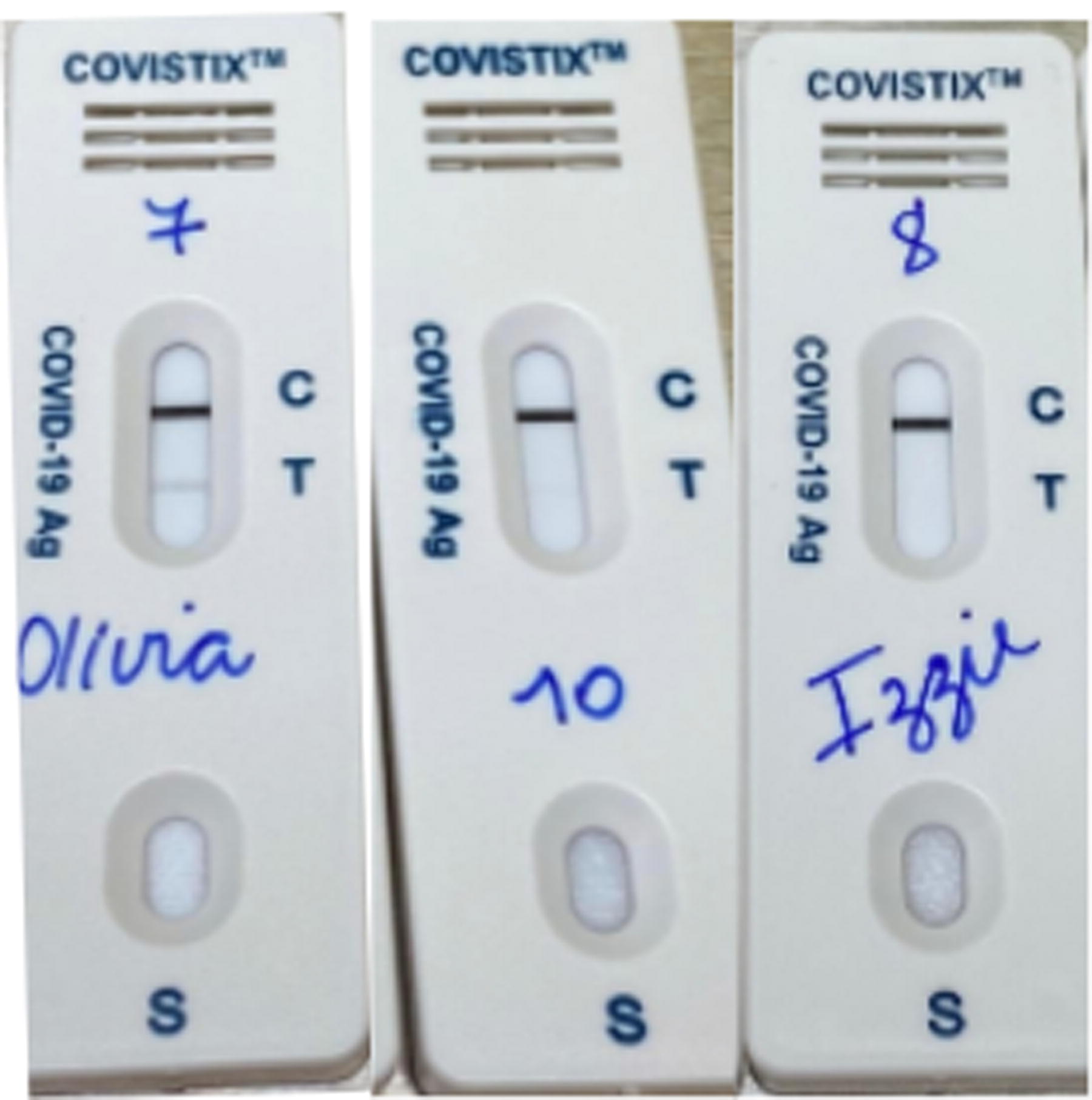
Result of the SARS-CoV-2 research by immunochromatography of positive samples in the molecular biology test in three felines (Felis silvestris catus) domiciled in the western region of the state of Paraná, Brazil, August–October, 2021. Legend: Samples considered positive (#7 and #10) and sample considered negative (#8).
Regarding the analysis of the phylogenetic position of these three SARS-CoV-2 samples, it was observed that they consistently cluster with the genome of the AY.101 lineage of the Delta variant (Fig. 4) with significantly high bootstrap support. These results provide strong evidence that the samples in question indeed belong to this specific genomic lineage.
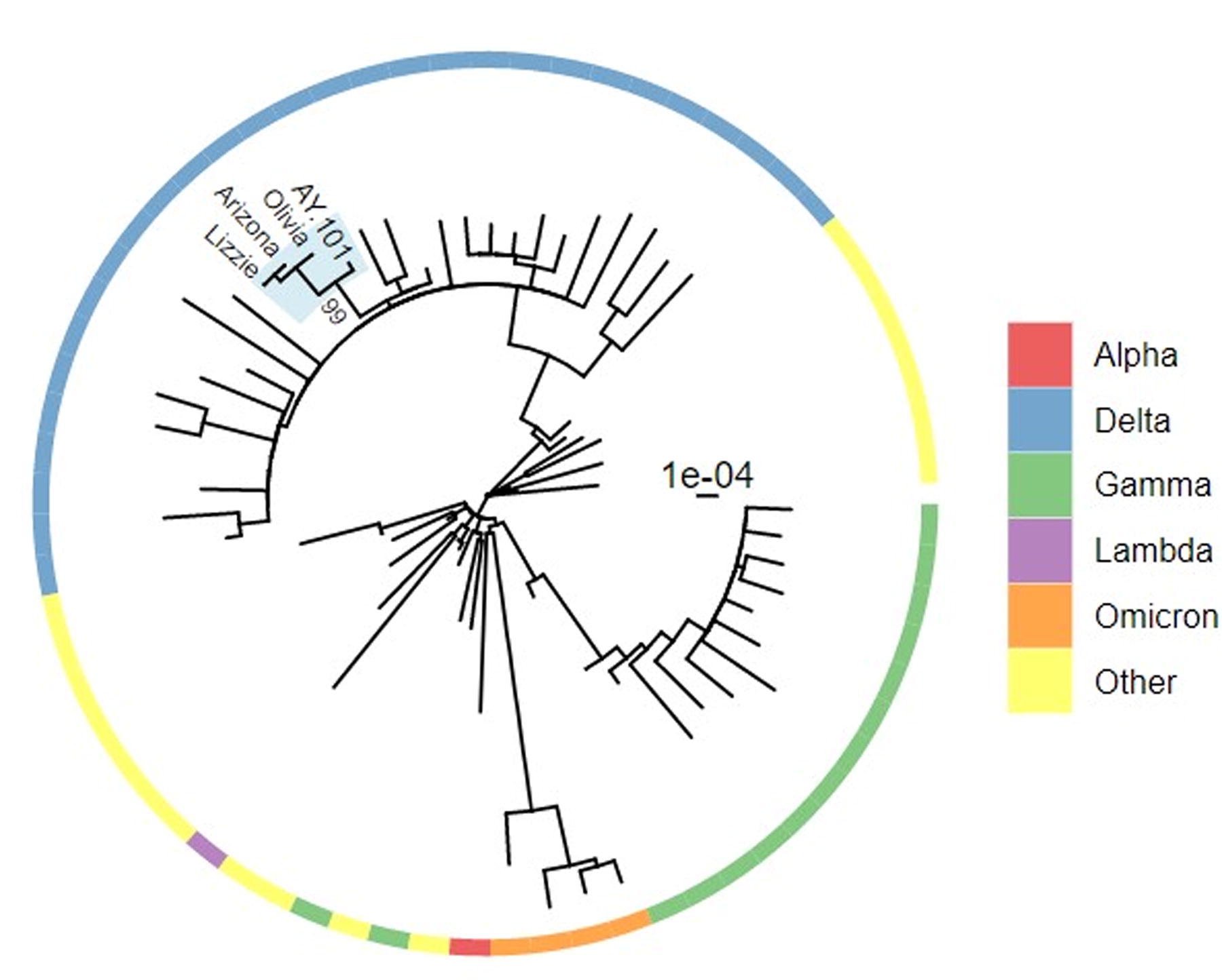
Phylogenetic position of the SARS-CoV-2 sequences sampled from cats. Labeled are the three newly sequenced genomes and the lineage AY.101, as well as the ultrafast bootstrap support for the node. Legend: Outer ring indicates the VOC/VOI classification of lineages. VOC, variant of concern; VOI, variant of interest.
Discussion
In the context of the SARS-CoV-2 pandemic, epidemiological studies on infectious diseases, particularly in border regions between countries, have proven to be extremely important. These studies help us understand the actual prevalence, modes of transmission, resulting physiological changes, and the factors that increase the risk of infection and mortality.
Using scientific information can help governments in the implementation of improved actions among health agencies, contributing to the establishment of more effective strategies and decision-making regarding prevention and health education actions for local communities.
In this work, no positive sample for SARS-CoV-2 was detected in the canine species; however, in another study, a 2-year-old asymptomatic male German Shepherd dog in Hong Kong was diagnosed with SARS-CoV-2 shortly after his owner (LOPES et al., 2020). In February 2020, in the same location, a 17-year-old male Pomeranian dog, also asymptomatic, was diagnosed as positive for this disease by the real-time PCR technique. In this case, the authors suggested the possibility of human–animal transmission because the owner for the animal had been previously diagnosed with COVID-19 (Almendros, 2020).
A study recently published in Chile by Agüero et al. (2024) revealed positive results for the BA.4.1 variant of COVID-19 Omicron in three dogs. Similarly, a study conducted in Switzerland by Kuhlmeier et al. (2023) also identified positive results in 3 dogs and 11 cats that shared a history of COVID-19 positivity with their owners. In addition, the strains detected were identified as the Delta variant, which was circulating in the region during the research period.
Kuroda et al. (2023) also obtained positive results in both dogs and cats from positive owners. Interestingly, these animals, although they did not show apparent clinical signs and symptoms, produced neutralizing antibodies against the disease.
Cardoso et al. (2023), in their research in the suburbs of Buenos Aires, found that cats were infected at a higher rate than dogs, with a seroprevalence of 7.1% and 1.68%, respectively. On the contrary, Yamayoshi et al. (2023) found much lower positivity rates in both dogs and cats between 2020 and 2021, leading to the conclusion that the seroprevalence of antibodies against SARS-CoV-2 in dogs and cats in Japan is low. This suggests that these animals are not a major reservoir for SARS-CoV-2.
The study by Cabrera et al. (2022) identified confirmed cases of natural infection in cats and dogs, suggesting an association with direct contact with COVID-19-positive human patients, although the virus’s virulence in animals and its ability to transmit between species are still not clear. On the contrary, Guo et al. (2023) provided a broader view of the molecular and serological prevalence in pets, noting that the presence of COVID-19-positive individuals in households can significantly increase the prevalence of SARS-CoV-2 in pets. Both studies agree on the need for global standards for virus detection in pets and the importance of preventive measures to protect the health of the entire family, including pets.
Although real-time PCR results were negative in the dogs in this study, we cannot rule out the possible exposure to the etiological agent, as it was not possible to perform a neutralizing antibody analysis to verify their exposure to SARS-CoV-2. In addition, studies have shown similarities between genetic sequencing analyses of viruses in dogs and their respective owners, indicating the possibility of virus sharing in the household environment and affecting dogs residing in the same residence (Lopes et al., 2020; Sit et al., 2020).
Of the three positive samples in the real-time PCR of this work, two of them (67%) were also positive in the rapid immunochromatographic test. A previous study has already indicated the detection of viral RNA and virus titration in samples of feline nasal turbinate, soft palate, tonsils, and trachea, which indicates a possible transmission of respiratory droplets (Shi et al., 2020).
Both techniques used in this study possess unique characteristics that merit consideration. Real-time PCR exhibits heightened sensitivity in contrast to immunochromatography owing to its ability to amplify a single molecule of the viral genetic material present in the sample billions of times. Conversely, immunochromatography quantifies the level of antibodies or antigens present in the sample. Although serology assesses the presence of specific antibodies against the virus in the animal’s blood, offering valuable insights into exposure and immune response development, it may not match the sensitivity of real-time PCR in directly detecting the virus (Perez-Lopez and Mir, 2021).
Therefore, a considerable amount of virus needs to be present in the material for a positive reaction to occur. As samples #7 and #10 in this study exhibited higher viral concentrations than sample #8, as interpreted based on Ct values, it is reasonable to infer a technical limitation owing to the lower viral load present in the last-mentioned sample (Perez-Lopez and Mir, 2021).
In this work, three (9%) SARS-CoV-2 samples obtained from the feline species, all females, were positive in the real-time PCR molecular analysis. In Belgium, in March 2020, a domestic cat had a confirmed diagnosis of SARS-CoV-2 in samples submitted to this same test. This animal developed some clinical signs suggestive of the disease, such as diarrhea and respiratory problems, whose owner also had the diagnosis confirmed for the disease (Lopes et al., 2020).
In Spain, an asymptomatic cat was also diagnosed with the new coronavirus (Ruiz-Arrondo et al., 2021). Despite evidence that cats can transmit COVID-19 to each other, it is believed that the highest probability is a human–animal transmission, with the main route of viral transmission to cats being the contact with people infected with the virus (Ristow et al., 2020).
Research has already reported that cats are more likely to contract SARS-CoV-2 than dogs. This is due to the similarity that exists between angiotensin-converting enzyme 2 (ACE2) and human and feline amino acids, and the lack of variation in ACE2 indicates that felines are susceptible, as they share highly homologous ACE2 proteins (Mathavarajah and Dellaire, 2020; Sit et al., 2020).
Direct contact between humans and pets, especially dogs and cats, has been increasing in recent years, so it is important to understand the susceptibility of these two species of animals to SARS-CoV-2 (Grisolio et al., 2017; Shi et al., 2020). The hypothesis that the virus can be disseminated through an infected person to the pets that live with the patient was evidenced in this work, as it is known that the owner of the infected animals did not interrupt domiciliary contact with them, even during the period in which the individual was socially isolated because of the disease. This fact is reinforced by research carried out by other authors who share the premise that animals that share a bed or other indoor environments with their infected guardians are more likely to be associated with SARS-CoV-2 (Lopes et al., 2020; Sit et al., 2020; Hannah et al., 2022).
The studies by Klaus et al. (2021) and Van Aart et al. (2022) contribute to understanding the prevalence of SARS-CoV-2 infection in cats and dogs during the COVID-19 pandemic. Klaus et al. (2021) highlights the low prevalence of infection in pet cat and dog populations, especially during the first wave of the pandemic, even in hotspot areas. This is corroborated by Van Aart et al.’s study (2022), which also identified a low infection rate in cats and dogs, especially in infected mink farms. Both studies suggest that the risk of zoonotic transmission to veterinary staff from pets appears to be low, but they emphasize the ongoing importance of surveillance and adoption of preventive measures to ensure the safety of all involved.
Regarding the Delta variant, it was first reported in India in October 2020, and in April 2021, the World Health Organization (WHO) classified it as a variant of interest. One month after disclosure, it was recognized as a variant of concern (VOC), meaning that it showed increased transmission and severity/change in the clinical presentation of the disease as the Delta variant contains molecular signatures capable of triggering phenotypic and epidemiological implications of global relevance. In July of the same year, the VOC Delta represented the most dominant strain in the SARS-CoV-2 pandemic profile in humans around the world, as it exhibited multiple molecular signatures in its Spike protein (WHO, 2021; Arantes et al., 2022; Yang and Shaman, 2022).
According to the Butantan Institute, the Delta variant was responsible for 90% of cases of COVID-19 in humans in Brazil, being registered in the 26 Brazilian states and the Federal District (Moreno, 2021). The research identified that the spread of the Delta variant was described in eight Brazilian states in the first half of 2021. Regarding community transmission, which was already reported in June 2021, these were detected in the states of Rio de Janeiro, Goiás, Maranhão, and Paraná (Boehm, 2021; Lamarca et al., 2022).
At the beginning of September 2021, the Delta variant was already the most dominant in the southern region of the country. The AY.101 lineage that emerged in the state of Paraná at the end of April of the same year was responsible for the largest fraction of genomes sampled from the southern region of the country (Arantes et al., 2022).
All the feline samples that tested positive in this study were collected from the western region of Paraná state, Brazil, between August and October 2021, a period during which the Delta variant was predominant among humans in the same region. This finding strongly suggests that these animals might have contracted the infection from their owners, who had previously been diagnosed with COVID-19. Studies of this nature play a crucial role in highlighting the potential health risks associated with the close bond between owners and their pets, illustrating the bidirectional transmission of diseases that can occur between humans and animals.
Conclusion
This study confirmed the presence of three positive feline samples in the real-time PCR test, with phylogenetic analysis supporting their classification within the AY.101 lineage, corresponding to the Delta variant. Although the real-time PCR results were negative in dogs, the potential exposure to the virus cannot be ruled out, as the tests performed detect the presence of the virus and not late exposure to it, given the lack of serological tests to confirm the presence or absence of antibodies against SARS-CoV-2.
Infection of dogs and cats with SARS-CoV-2 is deemed an incidental event with significant epidemiological implications, including the risk of transmission to other animal species. Further research is essential for understanding the disease’s epidemiology in dogs and cats, especially those kept as pets, and to fully comprehend the implications of these infections within the One Health framework. It is imperative to emphasize efforts in health education to increase awareness within the community, particularly among pet owners, regarding the potential transmission of SARS-CoV-2 to animals and the necessary precautions that need to be implemented.
Footnotes
Acknowledgments
The authors would like to thank all the authors and administrators of the GISAID database, which allowed this study of genomic epidemiology to be conducted properly. A full list acknowledging the authors’ publishing data used in this study can be found in the Supplementary Data S1. The authors would also like to thank the guardians of the animals for their trust and constant collaboration during this work.
Authors’ Contributions
Laisa Marina Rosa Rey—Writing—Original Draft and Investigation; Robson Michael Delai—Project administration; Aline Cristiane Cechinel Assing Batista—Writing—Review & Editing; Leonardo Ferreira—Investigation; Isabela Carvalho dos Santos—Investigation, Writing—Review & Editing and Conceptualization; Marco Aurélio Cunha Del Vecchio—Investigation; Ana Cláudia Souza Andrade—Investigation; Pedro Teles—Investigation; Ulisses de Pádua Pereira—Conceptualization; Alexandra Lehmkuhl Gerber—Investigation; Ana Paula de Campos Guimarães—Investigation; Luiz Gonzaga Paula de Almeida—Investigation; Alessandra Pavan Lamarca—Investigation; Ana Tereza Ribeiro de Vasconcelos—Investigation; Daniela Dib Gonçalves—Supervision, Project administration and Writing—Review & Editing.
Author Disclosure Statement
No competing financial interests exist.
Funding Information
This work was developed in the frameworks of Brazil, Russia, India, China and South Africa (BRICS) National Council for Scientific and Technological Development (CNPq) (440931/2020-7) and Corona-ômica-RJ Carlos Chagas Filho Foundation for Research Support in the State of Rio de Janeiro (FAPERJ = E-26/210.179/2020 and E-26/211.107/2021). A.T.R.V. is supported by CNPq (307145/2021-2) and FAPERJ (E-26/201.046/2022). A.P.L. is granted a postdoctoral scholarship Technological and Industrial Development (DTI-A) from CNPq. The authors acknowledge the support from the Rede Corona-ômica BR Ministry of Science, Technology and Innovation (MCTI) FINEP affiliated to RedeVírus/MCTI (FINEP 01.20.0029.000462/20, CNPq 404096/2020-4). The authors also thank CNPq CP (CNPq) CP 09/2020, Fundação Araucária 09/2021 (PDP2021071000001), and Instituto de Biologia Molecular do Paraná.
Supplementary Material
Supplementary Data S1
References
Supplementary Material
Please find the following supplemental material available below.
For Open Access articles published under a Creative Commons License, all supplemental material carries the same license as the article it is associated with.
For non-Open Access articles published, all supplemental material carries a non-exclusive license, and permission requests for re-use of supplemental material or any part of supplemental material shall be sent directly to the copyright owner as specified in the copyright notice associated with the article.
