
Abstract
Background:
Vancomycin-resistant enterococci (VRE) have become an increasing public health concern in the past few decades, being associated with serious multidrug-resistant (MDR) infections. This study was conducted to investigate the role of diarrheic pet animals as potential reservoirs for virulent extensively drug-resistant (XDR) VRE and their threat on human health.
Materials and Methods:
Rectal swabs were collected from 153 diarrheic pet animals (80 dogs and 73 cats). The collected swabs were cultured on CHROMagarTMVRE for the isolation of vancomycin-resistant Enterococcus faecalis and Enterococcus faecium, and then suspected colonies were identified as enterococci after Gram staining, conventional biochemical tests, and molecular techniques. VRE were basically identified using the disk diffusion method; however, molecular identification of vanA and vanB genes was carried out among confirmed VRE isolates. Moreover, three virulence genes (cytolysin A, cylA; enterococcal surface protein, esp; and hyaluronidase, hyl) were investigated in VRE isolates. Thereafter, VRE strains that harbored virulence genes were tested for antimicrobial susceptibility.
Results:
Eighteen out of 153 animals (11.8%) were positive for VRE, which were obtained from 15% and 8.2% of the examined dogs and cats, respectively. None of the obtained isolates carried the vanA gene, whereas the vanB gene was detected in E. faecalis (4/10) with a prevalence rate (40%). Of the obtained VRE isolates, five possessed esp and/or cylA, while all strains were negative for the hyl gene. Furthermore, four virulent VRE isolates exhibited an XDR pattern, and one isolate was MDR.
Conclusion:
Diarrheic pet animals could represent a potential zoonotic reservoir for virulent XDR vancomycin-resistant E. faecalis, which may have serious public health implications.
Introduction
Enterococci are gram-positive bacteria that colonize the intestinal tract of human and animals. For many years, enterococci were believed to be commensal intestinal microbiota that inhabit the gastrointestinal tract and oral cavity of mammals including human. However, in the past few decades, Enterococcus has emerged as one of the most important hospital and community-acquired pathogens worldwide (Wada et al. 2021). Enterococci are opportunistic pathogens associated with many life-threatening infections, such as urinary tract infection (UTI), endocarditis, and blood stream infections (Arias and Murray 2012). Till date, over 50 enterococcal species are currently recognized, including the most clinically relevant, Enterococcus faecalis and Enterococcus faecium, which usually represent the main species recovered from human cases and they are a leading cause of multidrug-resistant infections (Lebreton et al. 2013). Notably, enterococci can rapidly develop resistance against antimicrobial agents including newer ones such as quinupristin-dalfopristin, linezolid, daptomycin, and tigecycline (Niebel et al. 2015). However, vancomycin remains the last treatment choice in some cases (Fiore et al. 2019). Since the first reports of vancomycin-resistant enterococci (VRE) in the 1980s (Uttley et al. 1988), epidemiological studies have demonstrated serious health and economic impacts of VRE-associated infections in human medicine, being implicated in outbreaks of intensive care unit-hospitalized patients (Gilmore et al. 2013, Moosavian et al. 2018). Recently, the emergence of multidrug-resistant (MDR) and extensively drug-resistant (XDR) VRE strains is a worrisome issue due to very limited treatment options of clinical cases (Said and Abdelmegeed 2019). Importantly, as horizontal gene transfer is responsible for the elevated levels of resistance to different antibiotics, likewise vanA and vanB genes being transmissible genes for conferring vancomycin resistance, it is wise to underscore such genes (Freitas et al. 2016).
Beside antimicrobial resistance, enterococci are equipped with an array of virulence factors that contribute to illness development and surviving in hospital settings (Van Tyne and Gilmore 2014). For example, the enterococcal surface protein (esp) gene is considered a marker for nosocomial-epidemicity that aids in the acquisition of antimicrobial-resistant genes and the spread of VRE in hospitals (Ahmed and Baptiste 2018). Cytolysin A (cylA) is necessary for the expression of component A (Vankerckhoven et al. 2004), while hyaluronidase (hyl) encoded by chromosomal hyl has been implicated in the invasion of the nasopharynx and pneumococcal pneumonia (López et al. 2013). Human colonization by VRE of animal origin has been documented, including transconjugant transfer of van genes between enterococcal species within the human intestinal microflora (Lester et al. 2006). Several reports have focused on the investigation of VRE in swine, poultry, and food-producing animals (Wada et al. 2022, Grudlewska-Buda et al. 2023, Paschoalini et al. 2023), but there are limited reports regarding the prevalence of VRE among pets (Ortiz-Díez et al. 2020). Therefore, the current study was conducted to highlight the potential role of diarrheic dogs and cats in the transmission of virulent XDR VRE and their threat to human health.
Materials and Methods
Collection of samples
Rectal swabs were collected from 153 pet animals (80 dogs and 73 cats) suffering from diarrhea from veterinary hospital at the Faculty of Veterinary Medicine, Cairo University, and different private veterinary clinics. The swabs were placed into Cary–Blair transport medium tubes and transported to the Department of Microbiology, Faculty of Veterinary Medicine, Cairo University, in an ice box for immediate bacteriological processing.
Isolation and identification of vancomycin-resistant enterococci
The collected swabs were directly plated on CHROMagarTMVRE and incubated at 37°C for 48 h. Vancomycin-resistant Enterococcus faecalis and Enterococcus faecium produced pink to mauve-colored colonies (Vijaya et al. 2014). The identification of VRE was carried out using Gram staining and conventional biochemical tests (Facklam and Collins 1989), as well as resistance to vancomycin by the disk diffusion method, which was performed according to the Clinical and Laboratory Standards Institute (CLSI) (2021).
Molecular identification of virulent vancomycin-resistant E. faecalis and E. faecium
DNA extraction
DNA was extracted from VRE isolates that exhibited resistance to vancomycin using the QIAamp® DNA Mini Kit (QIAGEN, Germany) (Huys et al. 2004).
Molecular identification of E. faecalis, E. faecium and detection vanA/vanB genes
The extracted DNA was enrolled in a quadruplex PCR targeting genes encoding for E. faecalis, E. faecium, vanA, and vanB genes according to the protocol described by Kariyama et al. (2000). The quadruplex PCR was performed under the following conditions: Initial denaturation at 94°C for 5 min, followed by 30 cycles of denaturation at 94°C for 1 min, annealing at 54°C for 1 min, and extension at 72°C for 1 min, with a final extension at 72°C for 10 min.
Molecular investigation of virulence genes among VRE isolates
Multiplex PCR was conducted for the detection of three virulence genes (cylA, esp, and hyl) in VRE strains (Vankerckhoven et al. 2004). The PCR reaction mixture contained 12.5 μL of Cosmo PCR RED Master Mix (Willowfor, UK), 4 μL of template DNA, 1 μL of each (cylA) and (esp) primers (10 pmoL), and 0.5 μL of hyl primer (10 pmoL), as well as 3.5 μL of nuclear free water, making the total volume 25 μL. The PCR mixture was subjected to 5 min initial denaturation at 94°C, followed by 30 cycles of denaturation at 94°C for 1 min, annealing at 56°C for 1 min, and extension at 72°C for 1 min, with a final extension at 72°C for 10 min. Thereafter, the amplified amplicons were visualized via agarose gel electrophoresis and photographed to demonstrate the specific bands (Fig. 1).
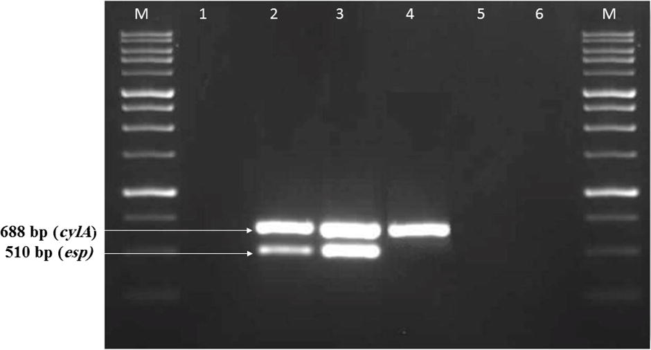
PCR amplification of virulence genes among vancomycin-resistant enterococci isolated from diarrheic dogs and cats. Lane M: DNA ladder (250 bp); lane 1: negative control; lanes 2, 3, and 4: positive strains showing specific bands; lanes 5 and 6: negative strains.
Antimicrobial susceptibility testing of the obtained virulent VRE strains
Antimicrobial susceptibility testing was performed for the obtained VRE isolates that possessed any virulence gene using the disk diffusion method according to the Clinical and Laboratory Standards Institute guidelines (CLSI 2021). The following antimicrobial agents were included: penicillin (P) (10 units), ampicillin (AMP) (10 μg), vancomycin (VA) (30 μg), erythromycin (E) (15 μg), tetracycline (TE) (30 μg), doxycycline (DO) (30 μg), ciprofloxacin (CIP) (5 μg), norfloxacin (NX) (10 μg), nitrofurantoin (NIT) (300 μg), rifampin (RIF) (5 μg), fosfomycin (FO) (200 μg), chloramphenicol (C) (30 μg), linezolid (LZ) (30 μg). The results were evaluated according to the guidelines of CLSI 2021. MDR was defined as non-susceptibility to at least one antimicrobial agent in three or more antibiotic classes, whereas XDR was defined as resistance to at least one agent in all but the bacterial isolate remains susceptible to only one or two antimicrobial categories (Basak et al. 2016).
Sequencing and phylogenetic analysis
PCR products of the esp gene from two vancomycin-resistant E. faecalis isolates retrieved from one dog and one cat were purified using the QIAquick purification Kit (Qiagen, Germany), and direct cycle sequencing was carried out on the ABI 3500 Genetic Analyser (Applied Biosystems, USA). The resulting sequences were blasted in the GenBank database to identify the most similar ones from human cases as well as animals. Multiple alignments were conducted using the ClustalW program of BioEdit software version (7.0.9), whereas, phylogenetic tree was built up through neighbor-joining approach in Mega7 software version 7.0.26, and a bootstrap consensus tree was obtained with 500 replicates (Fig. 2).
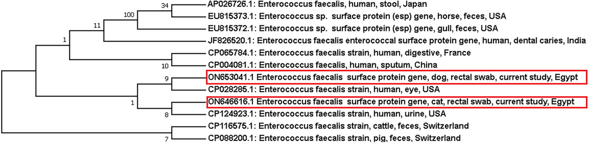
Phylogenetic bootstrap consensus tree was inferred via neighbor-joining approach using MEGA 7 software to show the evolutionary history and genetic relatedness between vancomycin-resistant E. faecalis esp gene partial sequences obtained in this study and those retrieved from GenBank records.
Nucleotide sequence accession numbers
The two partial sequences of vancomycin-resistant E. faecalis esp gene obtained in this study were deposited in GenBank under the following accession numbers: ON646616 for the cat sequence and ON653041 for the dog sequence.
Results
Prevalence of vancomycin-resistant Enterococcus faecalis and Enterococcus faecium among diarrheic dogs and cats
Eighteen out of 153 diarrheic pet animals yielded VRE (11.8%), where VRE strains were retrieved from 15% and 8.2% of the examined dogs and cats, respectively. Vancomycin-resistant E. faecalis was found in 10 animals, with a prevalence rate of 6.5% (dogs: 6.3%; cats: 6.8%), while 7 (8.8%) dogs and 1 (1.4%) cat were positive for E. faecium, with an overall prevalence of 5.2% (Table 1).
The Prevalence of Vancomycin-Resistant Enterococcus Faecalis and Enterococcus Faecium Among Diarrheic Dogs and Cats
Occurrence of vanA and vanB genes among vancomycin-resistant E. faecalis and E. faecium isolates
None of the E. faecalis or E. faecium had the vanA gene, while vanB was detected in 22.2% of enterococcal isolates, which was reported in E. faecalis only (40%). On the other hand, both genes were not found in 14 (77.8%) strains (E. faecalis: 6 strains, E. faecium: 8 strains), as shown in Table 2.
The Occurrence of Vana or Vanb Genes Among Vancomycin-Resistant E. Faecalis and E. Faecium Isolates
The distribution of virulence genes among the obtained VRE isolates
Of 18 VRE isolates, 5 vancomycin-resistant E. faecalis isolates possessed virulence genes (2 isolates carried esp and cylA genes, 2 have the esp gene, and one carried the cylA gene), while all strains were negative for the hyl gene (Table 3).
The Distribution of Virulence Genes Among the Obtained Vancomycin-Resistant E. Faecalis Strains
Antimicrobial susceptibility pattern of virulent VRE isolates
All five VRE strains harboring virulence genes were tested for antibiotic sensitivity. It was found that all five isolates were resistant to penicillin, ampicillin, vancomycin, erythromycin, tetracycline, and chloramphenicol, followed by rifampin and linezolid resistance, in four strains. Doxycycline and norfloxacin resistance were reported in three isolates, as well as two isolates exhibited non-susceptibility toward nitrofurantoin and fosfomycin, as detailed in Table 4. Furthermore, four isolates exhibited an XDE pattern, and one strain was MDR.
Antimicrobial Susceptibility Pattern of Virulent Vancomycin-Resistant Enterococci
P, penicillin; AMP, ampicillin; VA, vancomycin; E, erythromycin; TE, tetracycline; DO, doxycycline; CIP, ciprofloxacin; NX, norfloxacin; NIT, nitrofurantoin; RIF, rifampin; C, chloramphenicol; LZ, linezolid; FO, fosfomycin.
Discussion
The close relationship between pets and their owners has not only attracted more attention to their welfare, but also this intimacy has serious consequences, as it gives a lot of opportunity for acquiring and transmitting genes that confer antibiotic resistance to bacteria posing a serious threat to human being (Belas et al. 2020, Carvalho et al. 2020). The upsurge increases of studies on pet dogs and cats as a potential reservoir of antibiotic resistant pathogens (Samir et al. 2020, Das et al. 2023) prompted us to investigate the occurrence of virulent XDR VRE among diarrheic dogs and cats and its public health implication. In the present study, VRE were detected in 18 (11.8%) out of 153 examined diarrheic pet animals, where 15% and 8.2% of the examined dogs and cats were positive, respectively. Such result was higher than that obtained by Aslantaş and Tek (2019) who recovered VRE from one dog and two cats out of 276 dogs and 255 cats, but it was nearly similar to those detected by Bağcıgil et al. (2016) who isolated VRE from 14% and 11.3% of clinically healthy dogs and cats, respectively. Moreover, our findings showed no difference in the prevalence rate of vancomycin-resistant E. faecalis between diarrheic dogs (6.3%) and cats (6.8%) comparing with vancomycin-resistant E. faecium, which was higher in dogs (8.8%) than in cats (1.4%). It was noted that vancomycin-resistant E. faecium was the predominant Enterococcus species among diarrheic dogs which was agreeable with that of Herrero et al. (2004) who identified vancomycin-resistant E. faecium in 11 out of 15 VRE strains isolated from feces of dogs admitted at the Animal Hospital of the School of Veterinary Medicine in Madrid, Spain. The existence of VRE in dogs and cats may be attributed to the habits of some owners whose strong intimate relationships with household pets through activities such as licking, feeding, and cleaning gives an opportunity for bacterial transmission which is a potential threat to human contacts (Pillay et al. 2018).
Noteworthy, 4 (22.2%) out of 18 VRE strains carried vanB gene which was recorded in E. faecalis only while none of the enterococcal isolates harbored vanA gene as well as 14 isolates (77.8%) neither had vanA nor vanB. However, Gulhan et al. 2015 could not detect vancomycin resistance genes among enterococcal strains recovered from pet dogs and cats while being phenotypically vancomycin-resistant. On the contrary, some studies detected vanA gene in all VRE isolates retrieved from pets (Aslantaş and Tek 2019, Herrero et al. 2004, Iseppi et al. 2020
Enterococcus virulence genes facilitate the attachment and colonization of the bacteria in the host, enabling them to evade the host’s immune system and confirming the pathogenicity of these strains (Sava et al. 2010). In the current study, cylA and/or esp genes were detected in five VRE isolates while hyl gene was not recorded. Such virulence genes were reported only among Enterococcus faecalis strains which was similar to result of Aslantaş and Tek (2019) who found that all vancomycin-resistant E. faecium strains were negative for virulence genes. Conversely, Iseppi et al. (2020) could not find cylA and esp genes among VRE isolates recovered from dogs and cats. Cytolysin-producing E. faecalis are virulent strains causing animal and human infections and associated with increased severity of infection (Hällgren et al. 2009) while esp gene is frequently associated with nosocomial infections as it has an ability of adherence to epithelial surfaces and biofilm formation (Toledo-Arana et al. 2001) as well as it is related to strains that are mostly implicated in patients with UTIs (Sharifi et al. 2013) and endocarditis (Mohamed et al. 2004). On the other hand, the absence of hyl gene in all isolates of the current study was comparable with the findings of Gulhan et al. (2015). Accordingly, pets might act as a reservoir for enterococci carrying vancomycin resistance and virulence genes. The presence of VRE alone is a cause for concern because they limit therapeutic options (Cetinkaya et al. 2000), but resistance in combination with virulence genes is a problematic issue, given that each of the virulence genes may be associated with a different stage of infection (Sharifi et al. 2013).
In the current study, the antibiotic susceptibility pattern of vancomycin-resistant E. faecalis isolates that harbored esp and/or cylA genes is of great concern. Resistance level of 100% against ampicillin is troublesome given that it is the drug of choice for empirical treatment of enterococcal infections (Zacharopoulos et al. 2023) as well as ampicillin resistance is a marker for hospital-associated E. faecium (Damborg et al. 2009). Also, all strains were resistant to erythromycin, tetracycline and chloramphenicol. VRE isolates were generally resistant to different classes of antimicrobials such as macrolides and tetracyclines; the use of such antibiotics in pet animals might lead to coselection of VRE (Guardabassi et al. 2004). Detection of chloramphenicol resistant E. faecalis requires attention since this antimicrobial agent is used to treat human infections caused by VRE (Arias and Murray 2008). Interestingly, four out of five virulent vancomycin-resistant E. faecalis isolates in this work were resistant to linezolid, while Jackson et al. (2009) and Ghosh et al. (2012) found that all enterococcal strains were susceptible to linezolid. Oxazolidinone-linezolid was approved in 2000, and it was the medicine of choice for the treatment of VRE infections (Chen et al. 2018); however, the emergence of resistance has been associated with its therapeutic and long-term applications for VRE as well as vancomycin-susceptible enterococci cases (Ahmed and Baptiste 2018).
Seriously, in this study, four out of five virulent vancomycin-resistant E. faecalis isolates were XDR and one isolate was MDR. The level of XDR observed in this work, might be attributed to the fact that enterococci in addition to being intrinsically resistant and tolerant, are capable of rapidly gaining resistance to any antimicrobial agent put into clinical use (Oguttu et al. 2021). Ampicillin resistance was found in all XDR isolates. This was predicted because β-lactams are usually the first line of defense against enterococci (Miller et al. 2014). Moreover, the growing vancomycin and linezolid resistance underscores the challenge of limited treatment options caused by MDR and XDR enterococcal strains. As a result, antimicrobial susceptibility testing to all known antimicrobial drugs is critical for determining the proper treatment for VRE (Said and Abdelmegeed, 2019).
Importantly, the phylogenetic tree analysis of two partial esp gene sequences of virulent vancomycin-resistant E. faecalis isolates retrieved from dog and cat obtained in this study revealed that both dog sequence (Accession No. ON653041) and cat sequence (Accession No. ON646616) were grouped in the same clade with E. faecalis strains retrieved from human eye and urine samples, respectively, in USA, while Enterococcus strains isolated from horse and gull feces were included in another cluster as well as E. faecalis strains obtained from pig and cattle were considered out-group. Therefore, it is feasible that such virulent vancomycin-resistant E. faecalis may be of human origin and hence pets could represent a significant source of such strains to humans either through direct contact with their owners or contamination of households, as well as being able to contribute the horizontal spread of resistance genes among strains of animals and humans (Simjee et al. 2002).
Conclusion
The emergence of virulent XDR-vancomycin-resistant E. faecalis among diarrheic pet dogs and cats underscores their potential role as a possible reservoir for such strains to humans, which is of public health concern from One Health perspective. Consequently, antimicrobial stewardship strategies should be implemented for the appropriate use of antimicrobials in animals to limit the dissemination of VRE to human beings.
Footnotes
Ethics Approval
All the procedures in this study were accepted by the Institutional Animal Care and Use Committee (IACUC) of the Faculty of Veterinary Medicine, Cairo University, Egypt (Vet CU12/10/2021/377).
Authors’ Contributions
A.S., K.A.A-M., and H.M.Z. were responsible for study design and supervising the work, while A.A.S. handled sample collection and practical work. All authors have been included in writing the article.
Author Disclosure Statement
No competing financial interests exist.
Funding Information
No funding was received for this study.