
Abstract
Introduction:
The study presents renal manifestations in chronic filariasis, a substantial health concern in the eastern and north-eastern regions of India.
Materials and Methods:
The study is a retrospective analysis of a renal biopsy series of patients with chronic filariasis from a tertiary care hospital in Odisha. It involves eight cases of chronic filariasis.
Results:
Common indications of biopsy were nephrotic syndrome, chyluria, and unexplained renal failure. The mean duration from the diagnosis of filariasis to the onset of glomerular diseases was 15.75 years, SD ± 4.2 years. Patients were followed up for a minimum of 6 months. Renal histopathology revealed various patterns, including membranous nephropathy, minimal change disease, IGA nephropathy, and membranoproliferative glomerulonephritis.
Conclusion:
The study fills a critical gap in the literature by elucidating renal biopsy findings in chronic filariasis. The multifaceted nature of this disease underscores the need for continued research to understand kidney diseases due to filariasis, especially in endemic regions.
Introduction
India is responsible for about 40% of the global burden of filariasis, with a predominant distribution in the eastern and north-eastern states of the country (Bizhani et al., 2021). Odisha, a state in the eastern part of India, was formerly recognized as one of the regions with the highest prevalence of filariasis. Literature on renal involvement in filariasis is limited. Herein, we present a renal biopsy series of patients with chronic filariasis from a tertiary care hospital in Odisha.
Materials and Methods
All cases of chronic filariasis who had undergone renal biopsy in the department of nephrology from March 2021 to September 2021 were retrospectively reviewed. The demographic, clinical, and kidney histopathological data of patients were collected. The patients were followed up for 6 months after the diagnosis. Nephrotic syndrome (NS) was defined as 24-hour urinary protein excretion of more than 3.5 g per day with serum albumin <3.0 g/dL. Clinical response was determined as per the updated Kidney Disease Improving Global Outcomes (KDIGO) guidelines (Floege et al., 2019). Biopsies were reviewed by two independent renal pathologists.
Result
During the study period, a total of eight patients who underwent renal biopsies were diagnosed with chronic filariasis, which was characterized by either unilateral or bilateral swelling of the lower limbs, lymphedema with thickened skin thrown into folds, and dark pigmentation. All of these cases had received Diethylcarbamazine in the past after diagnosis based on either antigen positivity or filarial serology. None of the patients had a history of fever. The renal biopsy was performed in six patients due to NS, chyluria in one patient, and unexplained renal failure in two patients. Five (62%) were male. The mean age was 49.25 years, SD ±4.9 years. The mean 24-hour urinary protein, serum albumin, and serum creatinine at presentation were 7.4125 g/day, 2.51 g/dL, and 1.2 mg/dL, respectively. None of the patients had peripheral eosinophilia, blood smears, or antigen positivity for microfilaria. Serum complement levels were normal in all patients. Baseline characteristics are provided in Table 1. The mean duration from the diagnosis of filariasis to the onset of glomerular diseases was 15.75 years, SD ±4.2 years. The renal histolopathogical diagnosis showed that three cases had membranous nephropathy, two cases had minimal change disease (MCD), two cases had IGA nephropathy (IGAN), and one case had membranoproliferative glomerulonephritis (MPGN). The vascular compartment was normal in all the biopsies. Only the IGAN and MPGN patterns had a considerable level of interstitial fibrosis and tubular atrophy above 50%; in other biopsies, interstitium and vessels were unremarkable. (Detailed renal biopsies are provided in Table 1.) Electron microscopy was not done in any patient. None of the biopsy results showed microfilaria in the renal tissue. All patients, except for those with IGAN and MPGN, responded to immunosuppressive treatment. Figure 1 shows chronic filariasis in the right lower limb in a patient with IGAN.
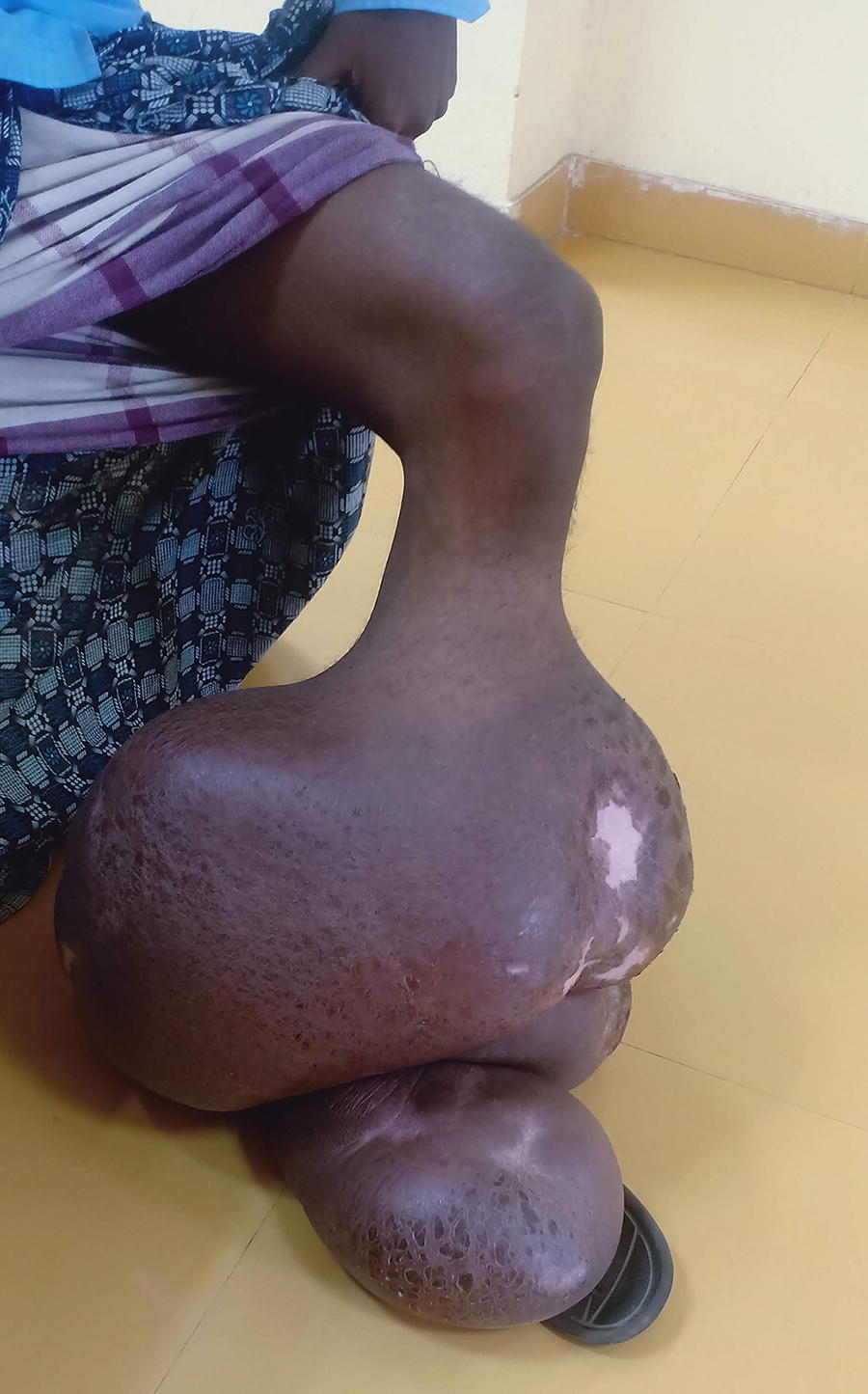
Chronic filariasis in the right lower limb in a patient with IGA nephropathy.
Baseline Characteristics of Patients
Discussion
Chronic filariasis, a neglected tropical disease, often eludes the understanding, especially when it comes to its renal manifestations. Lymphatic filariasis is attributed to the pathogens Brugia malayi and Wuchereria bancrofti, transmitted to humans through mosquitoes (Bizhani et al., 2021). Filariasis is well established to exhibit a spectrum of lymphatic and extralymphatic symptoms in clinical manifestations. In our retrospective study, we explored the epidemiological, clinical, and histopathological aspects of chronic filariasis. Renal manifestations in filariasis have been scarcely reported in the existing literature, with most of the available data originating from case reports (Chugh et al., 1978). Our findings align with the common renal presentations associated with filariasis, which include chyluria, microscopic hematuria, proteinuria, NS, and acute glomerulonephritis (AGN). Notably, all these manifestations were present in our patients, except for AGN (Langhammer et al., 1997; Rath et al., 1991). There is no gender predilection, the age of presentation varied widely, with patients ranging from 39 to 59 years, and a mean age of 49.25 years. This diversity in age at presentation is reflective of the complex nature of this disease and its interactions with individual immune responses and environmental factors. Renal injury in filariasis can be attributed to two primary mechanisms. First, due to mechanical injury to the glomeruli, possibly due to the presence of microfilariae in the circulatory system. The second mechanism involves the deposition of immune complexes within the glomerular basement membrane, facilitated by the presence of microfilariae antigens. In the previously published literature, histopathological analysis of renal tissue in chronic filariasis cases has revealed various patterns, including proliferative Glomerulonephritis (GN), MPGN, and eosinophilic predominant GN (Langhammer et al., 1997; Rath et al., 1991; Nayak et al., 2011). Additionally, AA-type renal amyloidosis has been reported in patients with chronic filariasis. Microfilariae have been identified in different parts of the kidneys, including renal arterioles, glomerular and peritubular capillaries, and the tubulointerstitial compartment (Shubham et al, 2018). These findings further emphasize the complex interplay of pathological processes in filariasis-associated renal disease. In addition to these findings, our analysis also showed the presence of IGAN and MCD. An important challenge in our study was the absence of microfilariae in the renal tissue of our patients. This finding can be attributed to the fact that the absence of microfilaria in the blood does not necessarily exclude filarial disease. Furthermore, microfilariae may not be detectable in the blood of individuals with filarial lymphedema, which can develop many years after infection. However, the histological diversity of renal lesions and the absence of microfilariae in the renal tissue make it challenging to establish a clear causal relationship. Nonetheless, in endemic areas, patients with renal and urinary abnormalities should be highly suspected of having a filarial infection.
Our study is one of the few that describes the renal biopsy findings in patients with chronic filariasis. There are merely a few pieces of literature available on the issue, and those are mainly from three decades ago. Only sporadic case reports are occasionally documented after that. In conclusion, our study provides valuable insights into the renal manifestations of chronic filariasis, shedding light on the rarity of biopsy findings in this context. The multifaceted nature of this disease, with its varied clinical and histopathological presentations, underscores the importance of further research and understanding in the field of nephrology. Further studies and collaborative efforts are needed to unravel the intricacies of renal involvement in filariasis, which can have far-reaching implications for patient care and public health in endemic regions.
Authors’ Contributions
P.M. and P.D. conceptualized and drafted the article. S.P. and A.G. curated the data. M.D. reviewed and edited the article. S.P. and P.A. interpreted the biopsy finding. All authors have read and approved the article.
Declaration
The article is submitted by agreement of all authors, and it is not under consideration for publication elsewhere and will not be submitted elsewhere.
Footnotes
Acknowledgements
Author Disclosure Statement
The authors declare that there is no conflict of interest.
Funding Information
No funding was received for this article.