




Abstract
Background:
Infection with tick-borne pathogens (TBPs) causes anemia, jaundice, and growth retardation in cattle. Many studies have conducted antigen (Ag) tests for major TBPs, such as Anaplasma spp. and Theileria spp., in Korean indigenous cattle (KIC); however, few studies have analyzed antibodies (Ab) against these pathogens.
Materials and Methods:
This study simultaneously tested 15 housed cattle raised indoor for over a year and 67 grazing cattle for Anaplasma spp. and Theileria spp. Ag using polymerase chain reaction analysis and Ab using enzyme-linked immunosorbent assay.
Results:
The A. phagocytophilum was detected in 3 housed cattle (20.0%) and 30 grazing cattle (44.8%), whereas the T. orientalis was detected in 3 housed cattle (20.0%) and 54 grazing cattle (80.6%). The positivity rates for Anaplasma spp. Ab did not differ significantly between housed and grazing cattle (4 out of 15 [93.3%] and 55 out of 67 [80.0%], respectively). The positivity rates for Theileria spp. Ab were higher in grazing cattle than in housed cattle (21 of 67 [31.3%] and 0 out of 15 [0.0%], respectively) (P < 0.01). No difference was observed between the frequency of grazing and the abundance of Ab against either pathogen. Meanwhile, as a result of comparing the detection of pathogen Ag and Ab, Anaplasma spp. showed the highest proportions of 73.3% and 47.8% in Ag (−)/Ab (+) for housed cattle and grazing cattle, respectively. Theileria spp. showed the highest rates of 80.0% in Ag (−)/Ab (−) for housed cattle and 52.2% in Ag (+)/Ab (−) for grazing cattle.
Conclusions:
This is the first study to determine the impact of antibody abundance against Anaplasma spp. and Theileria spp. on antigen prevalence in KIC.
Introduction
Korean indigenous cattle (KIC) are primarily raised indoors. However, housed cattle face various problems, including the rapid spread of the disease, escalated production, and feed costs. Grazing is an alternative to indoor housing; however, it also poses challenges in managing nutrition and disease. The exposure of cattle to ticks increases during grazing, which can lead to infection with tick-borne diseases. Tick-borne pathogen infections in cattle cause a variety of clinical symptoms, range from asymptomatic to death, depending on the pathogens and the severity of the infection, which has significant economic losses to farms. (Garcia et al., 2022; Kang et al., 2013; Singh et al., 2022). Economic losses associated with tick-borne diseases (TBDs) in Tanzania were estimated at 364 million USD per year, with 68% of losses attributed to Theileria spp. and 13% to Anaplasma spp. (Kivaria, 2006).
Anaplasma spp., which are members of the Anaplasmataceae family, are transmitted by Ixodes spp. Ticks, commonly known as hard ticks (Atif, 2015; Rymaszewska and Grenda, 2008). In cattle, the species A. bovis, A. centrale, A. marginale, and A. phagocytophilum cause infections (Battilani et al., 2017). A. phagocytophilum is a zoonotic bacterium that causes tick-borne fever. The first A. phagocytophilum infection in Korea was reported in Holstein cattle in 2018 (Han et al., 2018; Miranda et al., 2021). In KIC, Anaplasma spp. was detected in 8 out of 119 farms (6.7%), and A. phagocytophilum was detected in 2 farms (1.7%) (Cho et al., 2016). In addition, a higher Anaplasma spp. infection was confirmed in Korean cattle (5.3%) than in dairy cattle (1.2%) (Lee et al., 2020).
Theileriosis, a tick-borne intracellular protozoan disease predominantly transmitted by hard ticks, reduces livestock productivity. Among Theileria species, T. buffeli and T. orientalis are low-pathogenic parasites that cause hemolytic anemia, jaundice, and anorexia (McFadden et al., 2011; Morrison, 2015). Previous studies in Korea have shown infection rates for T. orientalis in 13% (7 of 53) of farmed cattle, 39% (13 of 33) of grazing cattle, and in 41.3% of asymptomatic cattle in another study (Choi et al., 2016; Park et al., 2017).
As the incidence of tick-borne diseases in Korea increases, various diagnostic methods are being investigated (Chae et al., 2009; Park et al., 2018; Seo et al., 2018a; Seo et al., 2018c). Detection of antigens (Ag) using polymerase chain reaction (PCR) allows early confirmation of infection and identification of an active infection at the time of testing (Hairgrove et al., 2015). Unlike Ag, antibodies (Ab) are produced in response to pathogens and persist for a certain period after infection (Brossard and Wikel, 1997). Past exposure to pathogens can be confirmed through Ab testing, and immune status can be confirmed by checking the titer of Ab. Detection of serum Ab using enzyme-linked immunosorbent assay (ELISA) can result in false negatives in the early stages of infection, and testing of Ag using PCR can result in false negatives if the amount of pathogens is below the threshold. Therefore, testing for Ag and Ab simultaneously is a good way to confirm past and recent infections. However, research investigating the serum Ab abundance against Anaplasma spp. and Theileria spp. in KIC is scarce (Seo et al., 2018c).
In our previous study, the infection rates of A. phagocytophilum and T. orientalis in grazing KIC were confirmed using PCR (Kim et al., 2024). The purpose of this study is to determine the abundance of Ab against Anaplasma spp. and Theileria spp. according to the living environment and frequency of grazing, and whether the Ag prevalence was affected by Ab abundance, which have not been elucidated so far in KIC. For the purpose, the prevalence of Anaplasma spp. and Theileria spp. Ag and Ab were simultaneously evaluated in housed and grazing KIC. To the best of our knowledge, this is the first study to determine the impact of Ab abundance against Anaplasma spp. and Theileria spp. on Ag in KIC.
Material and Methods
This study was approved by the Institutional Animal Care and Use Committee of the National Institute of Animal Science, Republic of Korea (JBNU IACUC No. NON2023-123).
Sample collection
A farm in Chungcheongnam-do, South Korea, which has indoor and pasture farms and raises approximately 3,500 cattle, was selected. Eighty-two female KIC aged 22–88 months were randomly selected from this farm, of which 15 were housed cattle and 67 were grazing cattle. The housed cattle raised had past grazing experience but had been raised indoors without grazing for over a year. On the other hand, grazing cattle are cattle that have been grazing in the pasture for the past 2 months. Of these, 52 are first-grazing cattle that have never accessed the pasture before, and 15 are second-grazing cattle that have grazing experience in the past. The indoor group was fed concentrate and roughage with a dry matter intake of 5.5 kg/d (10% crude protein), and the grazing group was fed forages of orchard grass and tall fescue.
Blood samples were collected indoors from both housed and grazing cattle. Blood (10 mL) was collected from the jugular vein of the 82 selected cattle, and samples were dispensed into blood sample EDTA-supplemented tubes (BD Vacutainer, Becton Dickinson and Company, Franklin Lakes, NJ, USA) and serum separation tubes (Vacutte serum tubes, Greiner Bio-One, Kremsmuster, Austria). All blood samples were refrigerated and transported to a laboratory within 1 hour. The serum-separating tubes were left standing for at least 1 hour, and they were centrifuged at 2599×g for 10 min. The serum was stored at −20°C until analysis.
DNA extraction and polymerase chain reaction
DNA was extracted from a 200 µL blood sample using the DNeasy Blood Kit (Qiagen, Valencia, CA, USA) according to the manufacturer’s instructions. The species-specific primers are used to amplify Theileria spp. (F-primer, 5′-GTTATAAATCGCAAGGAAGTTTAAGGC-3′; R-primer, 5′-GTGTACAAAGGGCAGGGACGTA-3′) and Anaplasma spp. (F-primer,5′-TACCTCTGTGTTGTAGCTAACGC-3′; R-primer, 5′-CTTGCGACATTGCAACCTATTGT-3′) (Choi et al., 2016). And PCR was performed under the following conditions: 98°C for 5 min; followed by 35 cycles of 10 s at 98°C; an annealing step at the appropriate temperature and time, 72°C for 1 min; and a final extension at 72°C for 5 min. All PCR products were purified using the AccuPrep PCR Purification Kit (Bioneer, Daejeon, Republic of Korea) according to the manufacturer’s instructions, and sequence analysis was performed (Bioneer).
Detecting serum Ab using ELISA
Cellular ELISA (cELISA) detects the presence of serum Ab using a monoclonal Ab that recognizes MSP5, a major surface protein of A. marginale, A. centrale, A. phagocytophilum, and A. ovis. (Hairgrove et al., 2015). A commercial Anaplasma Antibody Test Kit, cELISA V-2 (VMRD, Pullman, WA, USA), was used according to the manufacturer’s instructions to detect Anaplasma spp. Ab in cattle serum. The optical density was immediately analyzed at 630 nm using an ELISA plate reader (BioTek Instruments, Winooski, Vermont, USA), and the percentage of inhibition was calculated according to the manufacturer’s instructions.
A commercial kit, Bovine Theileria Antibody, Thi-Ab ELISA Kit (MyBIOSource Inc., San Diego, CA, USA), was used according to the manufacturer’s instructions to detect Theileria spp. Ab. This kit is based on a qualitative reverse-phase enzyme immunoassay technique. Optical density was analyzed at 450 nm, and Ab levels above the cut-off value were considered positive, according to the manufacturer’s instructions.
Statistical analysis
Statistical analysis was performed using the SPSS software (version 29.0; SPSS, Chicago, IL, USA). The Ab abundance results of Anaplasma spp. and Theileria spp. in housed and grazing cattle were analyzed using the Mann–Whitney U test according to the normality test results using the Kolmogorov-Smirnov test. The data are presented as medians (interquartile ranges). The Ag and Ab positivity rate of Anaplasma spp. and Theileria spp. was analyzed using the Chi-square test. The prevalence of Anaplasma spp and Theileria. spp Ag/Ab was compared with the use of McNemar’s test. P < 0.05 were considered statistically significant.
Results
Antigen and antibody positivity rates of grazing and housed cattle
In total, 3 of 15 housed cattle (20.0%) and 30 of 67 grazing cattle (44.8%) tested positive for the A. phagocytophilum Ag. There was no statistically significant difference between the grazing and housed cattle. The T. orientalis Ag was detected in 3 of the housed cattle (20.0%) and 54 of the grazing cattle (80.6%). The positivity rate was significantly higher in grazing cattle than in housed cattle (P < 0.01) (Fig. 1).
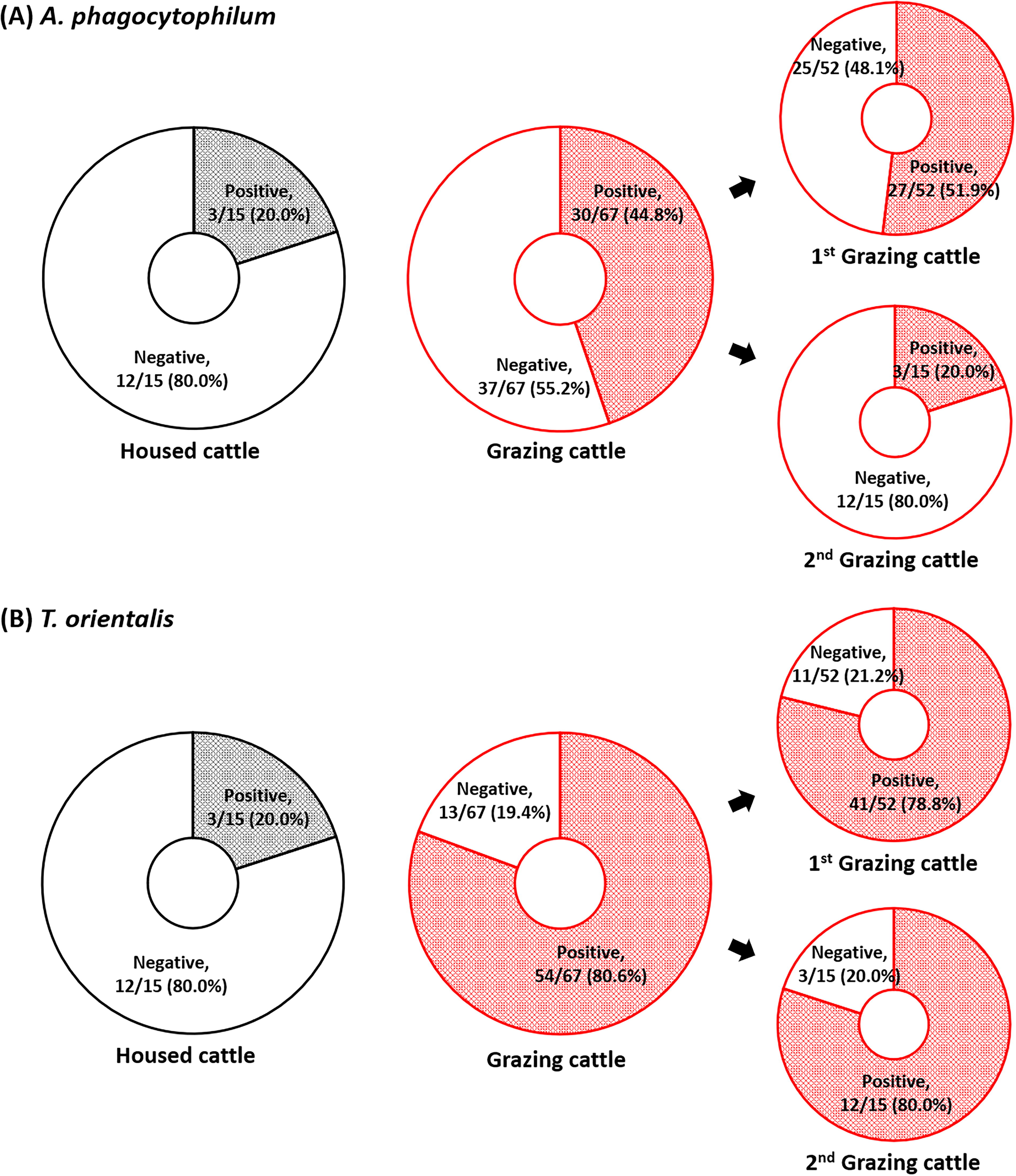
Detection of
The prevalence of serum Ab against Anaplasma and Theileria spp. was investigated using ELISA. The abundance of Anaplasma spp. Ab in serum was significantly lower among the grazing cattle than the housed cattle (P < 0.05; 59.4 (22.0) versus 64.5 (20.8). Anaplasma spp. Ab were detected in 55 grazing cattle (80.0%) and 14 housed cattle (93.3%), with no significant differences (P > 0.05). The serum abundance of Theileria spp. Ab was 7.9 (4.3) in the housed cattle and 18.3 (23.4) in the grazing cattle, a significant difference (P < 0.01). Theileria spp. Ab were not detected in the housed cattle but were found in 21 of the grazing cattle (31.3%), which was a significantly higher positive rate than that among the grazing cattle (P < 0.01) (Fig. 2).
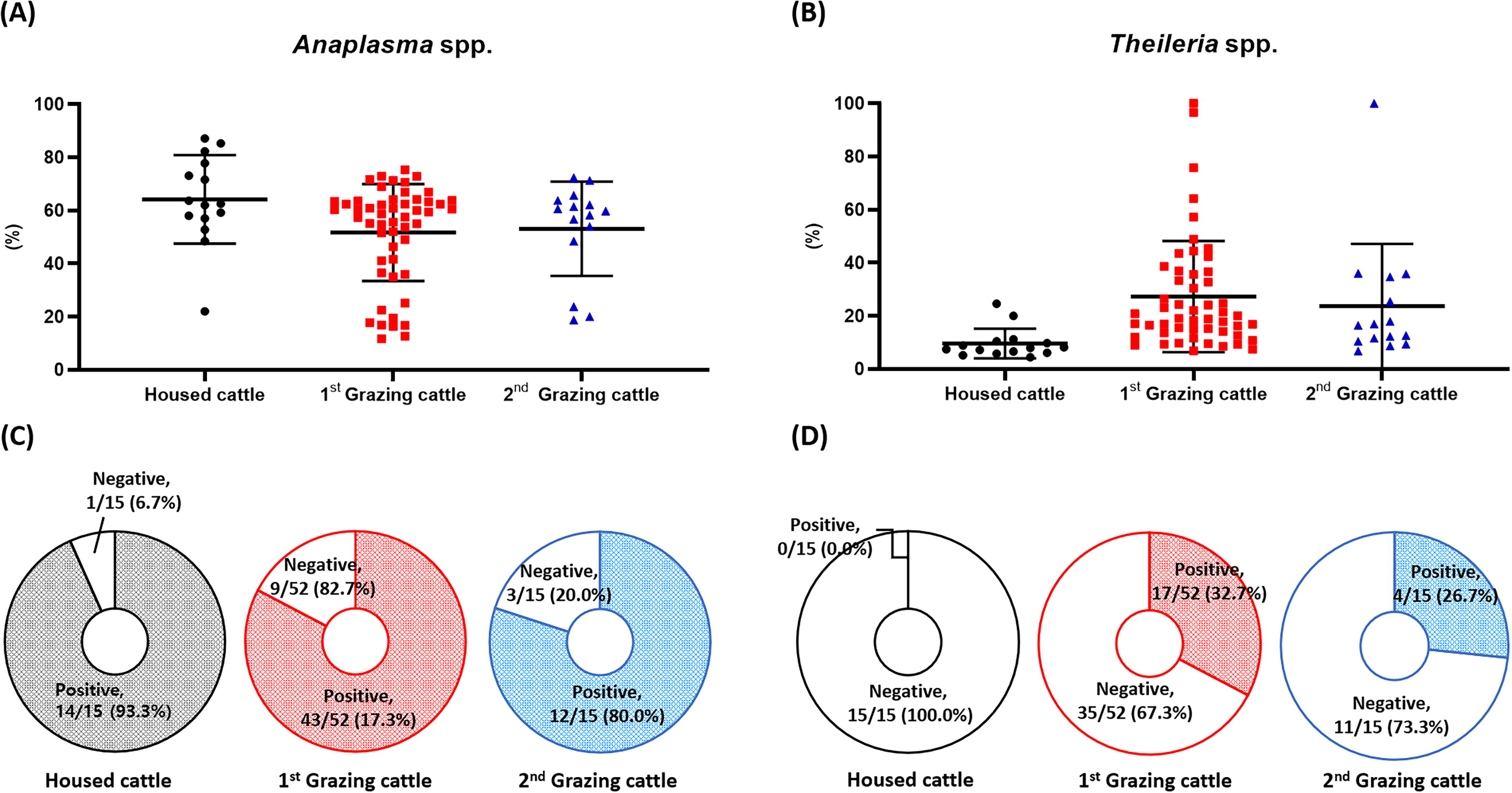
Serum antibody levels and prevalence in cattle according to the frequency of grazing.
Antigen and antibody prevalence according to frequency of grazing
The prevalence of Ag and Ab according to the frequency of grazing was compared in 67 grazing cattle (first grazing; n = 52, second grazing; n = 15). Significantly more first-grazing cattle (27 out of 52, 51.9%) tested positive for the A. phagocytophilum Ag than second-grazing cattle (3 out of 15, 20.0%) (P < 0.05). Regarding the T. orientalis Ag, 41 first-grazing cattle (78.8%) and 13 s-grazing cattle (86.7%) tested positive. However, this difference was not statistically significant (Fig. 1).
No significant differences regarding Anaplasma spp. or Theileria spp. Ab were found depending on the frequency of grazing. Anaplasma spp. Ab were detected in 43 of 52 first-grazing cattle (82.7%) and 12 of 15 s-grazing cattle (80.0%). The respective abundances were 59.1 (22.3) and 59.8 (15.4). Theileria spp. Ab were detected in first-grazing cattle (32.7%, 17/52) and second-grazing cattle (26.7%, 4/15). The abundances were 19.5 (22.6) and 16.5 (24.3), respectively (Fig. 2).
Comparison of antigen and antibody test results
By comparing Ag detection using PCR and Ab positivity in the serum using ELISA, Anaplasma spp. among the housed cattle had an Ag (+)/Ab (+) rate of 20.0% and Ag (−)/Ab (+) rate of 73.3%. The Ag (−)/Ab (+) rate was the highest. For the grazing cattle, the Ag (+)/Ab (+) ratio was 34.3% and the Ag (−)/Ab (+) ratio was 47.8%, exhibiting high Ag (+)/Ab (+) and Ag (−)/Ab (+) rates (Table 1). Theileria spp. among the housed cattle had an Ag (+)/Ab (−) rate of 20.0% and Ag (−)/Ab (−) rate of 80.0%, demonstrating a high rate of Ag (−)/Ab (−). Among the grazing cattle, the Ag (+)/Ab (+) ratio was 28.4% and the Ag (+)/Ab (−) ratio was 52.2%, This group had the highest Ag (+)/Ab (−) ratio (Table 1).
Prevalence of Anaplasma Spp. and Theileria Spp. of Antigens and Antibodies
ELISA, enzyme-linked immunosorbent assay; PCR, Polymerase chain reaction.
As a result of comparing the Ag and Ab positivity of Anaplasma spp. according to the frequency of grazing, the Ag (−)/Ab (+) rate was 42.3% and the Ag (+)/Ab (+) rate was 40.4% in the first-grazing cattle, and the Ag (−)/Ab (+) rate was 66.7% and the Ag (+)/Ab (+) rate was 13.3% in the second-grazing cattle. The risk difference for first-grazing cattle is 0.37 (95% CI: 0.16–0.53), and for second-grazing cattle, the risk difference is 0 (95% CI: −0.26–0.26) (Fig. 3). Theileria spp. had an Ag (+)/Ab (−) rate of 50.0%, Ag (+)/Ab (+) rate of 28.8%, and Ag (+)/Ab (+) rate of 28.8% in the first-grazing cattle, and an Ag (+)/Ab (−) rate of 60.0% and Ag (+)/Ab (+) rate of 26.7% in the second-grazing cattle, showing similar ratios regardless of the frequency of grazing (Table 1). The risk difference for first-grazing cattle is 0.12 (95% CI: −0.07–0.30), and for second-grazing cattle, the risk difference is 0.13 (95% CI: −0.19–0.45) (Fig. 3).

Risk difference between first-grazing cattle and second-grazing cattle in
Discussion
The tick-borne pathogens Anaplasma spp. and Theileria spp. infect various hosts, and their incidence is increasing in Korea. A. phagocytophilum and T. orientalis were detected at higher prevalence in grazing cattle compared to housed cattle. Our results were in agreement with those of previous studies showing that the risk of tick-borne diseases increases during grazing and indicated that A. phagocytophilum and T. orientalis are prevalent in Korea (Choi et al., 2016; Seo et al., 2011; Song and Sang, 2003). Although ticks were not collected and tested in this study, the infection was expected to be caused by ticks present in cattle. Grazing cattle are exposed to the natural environment and have more opportunities to come into contact with ticks, the vectors of pathogens; therefore, they have a higher prevalence compared to housed cattle. In addition, no clinical symptoms were observed in housed cattle in which Anaplasma spp. and Theileria spp. were detected, which is thought to be due to better management in housed cattle compared to grazing cattle (Fukushima et al., 2021).
In this study, A. phagocytophilum Ag were detected in 3 housed cattle (20.0%), Ab were present in 14 housed cattle (93.3%), and both Ag and Ab were detected in 3 housed cattle (20.0%). Previous studies have reported that the Ab status for A. phagocytophilum changed twice (from negative to positive or positive to negative) between spring and fall, and Ab, tested using an indirect immunofluorescence assay, persisted for up to 9 weeks after PCR positivity for A. phagocytophilum (Lempereur et al., 2012; Silaghi et al., 2018). However, our results showed a high prevalence of Anaplasma spp. Ab in cattle that had not been grazed for more than a year. It is unknown whether the Ab were vestiges of past infections while grazing or were produced due to new contact with ticks while housed. This finding suggested that the Anaplasma spp. Ab is unrelated to the presence or absence of Ag at the time of sample collection and could indicate a past Anaplasma spp. infection. In humans, serological diagnostic methods are most commonly used for diagnosing A. phagocytophilum, but these tests have been shown to have low sensitivity during the acute phase of infection. Additionally, a single positive titer result cannot distinguish between a current infection and evidence of previous exposure to this pathogen, as IgG Ab can persist in patients for several years after infection (Schotthoefer et al., 2013). A. marginale was not detected in our study; however, the possibility of Ab positivity due to infection with A. bovis and A. capra cannot be excluded as previously reported in KIC (Miranda et al., 2021; Park et al., 2018; Seo et al., 2018b) or false positives due to Ehrlichia spp. infection, as reported by Al-Adhami et al. (2011). In addition, regardless of the grazing frequency, high Anaplasma spp. Ab positivity was observed among the first- and second-grazing cattle; however, Ag detection decreased significantly among the second-grazing cattle compared with the first-grazing cattle. Therefore, Ab production (immunity) through past infections is anticipated to help prevent reinfection. Additional research on the titer of Ab and immunity generated by Anaplasma spp. infection.
For T. orientalis, the pathogenic stage occurs within erythrocytes, and the onset of the serological response coincides with the appearance of piroplasms in erythrocytes (Shimizu et al., 1988). Previous studies have shown that the Ikeda genotype of T. orientalis is associated with high infection intensity, which negatively correlates with packed cell volume, indicating that severe erythrocyte destruction and strong serological responses are likely. Conversely, the Chitose and Buffeli genotypes have shown weak or no correlation with MPSP ELISA ratio results. Since T. orientalis is a persistent infection, there was no difference in the prevalence between the first- and second-grazing (Kubota et al., 1996; Onuma et al., 1998). T. orientalis infections demonstrate a rapid decline in Ab titers following acute infection (Jenkins and Bogema, 2016). In our results, 35 out of 67 (52.2%) cattle were were Ag (+)/Ab (−) in Theileria spp., which may indicate lack of seroconversion or insufficient humoral response to detectable levels. Previous research has reported that in the absence of reinfection, Ab against T. parva generated in the primary immune response decline to negative levels within 6 months of infection (Magona et al., 2011). However, another study reported that T. orientalis Ab persist for 3–5 years without reinfection (Morzaria, 1998). The production and persistence of Ab following Theileria infection merits further investigation.
In Korea, Haemaphysalis longicornis is the dominant tick species, and their numbers increase with rising temperatures from April to September, peaking in September (Seo et al., 2021). Grazing was conducted in this study from April to June, which may have led to an underestimation of prevalence. Previous studies have suggested that factors such as immune status and sex of cattle, farm management, pasture length, and type of pesticide may influence the susceptibility to tick-borne diseases among cattle (Ghafar et al., 2021; Heylen et al., 2023; Kispotta et al., 2017; Kolte et al., 2017; Miyama et al., 2020; Ola-Fadunsin et al., 2020; Wondimu and Bayu, 2021). Since this study included only a small number of female KIC in one farm, there are limitations in generalizing the results. However, the Ag prevalence for Anaplasma spp. was higher in grazing cattle than in housed cattle, and as the frequency of grazing increased, the Ag prevalence decreased; however, the serum abundance of Ab was maintained. For Theileria spp., there was a difference in the positivity rates of Ag and Ab between grazing and housed cattle; however, there was no difference in the prevalence of Ag and Ab based on grazing frequency. Anaplasma spp. Ab are suspected to be long-lasting and help prevent reinfection, whereas Theileria spp. Ab are not thought to be associated with reinfection. These results will be useful to improve understanding of seroconversion after infection against Anaplasma spp. and Theileria spp. In KIC. Additionally, Ab abundance helps in establishing a vaccine strategy for the prevention of Anaplasma spp.
Footnotes
Authors’ Contributions
Investigation, formal analysis, and writing (original draft) by J.-Y.K. and Y.J. Formal analysis visualization by Y.K. Conceptualization, funding acquisition, and supervision by K.-S.C. and J.P.
Author Disclosure Statement
No conflicting financial interests exist.
Funding Information
This study was supported by the Korea Institute of Planning and Evaluation for Technology in Food, Agriculture, and Forestry (IPET) (grant No. RS-2024–00402276). This research was partially supported by ‘regional innovation mega project’ program through the Korea Innovation Foundation funded by Ministry of Science and ICT (Project Number: 2023-DD-UP-0031).