
Abstract
Background:
Rickettsia spp. are obligate intracellular bacteria of utmost importance for public health and the economy since their presence can generate significant losses in livestock production, affecting the health of animals, the quality of the meat, milk, and other products derived from affected animals. Therefore, prevention of the transmission of these pathogens, their early identification and timely treatment are essential to mitigate their impact on public health and animal production.
Materials and Methods:
In the present work, hard ticks were collected from infested Bos indicus cattle in Aldama, Tamaulipas, Mexico. They were identified by morphology using dichotomous keys and by sequencing and analyzing of a fragment of the mitochondrial 16S. PCR was performed using specific primers targeting Rickettsia sp. gltA. Phylogenetic analyses were performed, aligning the amino acid sequences with Muscle, and a phylogenetic tree was generated using PhyML.
Results and Conclusions:
Amblyomma mixtum and Rhipicephalus microplus ticks infesting the cattle were identified. Using molecular techniques, the presence of Rickettsia amblyommatis was identified in the cohort of analyzed ticks, suggesting a circulation of this pathogen in livestock in this region and granting further research in this area.
Introduction
After mosquitoes, ticks are considered worldwide the most important vectors involved in transmitting of pathogens and the most significant vector of pathogens for domestic and wild animals (Merino et al., 2020). Within this group, the genera Amblyomma, Dermacentor, and Rhipicephalus are regarded the most medically relevant, both for humans and domestic and wild animals, given they are associated with a wide range of zoonoses such as anaplasmosis, ehrlichiosis, and many of the rickettsiosis (Rosenberg et al., 2018; Álvarez-López et al., 2021 ; Montiel-Armendáriz et al., 2021). Rickettsioses constitute a group of great relevance among these diseases due to its emerging nature (Merino et al., 2020).
The Ricketssia species of greatest medical importance are in the spotted fever group (SFG) and the typhus group groups, which represent a significant risk to the health of both, humans and animals, as they are the cause of Rocky Mountain spotted fever, the murine typhus fever, epidemic typhus fever, and scrub typhus fever, pathologies associated with a fatality rate of up to 30% in the vulnerable populations such as children, elder adults, pregnant women, and people living in poverty (Álvarez-Hernández et al., 2024 ; Duncan et al., 2021; Estrada-Mendizabal et al., 2023).
In Mexico, rickettsioses are an emerging threat, particularly in the northern region, where most of the endemic outbreaks in humans and domestic, stray, and wild animals occur because the latter are considered the main host vectors of the causative agents (Alvarez-Hernandez et al., 2020; Zazueta et al., 2021; Estrada-Mendizabal et al., 2023). Moreover, rickettsial pathogens have been also documented in other regions nationwide (Vázquez-Guerrero et al., 2023). Furthermore, most of the data on tick infestation and its effects in Mexico come from livestock (cattle, goats, and sheep), given that they require grazing for their maintenance, and this exposes them to parasitism from multiple ectoparasites among which ticks stand out (Álvarez-Henrnández et al., 2024; Coronel-Benedett et al., 2018).
Here, we identified Rickettsia amblyommatis in ticks collected from cattle in the Aldama, Tamaulipas, a northeastern community in Mexico. We consider that these results help to gain an understanding of the geographical distribution of Rickettsia species in the northern region of Mexico.
Materials and Methods
Tick collection
Hard ticks (adults and nymphs) were collected from cattle (Bos indicus) in the Aldama region, Tamaulipas (23.320891, −97.851750) (Fig. 1), a region with a semiwarm and subhumid climate with summer rainfalls, a temperature ranging between 18–26°C, located in the northeast of Mexico. According with local authorities this region is the major livestock area within the state (INEGI, 2007). Overall, the cattle included in this study received periodic visits from a veterinarian for growth and health control and additionally they received vaccines and antibiotics if needed throughout their lives. The tick collection occurred from November 1−3, 2023, because a heavier presence of ticks in the study region was reported by ranch owners, despite not being the historical season in which they are observed.
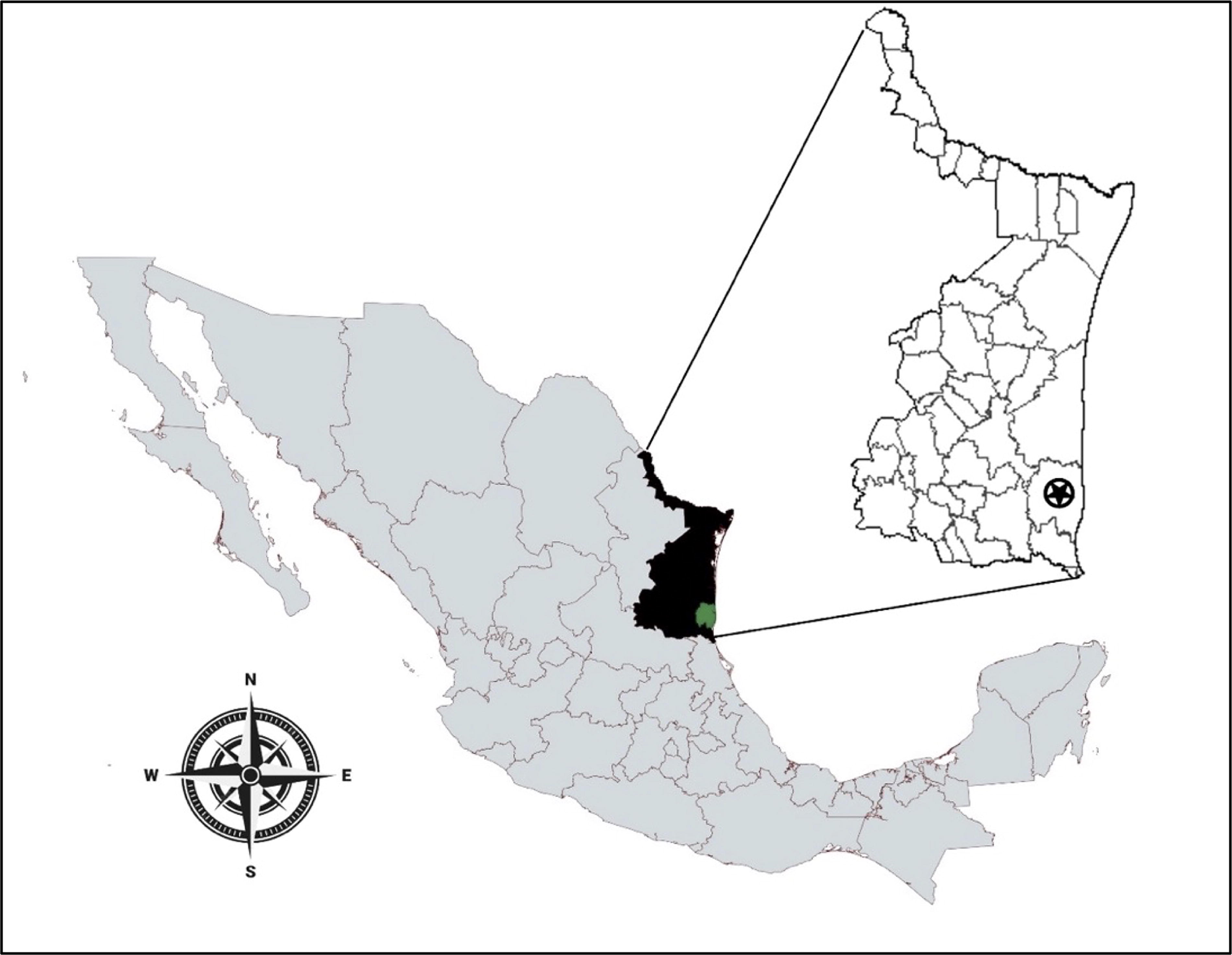
Location of the tick collection region. The location of Tamaulipas (colored black) in Mexico and the geolocation of the Aldama municipality (colored in green and with a circular star) are shown. The map was prepared using the MapChart platform based on geographic data from INEGI: https://www.inegi.org.mx/app/mapas.
In the collection procedures, the ticks were removed mechanically, using entomological tweezers as described before (Vázquez-Guerrero et al. 2023). All specimens were grossly identified and stored in a sterile empty urine collection container along with wet cotton swabs, to be later transferred and analyzed under laboratory conditions in Mexico City. The arthropods that managed to survive the trip were analyzed with the procedures described below, while the specimens that did not survive were stored in 70% ethanol and deep frozen at −70°C.
Morphological identification of ticks
Once in our laboratory specimens were observed on a VELAB VE-S5C stereoscopic microscope with a magnification of up to 40× and using the VELAB TCAPTURE software. Morphological identification was carried out following the dichotomous keys of Brinton and Beck, (1963a); Brinton and Beck, (1963b), Hoskins (1991), and Guzmán-Cornejo et al. (2011).
DNA extraction
Out of all the collected ticks, 16 live gorged female ticks were randomly selected from which as much blood as possible was extracted using a U-100 ultrafine insulin syringe (0.5 mL 31G × 6 mm), while the rest of the specimens were either macerated for subsequent DNA extraction or stored in the freezer as mentioned above. Samples were placed in a sterile 1.5 mL microtubes and the DNA was extracted as described before (Vázquez-Guerrero et al., 2023). Briefly, to obtain the DNA the protocol for the purification of total DNA from the specimens the QIAamp DNA Blood Mini Kit (QIAGEN) was used. During the arthropod maceration process, the sample was resuspended in 180 μL of phosphate-buffered saline (25 mM KH2PO4, 50 mM K2HPO4, 150 mM NaCl, pH 7.2). Subsequently, each sample was quantified with a NanoDrop 2000 spectrophotometer (Thermo Scientific).
Molecular identification of the ticks and Rickettsia spp
To molecularly characterize the collected ticks and determine the potential presence of Rickettsia sp. they could harbor, PCRs were used targeting the mitochondrial 16S gene rRNA gene for tick identification, and the gltA gene for the bacterium. For the molecular identification of the collected ticks, PCR were done for the mitochondrial 16S and 16S rRNA, using the primers 16 s + 1 (5′-CTGCTCAATGATTTTTTAAATTGC-3′) and 16 s-1 (5′-CCGGTCTGAACTCAGATCATGTA-3′). To determine the presence of bacteria in the purified metagenomic DNA oligos Eub338F (5′-ACTCCTACGGAGGCAGCAG-3′) and Eub518R (5′-ATTACCGCGGCTGCTGG-3′) were used to amplify a fragment of the bacterial 16S rRNA (Vázquez-Guerrero et al., 2023). In both cases, DreamTaq Green polymerase chain reaction (PCR) Master Mix (2×) (Thermo Scientific) was used, and the amplification conditions were those described by (Black and Piesman, 1994) and Guo et al. (2008), respectively. As positive controls, the metagenomic DNA of Ornithodoros turicatae and the DNA of Escherichia coli BL21 were used. In both cases, positive amplicons of the expected sizes 475 bp (for mitochondrial DNA) and 200 bp (for bacterial DNA) were obtained. The mitochondrial 16S gene fragment was sequenced using the same primers at Macrogen Inc.
To detect Rickettsia in the DNA samples CS78 (5′-GCAAGTATCGGTGAGGATGTAAT-3′) and CS323 (5′-GCTTCCTTAAAATTCAATAAATCAGGAT-3′) primers were used and the amplification conditions of Labruna et al. (2004) for amplification of gltA. A positive fragment obtained from a patient with confirmed infection by Rickettsia rickettsii was used as a positive control (Vázquez-Guerrero et al. 2023). Samples that were positive for the gltA fragment were Sanger sequenced using primers CS78 and CS323. Subsequently, a phylogenetic analysis was performed, aligning the amino acid sequences with Muscle (https://www.ebi.ac.uk/Tools/msa/muscle/), and a phylogenetic tree was generated using PhyML (Guindon et al., 2010) with maximum likelihood method using the Blosum62 amino acid substitution model. The tree was visualized with MEGA v11 (Tamura et al., 2021).
Results
Morphological classification of collected ticks
Hard ticks were collected from 10 cattle (B. indicus) dedicated to meat production at the study site. In total, 627 tick specimens were obtained and characterized (Table 1). Ticks were morphologically identified, and two different tick genera and species were identified: Amblyomma mixtum and Rhipicephalus microplus, among which the nymphal and adult stages were observed (Table 1). The main morphological characteristics used to discern R. microplus specimens were the base of the capitula dorsally hexagonal, the presence of eyes, very short palps, a rounded or oval spiracular plate, absence of festoons, furrowed dorsal, and laterally compressed. The main characteristics observed in A. mixtum were long palps, ornamented, ocelli, and festoons present, the base of the gnathosoma is variable in shape and pentagonal dorsal, presence of ventral shield and extended beyond the posterior margin of the festoons (in the male) and subtriangular stigma plates (Fig. 2). Together, these results showed that the two genera of ticks were present in the surveyed cattle in Aldama, Tamaulipas.

Representative specimens of the collected Rhipicephalus microplus and Amblyomma mixtum ticks were obtained from Aldama, Tamaulipas. Stereoscopic microscope images of a female R. microplus specimen:
Morphological Classification of Collected Ticks
The number in parenthesis are those of the adults, the rest were nymphs; ♀, females; ♂, males.
Molecular identification of the ticks and Rickettsia spp
Despite the fact that more specimens were collected, DNA extraction was performed only in 16 ticks (eight specimens from R. microplus and eight from A. mixtum) as described above because most of them arrived at our lab either dead or decomposing; out of these samples, six were selected for molecular analyses. Amplicons of 475 bp were obtained in all samples, corresponding to the mitochondrial 16S and sequenced to confirm the morphological identification made previously to all ticks. The obtained sequences confirmed the previous morphological identification of the specimens as follows: A. mixtum 98.71% to 99.71%, and R. microplus 99.77% to 100%. These same samples were positive for amplifying the gltA gene from Rickettsia spp.
Amplification of the gltA gene in the DNA from 16 ticks was positive, suggesting infection with Rickettsia spp. Unfortunately, given that during the collection process once they were grossly identified all the ticks were mixed and it is difficult to determine which cattle they came from. Five of the fragments, three from R. microplus and two from A. mixtum, were sequenced and analyzed with phylogenetic tools, indicating that all the samples corresponded to the species R. amblyommatis (Fig. 3). All generated sequences are available in the GenBank database with accession numbers: PP681179-PP681183. These results showed that hard ticks collected from B. indicus cattle were infected with R. amblyommatis.
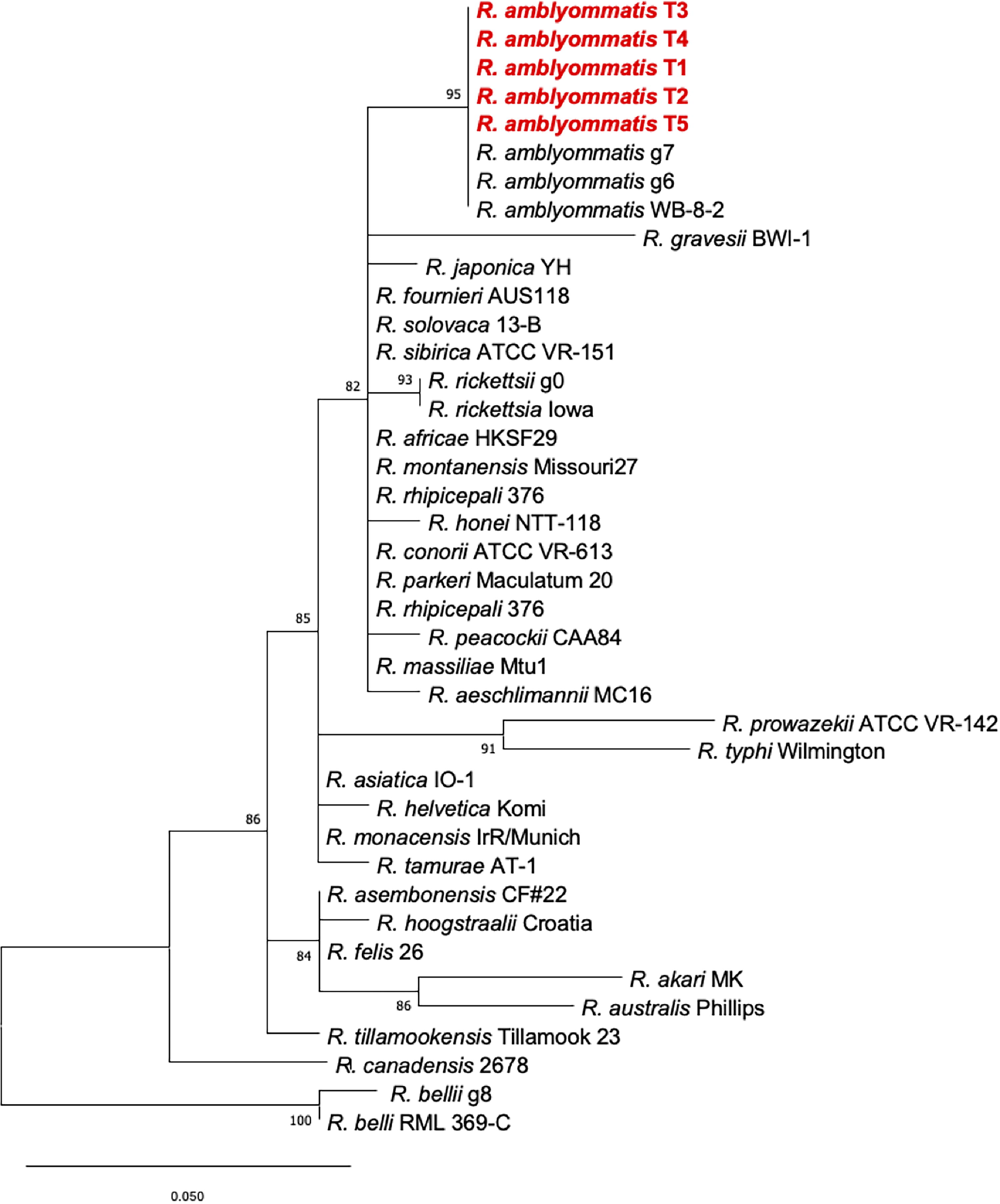
Phylogenetic analysis of gltA gene sequence of Rickettsia species. The phylogenetic tree was performed with maximum likelihood and the amino acid substitution model Blosum62. The sequences generated in this study are in red bold lettering (T1-T5). The outgroup was taken as the bacteria with the lowest identity percentage (R. conorii and R. massiliae). Numbers represent bootstrap support generated from 1000 replications. The bar represents 0.50 different nucleotides between sequences.
Discussion
Tick-borne diseases cause significant economic losses in livestock farming and represent a global threat to public health, a growing risk due largely to climate change, which alters transmission dynamics and the ticks’ geographical distribution (Richardson et al., 2023). Rickettsial diseases caused by Rickettsia spp. are increasing worldwide in recent decades, along with the diversity of species within this genus (Parola et al., 2013; Richardson et al., 2023). In Mexico, to date, at least 13 species of the Rickettsia genus have been reported: R. akari (Zavala-Castro et al., 2009), R. rickettsii, R. typhi (Estrada-Mendizabal et al., 2023; Merino et al., 2020), R. prowazekii (Burns et al., 2014), “R. lusitaniae” (Sánchez-Montes et al., 2016), R. amblyommatis (Merino et al., 2020), Candidatus R. andeanae, R. parkeri (Salomon et al., 2022), R. monacensis (Sánchez-Montes et al., 2021), R. bellii (Vázquez-Guerrero et al., 2023), R. asembonensis (Sánchez-Montes et al., 2020), R. felis (Ballados-González et al., 2023), and R. massiliae (Nieto-Cabrales et al., 2024). Identifications of these Rickettsia species have been greatly facilitated by the recent adoption of molecular techniques. Specifically, R. rickettsii was identified in two studies done in ticks isolated from animals and humans in the Mexican state of Tamaulipas (Merino et al., 2020; Guzmán-Cornejo et al., 2019). Here we report the identification of R. amblyommatis from ticks captured from cattle.
The finding of R. amblyommatis is important as this pathogen is part of the SFG with an ample distribution and prevalence in the United States (Chaparro-Gutiérrez et al., 2023) and recently documented in southeastern Mexico (López-Pérez et al., 2022). Although its pathogenicity in humans has not yet been clarified, recent research suggests it may be an opportunistic pathogen (Salomon et al., 2022). However, it is considered a species of veterinary importance (Richardson et al., 2023). Here, ticks captured from live cattle are infected with this species, representing a potential risk for humans and other domestic animals that coexist with these livestock in the region. It is remarkable that the number of ticks captured was very high and that those molecularly analyzed all were positive for gltA. Given that during the collection process the tick specimens were mixed it is complicated to determine whether all came from only one cattle or from multiple. Despite this latter, the presence of R. amblyommatis was corroborated in this region. These results coincide with previous reports in the region (Merino et al., 2020) about the presence of infected ticks collected from cattle but also from dogs and pigs and, in contrast to the results of the current study, they found R. amblyommatis only in six of the 110 collected ticks. They found that the most common species of these arthropods in this state corresponded to Rhipicephalus spp. and Amblyomma spp. In addition, we report the presence of a new vector previously not identified, R. microplus. Although it has previously been proposed that the latter acted as a vector of the bacteria (Richardson et al., 2023), our study marks for the first time the finding of R. amblyommatis in these vectors in the Tamaulipas region.
The fact that R. amblyommatis was identified in the small sample taken randomly may implicate a health problem involving both wildlife and domestic animals, as well as the human population, especially because of the interaction between persons and a great variety of animals that act as hosts and reservoirs for A. mixtum and R. microplus. These results grant further studies in this region to determine a more detailed study in both, animals and humans, to define possible transmission of this bacterium.
Footnotes
Acknowledgments
The authors would like to thank member of the Laboratorio de Genética Microbiana at ENCB-IPN for their invaluable support.
Authors’ Contributions
S.L.L.-R.: Conceptualization, methodology, formal analysis, investigation, resources, visualization, writing—original draft, and writing—reviewing and editing. E.V.-G.: Conceptualization, methodology, formal analysis, and investigation. E.D.-A.: Methodology and resources. A.C.-C.: Methodology and resources. R.H.-C.: Investigation and resources. G.A.-H: Supervision, validation, writing—reviewing and editing. P.E.-d.L.S.: Methodology, resources, software, and supervision. J.A.I.: Conceptualization, methodology, resources, supervision, validation, visualization; writing—original draft, writing—reviewing and editing.
Author Disclosure Statement
The authors have no relevant financial or nonfinancial interests to disclose.
Funding Information
This work was supported by Secretaría de Investigación y Posgrado del Instituto Politécnico Nacional (SIP-IPN) (grant numbers 2023-2722, 2024-0122, 2024-1496 to J.A.I.). S.L.L.-R. was provided by a scholarship by CONAHCYT (grant number CVU1026105).