
Abstract
The highly pathogenic avian influenza H5N1 viruses have become widespread and evolved into several clades. In our previous studies, the antigenic sites of the H5 hemagglutinin (HA) were characterized by selection and sequencing of escape mutants. In the present studies we analyzed the antigenic epitopes recognized by monoclonal antibodies against avian influenza A/Duck/Novosibirsk/56/05 (H5N1) virus isolated in western Siberia and belonging to subclade 2.2 of the H5N1 viruses. The analysis revealed several antigenically relevant positions of amino acid residues in the globular head of the HA not encountered earlier in the escape mutants of the H5 subtype. The newly recognized positions (113, 117, 118, 120, and 123, mature H5 numbering) are concentrated in an area adjacent to the region described in earlier studies as corresponding to site B in H3 HA, but extending far beyond this area. The amino acid positions recognized by the monoclonal antibodies against A/Duck/Novosibirsk/56/05 (H5N1) virus differ from the positions recognized by the monoclonal antibodies against H5N2 influenza viruses. The data suggest that the evolution of the HA of H5 avian influenza viruses is associated not only with the changes of antigenic epitopes recognized by antibodies, but also with a change in the dominance of the immunogenicity of different sites in the HA.
Introduction
The influenza virus HA is the target of neutralizing antibodies, and the peculiarities of the antigenic structure of its molecule are of primary importance for the understanding of the mechanisms underlying antiviral immunity. The first data on the location of antigenically relevant amino acid residues in the HA molecule of H5 subtype were reported by Philpott et al. (12). The authors detected six amino acid changes in the HA of escape mutants, and ascribed them to five epitopes. Since one, or at best two, amino acid changes were revealed for each epitope, only the locations of the antigenic sites could be deduced, not their fine structure. The x-ray crystallographic three-dimensional model was at that time available only for the H3 subtype (20), and thus the authors could not map the antigenic sites on the surface of the HA molecule of the H5 subtype.
In our previous studies, we selected a set of escape mutants and characterized the antigenic sites of H5 HA, first for an H5N2 avian influenza virus (5), and later for a human H5N1 isolate, A/Vietnam/1203/04 (6). Our studies revealed the fine structure of two antigenic sites, one of them similar to site A in H3 HA, and the other occupying a vast region at the surface of the HA globule and overlapping the sites described earlier as site B in H3 (19) and Sa in H1 (1). Our studies also revealed some differences in the antigenically relevant areas recognized by the monoclonal antibodies (mAbs) against the HA of the old H5N2 strains, and the area recognized by mAbs against the HA of a recent H5N1 strain (6).
The H5N1 strains isolated in Africa, Europe, and western Asia belong to subclade 2.2 (13). Antigenic variation within clade 2 viruses was revealed by comparison of HA sequences and immune cross-reactions of natural isolates (18). However, no attempts at revealing the epitopes of mAbs against HA of subclade 2.2 viruses using escape mutants have been reported.
In the present report we describe the results of the analysis of epitopes recognized by mAbs against avian influenza A/Duck/Novosibirsk/56/05 (H5N1) virus isolated in western Siberia and belonging to subclade 2.2 (9). In order to facilitate the comparison of the results with our earlier data, we used the strain A/Mallard/10218/84 (H5N2) as the wild-type virus for the selection of escape mutants. This strain was used previously in our studies for the antigenic mapping of H5 HA (5). The results reveal a diversity of the immunogenic activity of antigenically relevant areas of HA among H5 strains, and extend the knowledge of the fine structure of antigenic sites on the HA molecule of the H5 subtype.
Materials and Methods
Viruses
The mouse-adapted variant of avian influenza virus A/Mallard/Pennsylvania/10218/84 (H5N2) and its escape mutants m46(7), m46(8), m55(2), m58(1), and m24B9 have been described (5). The viruses A/Duck/Primorie/2633/01 (H5N3) and A/Ruddy Turnstone/Delaware/244/91 (H5N2) were obtained from the virus collection of the D.I. Ivanovsky Institute of Virology, Moscow. A reverse genetics-derived influenza virus containing the HA and NA genes of A/Vietnam/1203/04 (H5N1) virus in the genetic background of the high-growth master strain A/Puerto Rico/8/34 (H1N1) (VNH5N1-PR8/CDC-RG) was kindly provided by Dr. R. Donis (Centers for Disease Control and Prevention, Atlanta, Georgia). The viruses were propagated by growth for 48 h in the allantoic cavities of 10-day-old embryonated chicken eggs at 37°C and were stored at −80°C until use. The β-propiolactone-inactivated preparation of A/Duck/Novosibirsk/56/05 (H5N1) virus for the immunization of mice and for the competitive ELISA was kindly supplied by Dr. P.G. Deryabin (D.I. Ivanovsky Institute of Virology, Moscow).
Preparation of mAbs
Female BALB/c mice were immunized intraperitoneally four times at 2-wk intervals with 100 μg of purified β-propiolactone-inactivated virus mixed with an equal volume of Freund's adjuvant (complete adjuvant for the first immunization, and incomplete for the next three immunizations). Three days before cell hybridization the mice were boosted by intravenous injection of 100 μg of inactivated virus. Hybridization of the mouse splenocytes with the Sp2/0 myeloma cells was performed as previously described by Kohler and Milstein (7). Hybrid cells producing antiviral antibodies were selected by indirect ELISA as described in our previous communication (10), and by hemagglutination-inhibition (HI) test. The positive hybridomas were cloned by the method of limiting dilutions. mAb-containing ascites fluids were obtained by intraperitoneally inoculating BALB/c mice with 5 × 10⁶ hybridoma cells.
Selection of escape mutants
As described previously (5), virus was incubated with an excess of mAb for 1 h at 20°C. The mixture was inoculated into 10-day-old embryonated chicken eggs and incubated for 48 h at 37°C. Virus was harvested and used for limiting-dilution cloning in embryonated chicken eggs.
Competitive ELISA
The mAbs were purified by affinity chromatography on a protein A-Sepharose CL-4B column (Pharmacia, Uppsala, Sweden) according to the manufacturer's instructions. The protein concentration was measured using a Bio-Rad Protein Assay kit (Bio-Rad Laboratories, Inc., Hercules, CA), and the purified mAbs were conjugated with horseradish peroxidase (HRP) by the periodate method (11). The HRP-conjugated mAbs in dilutions were incubated in wells coated with virus together with purified non-labeled antibodies in a concentration 100 μg/mL.
The results were expressed as the degree of inhibition of the binding of the conjugated mAb in the presence of non-labeled mAb.
PCR amplification and sequencing
Viral RNA was isolated from virus-containing allantoic fluid. Reverse transcription and subsequent PCR was performed using primers specific for the HA gene segment (primer sequences are available upon request). PCR products were purified with the QIAquick PCR purification kit (Qiagen Inc., Valencia, CA). The DNA template was sequenced by using a DNA ABI Prism 3130 sequencer (Applied Biosystems, Foster City, CA) and BigDye Terminator v3.1 kit. DNA sequences were completed and edited by using DNASTAR sequence analysis software (DNASTAR Inc., Madison, WI).
Nucleotide sequence accession numbers
The nucleotide sequences obtained in this study have been deposited in the GenBank database (accession numbers GU183554 to GU183568).
Results
Hemagglutination-inhibition (HI) reaction of the mAbs against A/Duck/Novosibirsk/56/05 (H5N2) virus with influenza H5 viruses
Seven anti-HA hybridomas against A/Duck/Novosibirsk/56/05 (H5N1) virus were obtained and used to generate ascites fluid in mice. Their preliminary characterization with respect to immunoglobulin class/subclass, as well as their immune reactions with A/Novosibirsk/56/05 (H5N1) virus has been published (8). All the mAbs used in these studies were shown to belong to IgG2a subclass except 6F3, which was shown to belong to IgA class. We assayed the mAbs in an HI test with four H5 viruses. Unexpectedly, all the mAbs exhibited high HI titers in the reaction with A/Mallard/10218/84 (H5N2), close to the titers described for the reaction with homologous virus, whereas the reaction with the H5N1 virus VNH5N1-PR8/CDC-RG was either negative or very low (Table 1). The virus VNH5N1-PR8/CDC-RG contains the HA of A/Vietnam/1203/04 (H5N1) strain, which is genetically much closer to the HA of A/Duck/Novosibirsk/56/05 (H5N1) than to the HA of A/Mallard/Pennsylvania/10218/84 (H5N2). These data of HI testing indicate that the A/Vietnam/1203/04 (H5N1) and A/Duck/Novosibirsk/56/05 (H5N1) strains, while sharing the overall pattern of HA amino acid sequences, have differences in the antigenic sites of the HA. The result allowed us to use A/Mallard/Pennsylvania/10218/84 (H5N2) as the wild-type virus for the selection of escape mutants. This was fortunate, because this strain had been extensively characterized in our previous studies with respect to the structure of HA antigenic sites (5), so that we were able to use in the present studies the same strain that was used for selection of escape mutants before.
Reciprocals of HI titers.
Recombinant strain VNH5N1-PR8/CDC-RG containing the ΔHA gene of A/Vietnam/1203/04 (H5N1) was used.
Analysis of the mAbs in competition ELISA
The mAbs belonging to IgG class were purified, labeled, and used for competition ELISA with the whole mAb panel. The results revealed that the mAbs 4F11 and 7E11 reacted with an epitope having no overlapping area with the other epitope recognized by the mAbs 3G9, 6E2, 5F12, and 5G9 (Table 2).
+ = Competition over 50%.
− = Competition less than 25%.
HI test of the mAbs against A/Duck/Novosibirsk/56/05 (H5N1) virus with previously selected escape mutants of the A/Mallard/Pennsylvania/10218/84 (H5N2) virus
In our previous studies (5) we selected and characterized a set of escape mutants of A/Mallard/Pennsylvania/10218/84 (H5N2) virus using the panel of mAbs against A/Chicken/Pennsylvania/1370/83 (H5N2), A/Chicken/Pennsylvania/8125/83 (H5N2), and A/Turkey/Ontario/7732/66 (H5N9) viruses for selection. Since the amino acid changes in the HA of the escape mutants had been determined, it seemed appropriate to use the mutants for the characterization of the mAbs against A/Duck/Novosibirsk/56/05 virus. The data of the HI test (Table 3) revealed that the mAbs 4F11 and 7E11 failed to react with the escape mutant m24B9 carrying a mutation in position 140 (from here on the positions refer to the mature H5 sequence). This implied that the mAbs 4F11 and 7E11 recognized the antigenic site similar to site A in the H3 sequence (19). However, all the other mAbs of the panel reacted with all the mutants used for the HI reaction to the same extent as with the wild-type virus. This result suggested that the mAbs against A/Duck/Novosibirsk/56/05 virus do not recognize the site B-like area recognized by the mAbs against A/Chicken/Pennsylvania/1370/83 (H5N2) and A/Chicken/Pennsylvania/8125/83 (H5N2) (5).
The values are the differences in log2 units between the titers of mAbs in reactions with the wild-type virus and the escape mutants. < = The HI titer was at least 32-fold (5 log2) less than the titer with the wild-type virus.
Selection of escape mutants with the panel of mAbs against A/Duck/Novosibirsk/56/05 (H5N1) virus and their characterization by immune cross-reactions and sequencing
To select the escape mutants resistant to the mAbs against A/Novosibirsk/56/05 (H5N1) virus, we used the mouse-adapted A/Mallard/Pennsylvania/10218/84 (H5N2) strain as wild-type virus. Six mAbs against A/Novosibirsk/56/05 (H5N1) virus were used for selection, and two, three, or four escape mutants were selected with each mAb. The mutants were cloned by limiting dilution passages and used in HI cross-reaction with the panel of mAbs (Table 4). The results indicated that the mAbs could be distributed into groups operationally defining two antigenic epitopes, one recognized by mAbs 4F11 and 7E11, and the other reacting with mAbs 6F3, 3G9, 6E2, 5F12, and 5G9. The sequencing revealed that the mAb 4F11 selected escape mutants carrying amino acid substitutions in HA1 in positions 139 and 141 in the numbering based on the mature H5 sequence (4), that is, in the area corresponding to antigenic site A in H3 HA (19). The mAb 7E11 reacted in exactly the same way as the mAb 4F11. The mAbs belonging to the group reacting with the other operational epitope, that is, 6F3, 3G9, 6E2, 5F12, and 5G9, selected escape mutants with single amino acid substitutions in positions 113, 115, 117, 118, 120, 121, 123, and 162 (Table 4). Two escape mutants, m5F12(7) and m6F3(15), carried respectively double and triple mutations, with additional amino acid substitutions in positions 181, 218, and 226. However, no evidence was obtained indicating that the mutations in positions 181, 218, and 226 produced any changes in the antigenic specificity. The single mutant m6F3(11), carrying the amino acid change S123P, reacted similarly to the triple mutant m6F3(15) carrying the S123P, P181S, and K218N substitutions; and the single mutants carrying the substitution R162W reacted in the same way as the double mutant m5F12(5), having R162W and M226T substitutions. Thus among the amino acid substitutions in the HA of newly selected escape mutants, those in positions 113, 115, 117, 118, 120, 121, 123, 139, 141, and 162 may be regarded as antigenically relevant.
The values are the differences in log2 units between the titers of mAbs in reactions with the wild-type virus and the escape mutants. < = The HI titer was at least 32-fold (5 log2) less than the titer with the wild-type virus.
The comparison of the data presented in Tables 3 and 4 revealed that the amino acid changes in the escape mutants selected by the mAbs against A/Novosibirsk/56/05 (H5N1) virus differed from those selected in our earlier studies with the mAbs against the older strains A/Chicken/Pennsylvania/1370/83 (H5N2) and A/Chicken/Pennsylvania/8125/83 (H5N2). Fig. 1 shows the positions of both sets of mutations in the three-dimensional structure of the HA molecule of A/Duck/Singapore/3/97 (H5N1) (4). The presentation revealed that two sets of amino acid changes, except those in the lateral loop, are grouped in two different areas of the globular head of the HA, which are adjacent but not overlapping.
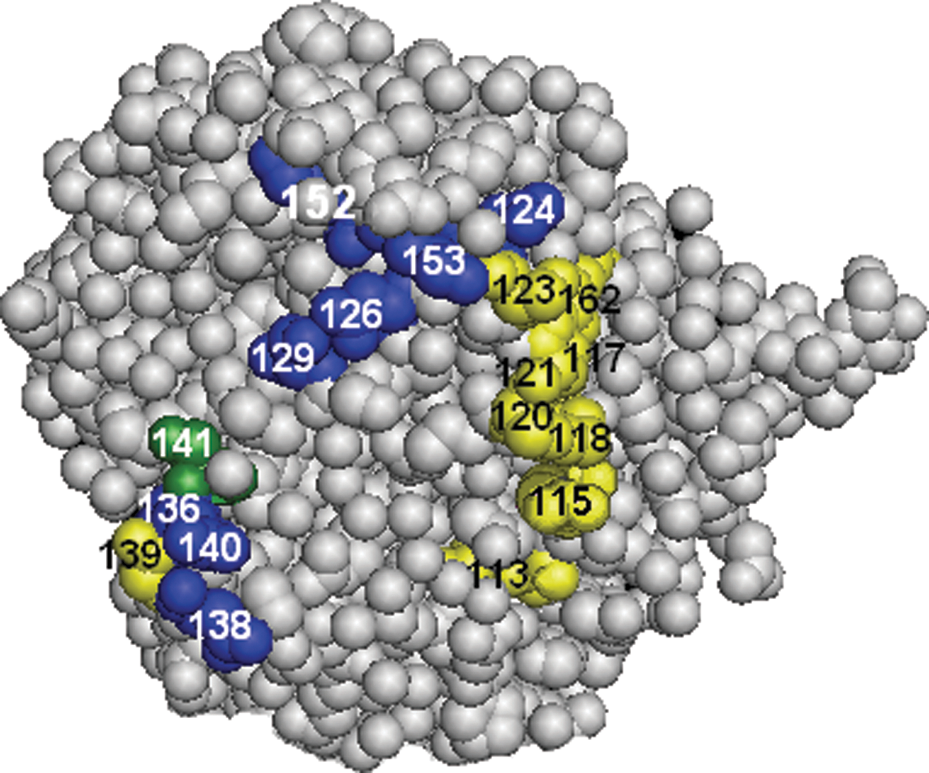
Antigenic sites on the globular head of the HA of A/Mallard/Pennsylvania/10218/84 (H5N2) influenza virus. Amino acid residues recognized by mAbs against A/Chicken/Pennsylvania/1370/83 (H5N2) and A/Chicken/Pennsylvania/8125/83 (H5N2) viruses (5) are marked in blue. Amino acid residues recognized by mAbs against A/Novosibirsk/56/05 (H5N1) virus are marked in yellow. The amino acid residue recognized by both panels is marked in green. The images were created with RasMol 2.6, and the HA structure was obtained from the Protein Data Bank (PDB accession number 1JSM). Amino acid positions are designated in mature H5 numbering (4).
Discussion
The antigenic mapping of the HA molecule of influenza A virus belonging to subtype H5 was started by the pioneering work of Philpott et al. (12). At that time the three-dimensional structure of H5 HA was not yet reported, and the authors used the x-ray crystallographic model of H3 HA (20). The authors selected six escape mutants of A/Turkey/Ontario/7732/66 (H5N9) virus and mapped the location of antigenically relevant amino acid substitutions (positions 36, 43, 115, 141, 152, and 189 in the mature H5 molecule). On the basis of the locations of the amino acid substitutions, the authors identified five neutralizing epitopes. The location of four of them was considered to be within the antigenic sites described for the H3 subtype (19), or close to them. Position 141 belonged to site A, positions 152 and 189 to site B, position 43 to site E, and position 36 was close to site C. Position 115, however, had no obvious relation to any of the sites described for the H3 molecule.
In our previous studies (5) we attempted to analyze not only the location, but also the extent and fine structure of the antigenic sites on the H5 HA molecule. The selection of escape mutants of the A/Mallard/Pennsylvania/10218/84 (H5N2) strain with mAbs against the A/Chicken/Pennsylvania/1370/83 (H5N2), A/Chicken/Pennsylvania/8125/83 (H5N2), and A/Turkey/Ontario/7732/66 (H5N9) viruses resulted in the identification of positions 124, 126, 129, 136, 138, 140, 141, 152, and 153 as antigenically relevant. Positions 136, 138, 140, and 141 were concentrated in the lateral loop corresponding to site A in the H3 molecule, and they included position 141 as described by Philpott et al. (12). The other positions (124, 126, 129, 152, and 153) were located at the top of the HA globule in the area coinciding partly with site B in H3, and partly with site Sa in H1 (1). This series of mutations included position 152 described by Philpott et al. (12). In our studies of the HA of A/Vietnam/1203/04 (H5N1) strain (6), the same approach, that is, the selection of escape mutants, was applied. The analysis allowed us to add positions 121 and 162 to the list of antigenically relevant amino acid residues. Besides, the mAbs against A/Vietnam/1203/04 (H5N1) recognized positions 140, 141, and 152, identified previously in our work with the A/Mallard/Pennsylvania/10218/84 (H5N2) strain (5) as antigenically relevant. However, neither of the mAbs used in our previous studies ever selected the mutants having amino acid changes in the areas of the antigenic site C, or the site E, or near the separate site containing position 115 (12).
In the present report we describe the application of escape mutant analysis to the mAbs raised against a subclade 2.2 virus, A/Novosibirsk/56/05 (H5N1). The studies had two goals. First, to extend our knowledge of the antigenically relevant sites on the HA globule of the H5 subtype, and second, to reveal the differences, if any, between the predominant epitopes recognized by the mAbs to recent H5N1 strains, and the mAbs raised against earlier H5 strains. We used the strain A/Mallard/Pennsylvania/10218/84 (H5N2) as the wild-type virus. The use of a strain different from the one used for the production of the mAbs was justified in this case, as it allowed us to compare the results with our previous data (5) without any regard to a possible effect of different wild-type viruses used for the selection of escape mutants. The mAbs against the HA of A/Novosibirsk/56/05 (H5N1) virus reacted with A/Mallard/Pennsylvania/10218/84 (H5N2), although they failed to react with a genetically much closer A/Vietnam/1203/04 (H5N1) virus. Most likely, this was due to amino acid differences in the antigenically relevant areas of HA between the H5N1 viruses belonging to the clade 1 and the subclade 2.2 (6). An extensive antigenic variation of the HA of H5N1 viruses, due in part to the vaccination of fowl, has been described (14).
The analysis revealed amino acid changes in HA of escape mutants in positions 113, 115, 117, 118, 120, and 123, which were not encountered in our earlier publications on the escape mutant analysis of H5 HA (5,6). The amino acid changes are concentrated in an area closely adjacent to the region recognized in earlier studies as corresponding to site B in H3 HA, but extending far beyond this area. The part occupied by these positions engulfed the amino acid residue 115, presumed by Philpott et al. (12) to be a part of a separate antigenic site (Fig. 1).
Interestingly, the areas recognized by the mAbs to the old Pennsylvania strains (A/Chicken/Pennsylvania/1370/83 and A/Chicken/Pennsylvania/8125/83), and the area recognized by the mAbs against A/Novosibirsk/56/05 (H5N1) virus, although adjacent, do not overlap (Fig. 1). On the other hand, the epitopes recognized by the mAbs against A/Vietnam/1203/04 (H5N1) virus partially overlaps both areas (6). The data may suggest that the evolution of H5 HA leads to a change in the dominance of the immunogenicity of specific parts of antigenically active regions on the HA molecule. However, more mAbs have to be analyzed in order to reach a definite conclusion.
In a recently published report (17), the epitopes recognized by two human recombinant antibodies against the HA of an H5N1 virus were characterized. The epitopes were analyzed by site-specific mutagenesis, without the selection of escape mutants. Two epitopes were described, partially coinciding with the areas delineated in our present studies. This may be regarded as evidence for the relevance of the data on epitope recognition by mouse mAbs for the immune reactivity of humans to the H5 hemagglutinin. Since only two human mAbs were used, it is premature to speculate whether the dominant immunogenicity of the 113–123 area revealed in our studies is characteristic for the human immune response to the HA of H5N1 strains. This region was recognized by the human antibody as a linear epitope (17). Interestingly, among the mAbs used in our studies, the mAbs 6F3 and 4G10 recognized this region as a linear epitope, whereas the mAbs 6E2 and 5F12 failed to react in a Western blot (8). Most likely, the amino acid residues in this region, besides being a part of a linear epitope, are also participating in the formation of one or more conformational epitopes.
The antigenic evolution within H5N1 sublineages resembles, to a certain extent, the antigenic drift observed in human influenza viruses. The amino acid substitutions in natural isolates (3,15,22) often occur within the antigenic sites outlined here and in our earlier publications (5,6). The effect of amino acid substitutions on the antigenic specificity of HA in natural strains was recently confirmed by site-specific mutagenesis (22). The increase of our knowledge of the extent of the parts of the HA molecule involved in the interaction with neutralizing mAbs is important for better understanding the molecular basis of the antigenic evolution of HA. The differences in the epitopes preferably recognized by the mAbs to different H5N1 strains (Fig. 1) may be equally important. This observation may be regarded as an indication of the changes in the immunogenicity of different antigenic sites or their parts in the course of the evolution of influenza virus HA.
Footnotes
Acknowledgments
The work was supported by grants 07-04-00005 and 08-04-12055 of the Russian Foundation for Basic Research (RFBR). The authors are grateful to Dr. P.G. Deryabin for the gift of purified β-propiolactone-inactivated preparation of A/Novosibirsk/56/05 (H5N1) virus.
Author Disclosure Statement
No competing financial interests exist.