
Abstract
The resurgence of Chikungunya (CHIK) virus in the form of an explosive, unprecedented epidemic with high virulence and unusual numbers of fatalities has created an immense public health concern in recent years. In the absence of an effective vaccine and specific antiviral therapy, early accurate diagnosis is essential for the best patient management. The present study describes the production and characterization of high-affinity and selective monoclonal antibodies (Mabs) against recombinant E2 protein (rE2) of the CHIK virus. The reactivity of Mabs for rE2 protein was demonstrated using ELISA. The specificity of the generated Mabs for rE2 was demonstrated by Western blot and indirect immunofluorescence. The application of this CHIK virus E2-specific monoclonal antibody in early clinical diagnosis was demonstrated by various analytical methods, such as immunoblotting, indirect immunofluorescence assay (IFA), and antigen-capture ELISA (AC-ELISA), for the detection as well as the identification of the novel ECSA genotypes of CHIK virus. These findings suggest that the high-affinity E2-specific monoclonal antibodies reported in this study will be useful for early clinical diagnosis and epidemiological studies of CHIK virus in developing countries.
Introduction
The resurgence of the CHIK virus in the form of an explosive epidemic with unusually high case fatality rates on Réunion Island and India after 2005 has been attributed to the ECSA genotype (3). During these recent epidemics severe clinical signs such as neurological signs and fulminant hepatitis were documented (18,20). CHIK has therefore emerged as an important public health problem in the Indian Ocean islands and India. On the Indian subcontinent the outbreak severely affected coastal states like Andhra Pradesh (AP), Kerala, Karanataka, Tamil Nadu, and other states of Central India in 2006. By the end of 2006, it had spread to 15 other states, including Kerala, before subsiding (3,24).
The routine laboratory diagnosis of CHIKV infection is based on culture and serology followed by identification of the viral genome through reverse transcription polymerase chain reaction (RT-PCR) (14,17,19). Serologically, CHIKV infection can be detected by immunoglobulin M (IgM) and IgG capture enzyme-linked immunosorbent assay (ELISA) (5,22). RT-PCR is the method of choice for the early detection of CHIKV replication in supernatants, clinical samples, or for epidemiological surveys (7). In addition to conventional RT-PCR, there are more rapid, sensitive, and real-time PCR-based assays such as TaqMan RT-PCR and loop-mediated isothermal amplification (LAMP) methods, which are currently under extensive evaluation with clinical samples (11,13).
The PCR-based methods, although useful for early confirmatory diagnosis, are very expensive due to the requirement for a thermal cycler and expensive reagents. Thus PCR-based methods exhibit several intrinsic disadvantages, requiring either a high-precision instrument for amplification or an elaborate method for detection of amplified products (11). On the contrary, the antigen and antibody based immunoassays are cost-effective and can be used as rapid screening tests for large numbers of samples during epidemics.
The sensitivity and specificity of the antigen-capture ELISA depend on the quality of the antigen and antibody used in the experimental protocol. In addition, the extensive cross-reactivity among the closely-related members of the Alphavirus genus demand the use of highly selective and specific antibodies for the early clinical diagnosis of Chikungunya infection with a high degree of accuracy. Monoclonal antibodies (Mabs) are therefore the most reliable option for the development of optimal diagnostic assays. The structural protein E2 is the major immunodominant antigenic protein of the CHIK virus. Thus the structural protein is the most appropriate target for use in the diagnosis of Chikungunya infection. The main objective of our study was to generate and characterize Mabs against the recombinant envelope (rE2) protein of the CHIK virus. In this study, four Mabs against the rE2 protein of CHIKV were characterized by isotyping and Western blotting. Further applicability of the high-affinity and specific Mab for detection and identification of CHIKV was checked using various analytical methods, such as immunoblotting, indirect immunofluorescence assay (IFA), and antigen-capture ELISA (AC-ELISA).
Materials and Methods
Materials
An Indian strain of CHIK virus, CHIKV ISW HYD06, isolated from a febrile patient (GenBank accession no. EF 210157) during the 2006 epidemic in Hyderabad, A.P., India was used in the present study. Other agents used in this study include Iscove's modified Dulbecco's medium (IMEM), hypoxanthine, aminopterin, and thyimidine (HAT), hypoxanthine and thyimidine (HT), antibiotics (gentamicin, penicillin, and streptomycin), fetal bovine serum (FBS), polyethyleneglycol (PEG), anti-mouse horseradish peroxidase (HRP) conjugate, anti-mouse FITC conjugate, 30% hydrogen peroxide, diaminobenzidine (DAB), 3,3,5,5-tetramethyl-benzidene substrate (TMB), a mouse monoclonal antibody isotyping kit (all from Sigma-Aldrich, St. Louis, MO), and the Immunopure G IgG purification kit (Pierce Biotechnology, Rockford, IL). SigmaStat software was used for data analysis (Systat Software, San Jose, CA).
Selection of patients
All patients between the ages of 5 and 85 y with symptoms considered to be the clinical features of CHIKV infection were included in this study. These symptoms included fever, headache, myalgia, joint pain with or without swelling, and the presence or absence of a rash on the body. A total of 200 serum/CSF samples comprising 160 serum and 40 CSF samples collected from CIIMS at Nagpur, and from KMC, Mangalore, in 2005 and 2007 were evaluated in this study. A slight majority of the subjects (122; 59%) were male. The median age was 45 y (range 5–85 y). All patients gave oral consent. Written consent was deemed unnecessary since all diagnostic tests are routinely used in clinical practice. Clinically, the patients were divided into the categories of confirmed Chikungunya infection and suspected Chikungunya infection. Those patients having high-grade fever, headache, maculopapular rashes, myalgia, and joint pain with and without swelling were considered to have clinically confirmed Chikungunya infection. Those patients with pyrexia of unknown origin (PUO) having low-grade fever, rash, and headache with nausea and vomiting were grouped as having suspected Chikungunya infection.
Preparation of recombinant CHIKV E2 protein
Viral RNA was extracted from 140 μL of culture supernatant of CHIK virus (ISW HYD06 strain) using a QIAamp viral RNA Mini Kit (Qiagen, Hilden, Germany), according to the manufacturer's instructions. To amplify the genomic region coding for the E2 protein, a forward primer E2: F-5′
Production and purification of Mabs
Six BALB/C mice aged 4 wk were primed intraperitoneally with 50 μg rE2 protein mixed with complete Freund's adjuvant. Two boosters were given at days 14 and 28 with 50 μg rE2 mixed with incomplete Freund's adjuvant. Three days after the last booster, the titer of polyclonal antiserum was assessed using an indirect ELISA (described below), with rE2 as antigen. The mouse with the highest titer was chosen to harvest splenocytes. Separated splenocytes were fused with SP2/0 myeloma cells at a ratio of 5:1 using 50% (w/v) PEG, according to a previously described protocol (10). The hybridoma cells were obtained and subsequently cloned by limiting dilution. The cell lines that produced specific antibodies were sub-cloned successively 3–5 times by limiting dilutions to ensure monoclonality and stability. Positive clones that secreted high-titer E2-specific antibodies in the indirect ELISA were further identified. The immunoglobulin subclass was determined using a mouse monoclonal antibody isotyping kit. A high-affinity clone was used to generate ascites in BALB/C mice, and the Mab was purified by protein A chromatography according to the manufacturer's protocols. The concentration of purified Mabs was determined by a bicinchoninic acid protein assay (1). Briefly, 25 μL of the protein sample was added to 200 μL BCA working reagent (1:8) on 96-well plates. Standards and blank wells were used as controls. The samples were incubated at room temperature for 30 min and absorbance was recorded at 562 nm.
Western blot analysis
Western blot analysis was performed to confirm the specificity of the clones using purified rE2 and E. coli extract as antigens on a 10% SDS-PAGE, and was transferred electrophoretically to nitrocellulose membranes for 2 h at 35 volts (23). The supernatant of hybrid cells was used as primary antibody (45 min at 37°C), and anti-mouse HRP conjugate at a dilution of 1:2000 (45 min at 37°C) as secondary antibody. The reaction was revealed using DAB chromogen.
Indirect ELISA
All ELISAs were carried out in 96-well microtiter ELISA modules. Titers of hybridoma-cell-secreted Mabs were detected by indirect ELISA per the standard protocol (4). Briefly, the wells were coated overnight at 4°C with 300 ng/well of purified E2 protein, diluted in 50 mM carbonate saline (pH 9.6). After blocking for 1 h at 37°C with PBS containing 3% BSA (PBSA), the wells were washed 4 times with 1×PBS. Serial twofold dilutions of Mabs were added to each well and incubated for 1 h at 37°C. After that the wells were washed 4 times with PBS containing 0.05% Tween-20 (PBST), then HRP-conjugated anti-mouse IgG (1:2000) was added to each well and incubated at 37°C for 1 h, then washed again. Antibody binding was visualized by the addition of TMB chromogen. After incubation for 15 min at 37°C, the reaction was stopped by the addition of 0.1 M H2SO4, and absorbance was read at 490 nm in a ELISA reader.
Immunofluorescence assay (IFA)
Binding of Mab to CHIKV-infected cells was determined by IFA. Sub-confluent Vero cells, which were grown in 24-well microplates, were infected with CHIKV (ISW HYD06 strain) at a multiplicity of infection of 0.1. After incubation for 3 d, high-affinity Mab was added to CHIK virus-infected Vero cells. After incubation at room temperature for 1 h, the wells were washed 3 times with PBS, and FITC-labeled anti-mouse IgG was added at a dilution of 1:1000. The wells were washed again after 1 h of incubation, and observed under a fluorescence microscope at 200×magnification. Cells showing strong green fluorescence were recorded as positive. The normal mouse serum was used as a negative control in CHIK virus-infected cells.
Affinity analysis by surface plasmon resonance (SPR)
The affinity between Mab and purified E2 was determined by a surface plasmon resonance (SPR) system (Autolab ESPRIT; Ecochemie B.V., Utrecht, The Netherlands). First, recombinant E2 protein was immobilized on the surface of a carboxymethyldextran-modified gold chip by amine coupling, and then used to capture purified Mab. Analysis was performed at 25°C at a constant flow rate of 30 μL/min, using 10 mM PBS (pH 7.4), as a running buffer. To determine the association rate, dissociation rate, and affinity constant (KD), a concentration series from 0.4 to 31.3 nM of purified Mab was injected (50 μL, associated for 500 sec, and then dissociated for 400 sec). The E2 surface was regenerated by injection of 10 mM HCl before each E2 injection. Binding curves and kinetic parameters were analyzed with a global fit 1:1 binding algorithm with drifting baseline by SPR kinetic evaluation software version 5.0 (Ecochemie B.V.).
Antigen-capture ELISA (AC-ELISA)
A double antibody sandwich ELISA test system was optimized by employing rabbit hyperimmune sera (HIS) and mouse Mab as capture and detector antibody, respectively. The wells of ELISA plates were coated with 100 μL of a 1:1000 dilution of rabbit HIS and incubated at 4°C overnight. Following washing with 1×PBS, the wells were blocked with 3% BSA, followed by incubation at 37°C for 2 h. After washing with 1×PBS, 100 μL of 1:100/1:10 dilution of patient sera/CSF samples were added to the wells and incubated at 37°C for 1 h. The plates were incubated with 100 μL of a 1:500 dilution of high-affinity mouse Mab, followed by washing steps. After washing, the wells were incubated with anti-mouse HRP conjugate at a dilution of 1:2000 in 3% BSA. The reaction was developed with 100 μL of TMB substrate and kept at room temperature for 5 min. The peroxidase reaction was stopped with 100 μL of 1 N H2SO4, and the absorbance was read at 490 nm in an ELISA reader. Further cross-reactivity of all the Mabs was also checked against closely-related circulating flaviviruses (dengue and Japanese encephalitis virus [JEV]), West Nile virus (WNV), Yellow fever virus (YFV), St. Louis encephalitis virus (SLEV), and alphavirus (Ross River virus [RRV]), by using culture supernatant as antigen in antigen-capture ELISA assays.
Statistical analysis
The results are expressed as the mean±standard deviation (SD). Data were analyzed by one-way analysis of variance (ANOVA), followed by Dunnett's test, using SigmaStat software. A p value <0.05 was considered significant.
Results
Generation and purification of Mabs against CHIKV rE2 protein
The positive-fused cell clones were screened using indirect ELISA with rE2 as antigen. The hybridomas with higher ELISA titers were selected for screening, and four Mabs (3A1, 5A9, 4D5, and 6G3) were finally isolated and cloned. Out of these 4 clones, the 6G3 clone was found to have high affinity toward the rE2 antigen. Further ascites was produced in BALB/C mice by the high-affinity clone. The heavy-chain subclasses of Mabs were determined to be IgG2a (3A1 and 5A9) and IgG1 (6G3 and 4D5), and the light chains of all of these were kappa isotype.
Reactivity and purification of Mabs with rE2 protein
An indirect IgM ELISA was performed to check the reactivity of all the Mabs against rE2. Out of the 4 clones, clone 6G3 showed higher affinity against rE2 with respect to OD values. The reactivity of purified Mabs with E2 is shown in Fig. 1. High-affinity Mab 6G3 was efficiently purified by protein G chromatography. The concentrations of purified Mabs were determined as 10–18 mg/mL.
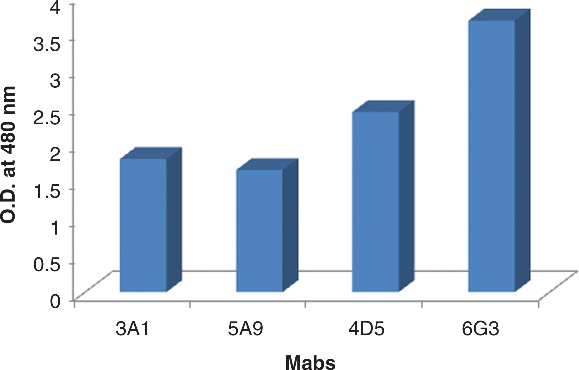
The reactivity of monoclonal antibodies against recombinant E2 (rE2) protein of Chikungunya virus in terms of optical density (OD) by indirect enzyme-linked immunosorbent assay. Color images available online at
Specificity of the Mabs
Immunofluorescence assay (IFA)
Immunofluorescence assay was performed to further analyze whether the Mab recognized the endogenously-produced E2 protein in CHIKV-infected Vero cells. The high-affinity clone showed strong reactivity with CHIKV-infected cells. Normal mouse serum did not show reactivity with CHIKV-infected cells (Fig. 2A).

(
Western blot analysis
The binding specificity of the Mabs against E2 protein were determined by Western blot analysis. All four Mabs reacted at 48 kDa with recombinant E2 protein. The irrelevant Mab (E7) against JEV did not bind to recombinant E2 antigen of Chikungunya virus (Fig. 2B).
ELISA
The cross-reactivity of the Chikungunya E2-specific high-affinity Mab (6G3) against the closely-related circulating flaviviruses (dengue and JEV), WNV, YFV, SLEV, and alphavirus (RRV) was checked by ELISA as described above. The Mab showed reactivity only with the Chikungunya antigen and no cross-reactivity was observed against any of the flaviviruses or alphaviruses as demonstrated by ELISA OD values (Fig. 3).
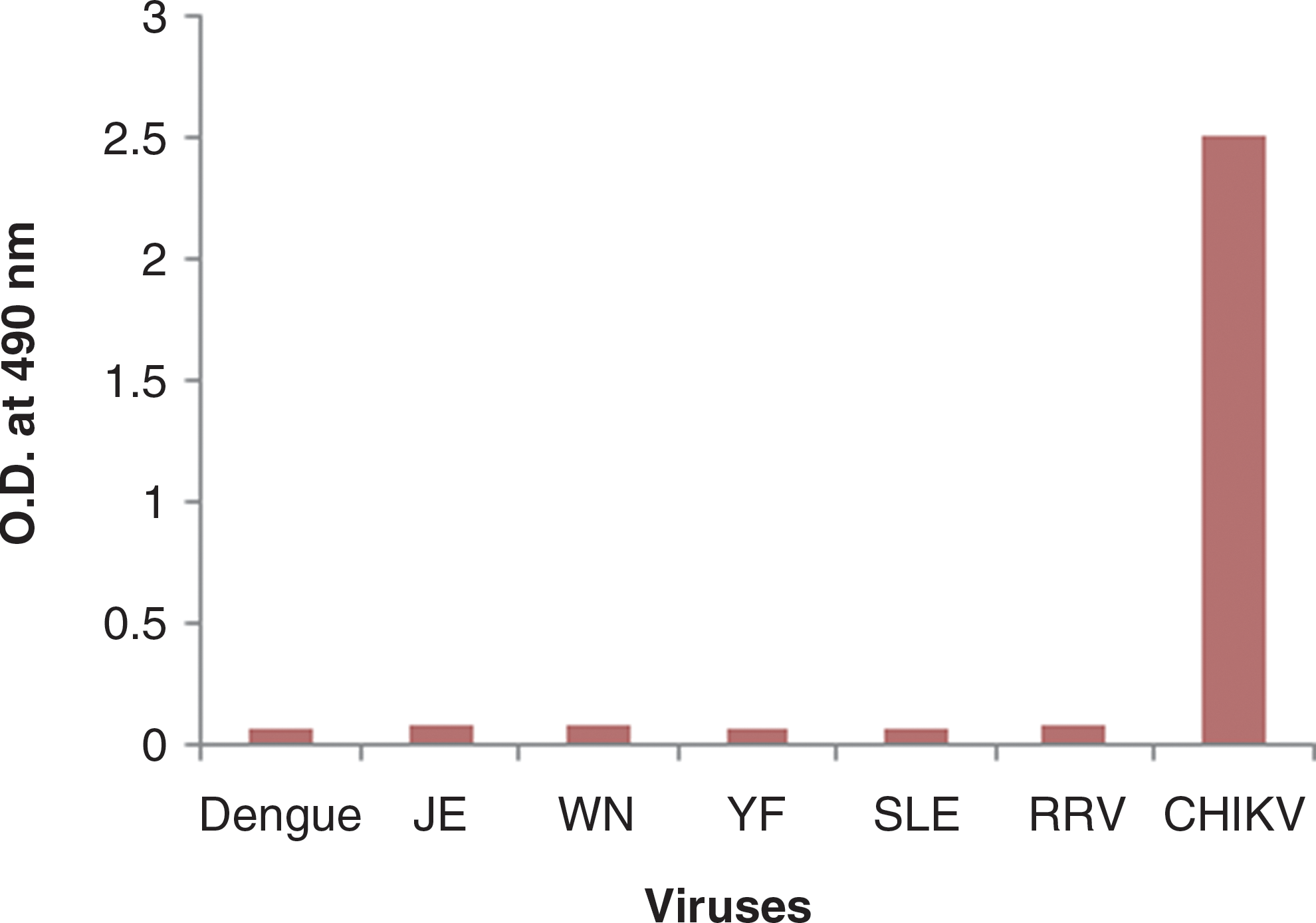
Specificity testing of Chikungunya E2-specific high-affinity monoclonal antibody 6G3 against closely-related members of the flavivirus and alphavirus groups (JEV, Japanese encephalitis virus; WNV, West Nile virus; YFV, Yellow fever virus; SLEV, St. Louis encephalitis virus; RRV, Ross River virus; CHIKV, Chikungunya virus). Color images available online at
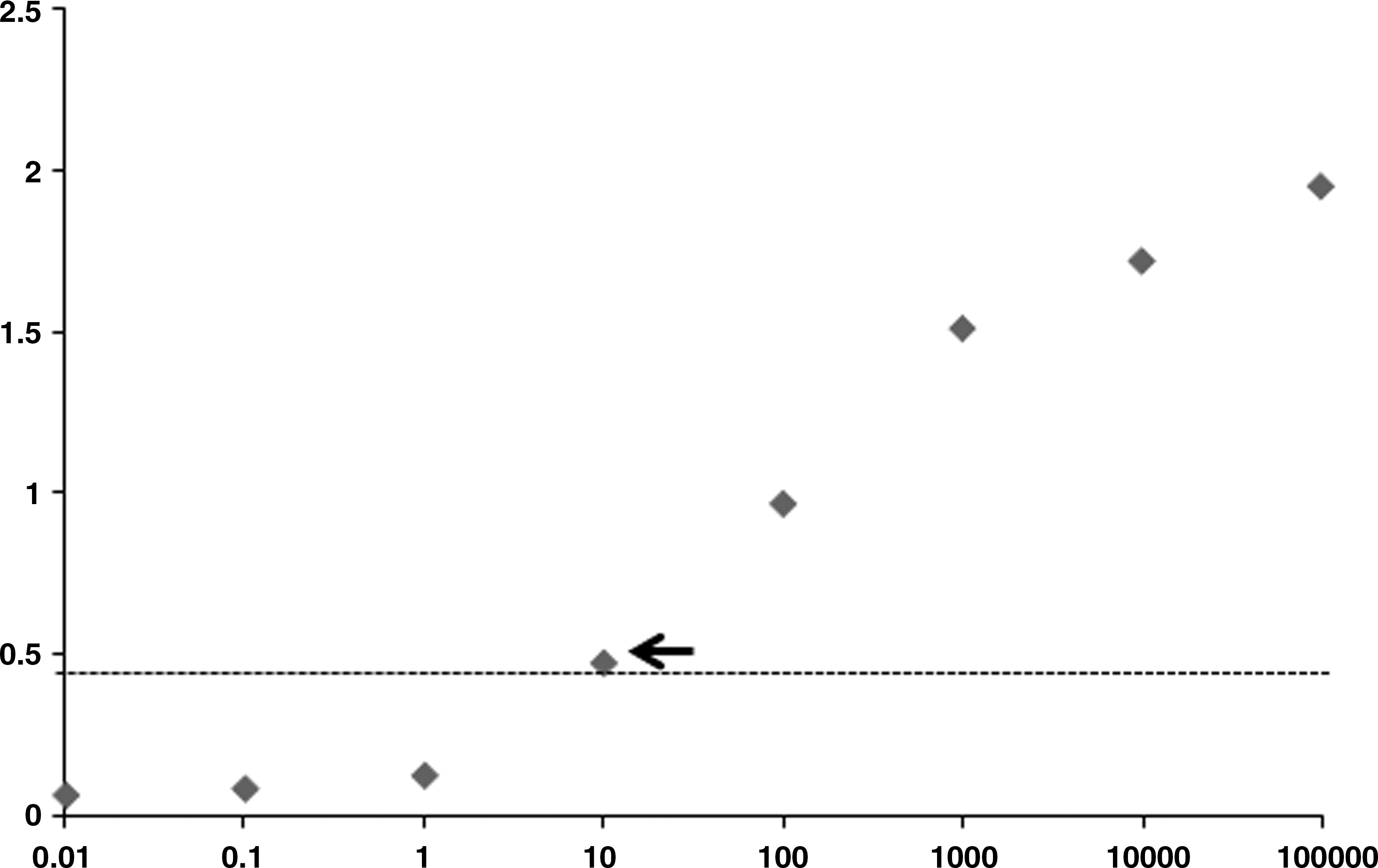
Binding affinity between purified Mabs and recombinant E2 protein
The binding affinity between recombinant E2 protein and purified high-affinity Mab was analyzed by SPR in the solid phase. The sensorgrams for the interaction of different concentrations of E2 Mabs (0.46–31.3 nM) with the E2 antigen-immobilized sensor chip are shown in Fig. 4. The affinity measurements for the interaction of E2 antigen with immobilized antibody were characterized by calculating the KD (affinity constant) using kinetic evaluation software, and the value was found to be 1.27 nM.

Surface plasmon resonance sensor response for the interaction of different concentrations of E2-specific monoclonal antibodies: (
Properties of Mabs against CHIKV E2 protein
Four Mabs against CHIKV E2 protein were identified by isotyping reactivity with recombinant E2 antigen by affinity assay, Western blot analysis, AC-ELISA, and IFA. The properties of these Mabs are summarized in Table 1.
++ and +++, positive; ++++, strong positive; ELISA, enzyme-linked immunosorbent assay.
Clinical samples
Of the total of 200 patients, 90 patients were in the category of individuals with confirmed CHIKV infection, and 110 were in the category of individuals with PUO. Dengue- and JEV-positive and healthy serum samples were also included in this study as negative controls. The majority of patients (81%) in all diagnostic categories were between the ages of 20 and 60 y. Only 5 patients (2%) were below the age of 10 y. Fever, arthralgia without swelling, myalgia, and skin rash, were the most common symptoms in individuals in nearly all of the diagnostic categories.
Antigen-capture ELISA (AC-ELISA)
In order to establish a sensitive AC-ELISA for CHIKV detection, the rabbit HIS and high-affinity mouse Mab (6G3) were used as capture and detector antibody, respectively. Out of 90 clinically-confirmed samples, 71 serum and 9 CSF samples were found to be positive for antigen by sandwich ELISA. The positivity for CHIK antigen among the serum and CSF samples were found to be 45% and 22%, respectively. A sample was considered positive if the OD was greater than twice the mean value of the negative controls. Using these criteria, the limit of CHIKV AC-ELISA was found to be 10 ng/mL (Fig. 5). No cross-reactivity was observed with any of the healthy persons and serum samples from patients with dengue and JEV. The comparative OD values of different categories of samples are depicted in Fig. 6. The mean absorbance value for CHIKV-positive samples was 1.15±0.12 (p<0.05), which was significantly higher than that for the PUO cases (0.175±0.01), dengue/JEV samples (0.057±0.007), and healthy samples (0.038±0.008). The sensitivity and specificity of the assay were found to be 89% and 100%, respectively.
Antigen-capture enzyme-linked immunosorbent assay (AC-ELISA) with purified Chikungunya virus recombinant E2 protein. The purified E2 recombinant protein was serially diluted in phosphate-buffered saline. Data represent the mean optical density values for three replicates. The cut-off value was set at twice the average value of the negative controls.
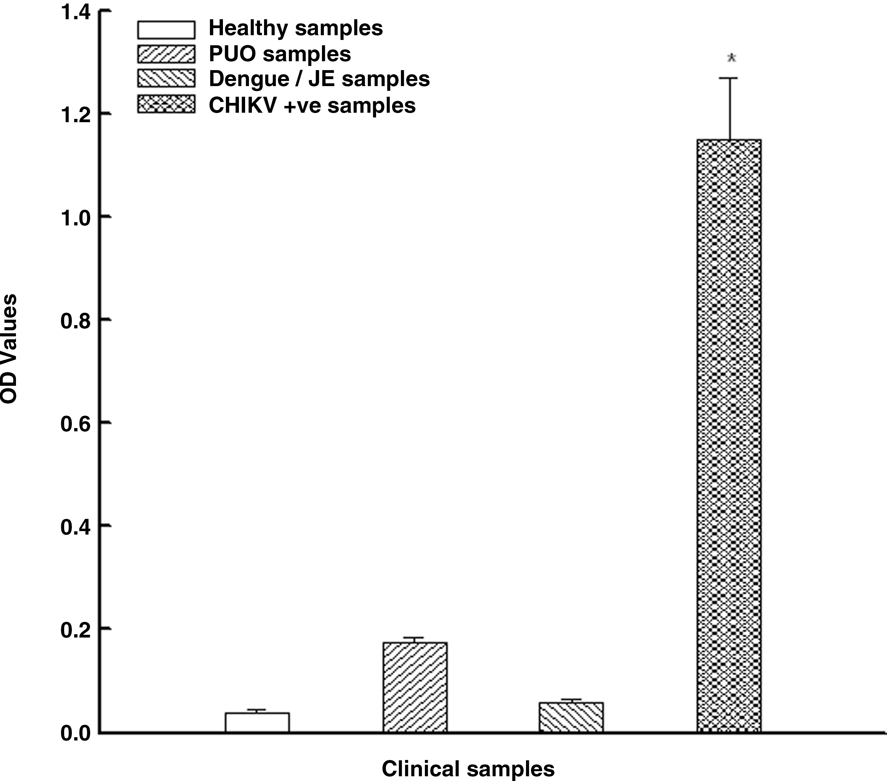
The optical density (OD) profile of different kinds of human patient samples including healthy controls as obtained through the Chikungunya-specific antigen-capture enzyme-linked immunosorbent assay (JEV, Japanese encephalitis virus; PUO, pyrexia of unknown origin; CHIK, Chikungunya virus).
Discussion
Chikungunya is a mosquito-borne arboviral infection that has caused a great deal of public health concern in recent years due to epidemics of unprecedented magnitude with high case fatality rates and unusual clinical signs (12). Three main laboratory tests are used for diagnosing CHIKV infection: virus isolation, serological tests for demonstration of virus-specific antibodies, and genomic detection by PCR-based methods. Virus isolation is the most definitive test and is considered to be the gold standard. However, the isolation process is time-consuming and the degree of success is dependent on a number of complicating factors, including the time of collection, transportation and maintenance of the cold chain, and the storage and processing of samples. However, the RT-PCR test is time-consuming and labor-intensive and has a very high risk of contamination. Serum IgM antibodies can be detected in CHIKV-positive patients as early as 3–5 d after the onset of fever, and generally persist for 30–90 d, although detectable levels may remain up to 8 mo post-infection.
Early and specific detection is critical in the management of CHIK patients. Various serological tests such as the serum neutralization (SN) test and hemagglutination inhibition (HI) test have been used to detect CHIKV-specific antibodies in patient sera and CSF samples (2,7). Serodiagnosis of CHIKV relies on the demonstration of a fourfold increase in Chikungunya IgG titers between acute and convalescent phase sera. However, obtaining paired samples is usually impractical. Alternatively, the demonstration of IgM antibodies specific for CHIK in acute phase sera is used in instances in which paired sera cannot be collected. The most commonly used test is the IgM capture (MAC)-ELISA. Cross-reaction with other alphavirus antibodies such as O'Nyong Nyong virus and Semliki Forest virus usually limits the application of MAC-ELISA as a confirmatory test. Furthermore, the co-circulation of dengue along with Chikungunya draws attention to the need for early and specific diagnosis for differentiation and better patient management (15). It has been demonstrated that CHIKV produces a very high level of viremia that persists for up to 8–10 d. Therefore, antigen detection is a viable proposition for early diagnosis of Chikungunya virus. Considering the fact that no antigen detection system is available for Chikungunya virus, and the limitations associated with the commercially available antibody detection systems, the Mab-based antigen-capture ELISA test reported in the present study has value for the clinical diagnosis of Chikungunya viral infection.
The Mabs generated in this study could be used for the development of immunoassays like ELISA to detect IgM and IgG immunoglobulins in the sera of suspected patients. Hundekar et al. (8) have developed a Mab-based ELISA system to detect Chikungunya virus antigen in mosquitoes. Greiser-Wilke et al. (6) have developed a Mab-based genus-specific antigen-capture immunoassay to detect Alphavirus antigen in clinical samples. Assays using Mabs are more specific for the detection of Chikungunya virus. Also, capture enzyme immunoassays using Mabs have higher sensitivity than direct EIA without Mabs, mainly because of the lower non- specific reaction of the Mabs to control antigen.
In conclusion, four Mabs specific to rE2 protein were produced and characterized. These Mabs could be used in immunoblot assay, IFA, and AC-ELISA. There was no cross-reactivity of any of the Mabs with flaviviruses and alphaviruses. The extensive evaluation of the Mab-based AC-ELISA on larger numbers of serum and CSF samples should show that it is useful for the early detection of Chikungunya with a high degree of sensitivity and specificity.
Footnotes
Acknowledgments
The authors wish to thank Dr. R. Vijayaraghavan, Director, Defence Research and Development Establishment (DRDE), Ministry of Defence, Government of India, for his support, constant inspiration, and help in providing the necessary facilities for this study. The authors are also thankful to the directors of CIIMS Hospital, Nagpur, and KMC, Mangalore, for the clinical samples.
Author Disclosure Statement
No competing financial interests exist.