
Abstract
A recombinant vaccinia virus harboring the full length hemagglutinin (HA) gene derived from a highly pathogenic avian influenza A/Thailand/1(KAN-1)/2004 (H5N1) virus (rVac-H5 HA virus) was constructed. The immunogenicity of the expressed HA protein was characterized using goat antiserum, mouse monoclonal antibody, and human sera. The expressed HA protein localized both in the cytoplasm and on the cytoplasmic membrane of the thymidine kinase negative cells infected with the rVac-H5 HA virus, as determined by immunofluorescence assay. Western blot analysis demonstrated that the rVac-H5 HA protein was post-translationally processed by proteolytic cleavage of the HA0 precursor into HA1 and HA2 domains; and all of these HA forms were immunogenic in BALB/c mice. The molecular weight (MW) of each HA domain was the same as the wild-type H5 HA produced in Madin-Darby canine kidney cells infected with the H5N1 virus, but was higher than that expressed by a baculovirus-insect cell system. Sera from all H5N1 survivors reacted to HA0, HA1, and HA2 domains; whereas sera from H5N1-uninfected subjects reacted to the HA2 domain only, but not to HA0 or HA1, indicating that some cross-subtypic immunity exists in the general population. There was a lot-to-lot variation of the recombinant HA produced in the baculovirus-insect cell system that might affect the detection rate of antibody directed against certain HA domains.
Introduction
HA is a type I integral membrane glycoprotein and is a major component of the viral envelope where it exists as noncovalent homotrimers. Initially, it is synthesized as a precursor polypeptide (HA0) that further undergoes post-translational modifications including glycosylation, proteolytic cleavage, and signaling peptide removal (8 –10). SDS-polyacrylamide gel electrophoresis of purified virus particles has demonstrated that the monomeric HA0 has a molecular weight (MW) of approximately 80 kilodaltons (kDa) and is cleaved into two glycosylated functional subunits, HA1 (MW 56–58 kDa) and HA2 (MW 25–26 kDa) which are connected together by disulfide linkages (11 –13). The cleavage of the HA0 precursor into HA1 and HA2 is necessary for viral infectivity (9,14). The HA1 is a hypervariable domain, which shares only 34%–59% amino acid sequence identity between influenza virus subtypes (15). This domain is responsible for binding to host cellular receptors and the antibody against HA1 is relatively strain or subtype specific (13). On the other hand, HA2 mediates fusion between the viral envelope and the endosomal membrane during the uncoating step of the viral replication cycle (13). It is more conserved than HA1, sharing 51%–80% amino acid sequence identity between subtypes and inducing heterosubtypic immunity (15,16). Neutralizing antibody against HA is the most important correlate of protection against influenza virus infection and is used for assessing vaccine immunogenicity. In addition, Western blot assay for antibody to H5 HA has been recommended by the World Health Organization (WHO) as the confirmatory test for serodiagnosis of patients infected with HPAI H5N1 virus (17).
Several protein expression systems have been employed to express the recombinant H5 HA for use as vaccine candidates, in studying host immune responses, and for disease diagnosis. These expression systems include: the infection of mammalian or avian cells with recombinant vaccinia viruses (18 –22) or pseudotype lentiviral viruses (23), the infection of insect cells with recombinant baculoviruses (24 –26), and the infection of plant cells with recombinant bacteria (27,28). In addition, the expression of H5 HA in yeast or bacterial cells transformed with recombinant plasmids have also been reported (29,30). Among these expression systems, the vaccinia virus vector provides a powerful tool for production of antigenically and biologically active proteins. Several seasonal influenza proteins including HA, NA, NP, M, NS, PB1, PB2, and PA have been expressed in the recombinant vaccinia viruses (31 –34) and used to demonstrate influenza subtype-specific and cross-reactive immune responses (35,36).
Even though the rVac-H5 HA has been expressed by several groups of investigators (18 –22), their immunobiological activities have only been investigated in animal models and never with human samples. Herein, we constructed a recombinant vaccinia virus harboring the complete influenza HA gene derived from an HPAI H5N1 virus. Localization of the rVac-H5 HA in the infected cells was visualized by immunofluorescence microscopy; its antigenicity was determined by Western blot assay using serum samples from H5N1 survivors and H5N1-uninfected subjects. The use of rVac-H5 HA as the test antigen was compared with the H5 HA produced from Madin-Darby canine kidney cells (MDCK) infected with wild-type H5N1 virus, and with the recombinant H5 HA expressed in a baculovirus-insect cell system.
Materials and Methods
Ethical issues
This study has been approved by the Institutional Review Boards of the Faculty of Medicine Siriraj Hospital, Mahidol University.
Human subjects and blood specimen
A total of 20 serum samples employed in this study comprised 11 anonymously archived samples, 3 from HPAI H5N1 survivors, 8 from influenza A/H3N2-infected patients, and 9 serum samples from healthy individuals with informed consent. All patients were diagnosed by real-time reverse transcription polymerase chain reaction (real time RT-PCR) using the protocols established by the U.S. Centers for Disease Control and Prevention. Virus isolation and/or serodiagnosis were also performed in most of the patients. The acute and convalescent blood samples were collected from H3N2 patients at approximately 3 weeks apart, while single blood samples were collected from healthy individuals. Serum and plasma samples were kept frozen at −20°C until used.
Cell lines and viruses
Madin-Darby canine kidney (MDCK) cells were grown in Eagle's minimum essential medium (EMEM) (Gibco, NY) supplemented with 10% fetal bovine serum (FBS) (Gibco) and antibiotics. Influenza A viruses: A/Thailand/Siriraj-Rama-TT/2004 [A/New Caledonia/20/1999 (H1N1)-like virus], A/Siriraj ICRC/SI-154/2008 [A/Brisbane/10/2007 (H3N2)-like virus], and A/Thailand/1(KAN-1)/2004 (H5N1) clade 1 virus (KAN-1 virus) were propagated in MDCK cell monolayers maintained in EMEM in presence of trypsin-tosylphenylalanyl chloromethyl ketone (trypsin-TPCK) (Sigma-Aldrich, St. Louis, MO) and antibiotics, and without FBS supplement. The culture supernatants were harvested, centrifuged, aliquoted, and kept as virus stocks.
TK- (thymidine kinase negative) cells were grown in Dulbecco's modified Eagle medium (DMEM, Gibco) supplemented with 10% FBS and antibiotics. The culture media was replaced with DMEM supplemented with 2% FBS for virus propagation and titration. Vaccinia virus vaccine strain Lister, kindly provided by the Thai Government Pharmaceutical Organization, was used as the gene vector for construction of the recombinant virus harboring H5 HA gene insert. The virus infected cell cultures were harvested, frozen, and thawed three times, followed by treatment with 0.1 volume of 0.25% trypsin (Gibco) for 15 min at 37°C in order to break the aggregates of the viral progenies and facilitating the viral release. Subsequently, a 0.1 volume of FBS was added to terminate the trypsin activity. The cell lysates were centrifuged, aliquoted, and kept as the virus stocks.
Construction of recombinant vaccinia virus carrying H5 HA gene insert
Total RNA was extracted from MDCK cells infected with the KAN-1 virus using a QIAamp® viral RNA mini kit (Qiagen GmbH, Hilden, Germany). The complete HA genomic segment was amplified by One-step RT-PCR kit (Qiagen) using universal primers: Bm-HA-1 (5′-TATTCGTCTCAGGGAGCAAAAGCAGGGG-3′), and Bm-NS-890R (5′-ATATCGTCTCGTATTAGTAGAAACAAGGGTGTTTT-3′) (37). The PCR product of 1,807 base pairs (bp) in length was purified by using QIAquick® gel extraction kit (Qiagen) and cloned into pGEM®-T Easy vector (Promega Corporation, Madison, WI) by using T4 DNA ligase (Promega) before transforming into E. coli JM109 cells. The NotI fragment containing complete HA gene with sticky ends derived from the cutting product of the recombinant plasmid was repaired by using klenow DNA polymerase (New England Biolabs Inc., Ipswich, MA) in order to generate blunt end DNA strands. The DNA product was subcloned into a pSC11 expression vector kindly provided by Prof. Bernard Moss, National Institute of Allergy and Infectious Disease, Maryland, USA. This vector contains the SmaI insertion site located downstream of vaccinia virus p7.5 promoter, together with the E. coli lacZ gene which encodes for β-galactosidase under a p11 promoter, and is flanked with thymidine kinase sequences (TKR and TKL). E. coli JM109 cells were transformed with the recombinant pSC11 containing HA gene insert (pSC11/HA) and plated on LB agar containing 5-bromo-4-chloro-3-indolyl-β-D-galactopyranoside (X-gal) (Promega), plus ampicillin as the selective marker. The cloned bacterial colonies were cultured in LB broth and the plasmids were extracted by a QIAprep® Spin mini kit (Qiagen). The purified plasmids were cut by the enzyme PstI in combination with XhoI (New England Biolabs) in order to check for the presence of the HA gene insert with correct orientation. DNA sequencing was performed in order to determine the in-frame translation of the HA insert using a set of 4 sequencing primers comprising pSC11 P7.5F (5′-GCACGGTAAGGAAGTAGAATC-3′), HAH5F (5′-ACTCCAATGGGGGCGATAAA-3′), HAH5R (5′-CAACGGCCTCAAACTGAGTGT-3′) and pSC11R (5′-CATCGAGTGCGGCTACTATAAC-3′). A mixture of pSC11/HA recombinant plasmids and DMRIE-C transfection reagent (Invitrogen, Carlsbad, CA) in DMEM was transfected into the TK- cells that had been pre-infected with vaccinia vaccine virus at the multiplicity of infection of 0.01 plaque forming unit (pfu)/mL for 2 h. The HA sequence flanked with TKR and TKL was inserted into the parental vaccinia viral genome by homologous recombination with the tk gene. As a result, the recombinant virus harboring the HA gene insert lost the ability to produce thymidine kinase enzyme (TK- phenotype). The transfected culture was further incubated for 2 days to allow virus replication. The recombinant vaccinia virus was distinguished from the parental TK+ vaccinia virus by plaque selection on the TK- cell monolayer in the presence of 5-bromo-2′-deoxyuridine (BrdU) (Sigma Aldrich) and X-gal in which the plaques produced by cells infected with the recombinant vaccinia virus appeared blue. Plaque purification of the recombinant virus was performed three times using low melting point agarose containing BrdU and X-gal. In parallel, the recombinant vaccinia virus containing pSC11 vector (rVac-pSC11) was constructed for use as the vaccinia virus control.
Immunofluorescence assay
TK- cells infected with recombinant vaccinia viruses containing the HA gene insert were investigated for expression and localization of HA protein by immunofluorescence assay (IFA). Goat antiserum against HA from A/Vietnam/1203/04 (H5N1) (VN1203) kindly provided by Prof. Robert G. Webster and Dr. Richard Webby, St. Jude Children's Research Hospital, Memphis, TN, and a mouse monoclonal antibody raised against purified VN1203 HA (US Biological, Swampscott, MA) were used as the primary antibodies. Fluorescein isothiocyanate (FITC) conjugated-rabbit anti-goat Ig (Dako Cytomation, Glostrup, Denmark) or FITC conjugated-goat anti-mouse Ig (Light Diagnostics™, Temecula, CA) was used as the secondary antibody. The slides were counterstained with trihydrochloride trihydrate (Hoechst 33342- Invitrogen, Eugene, OR) together with 0.5% Evan blue's dye and examined for the presence of fluorescent cells under laser scanning confocal microscopes (LSM 510 Meta, Zeiss, Jena, Germany).
Western blot assay for detection of anti-H5 HA antibody
The Western blot (WB) assay was performed for two purposes in this study. Firstly, to investigate the expression, MW, and post-translational proteolytic cleavage of HA protein in TK- cells infected with either rVac-H5 HA or rVac-pSC11 virus in comparison with the KAN-1 virus infected MDCK cells and recombinant HA expressed in a baculovirus-insect cell system. Second, the assay was used to detect specific and cross-reactive antibodies to HA antigen in H5N1 survivors and H5N1-uninfected subjects.
The H5 HA antigens used in the WB assay were derived from three sources: the lysates of TK- cells infected with rVac-H5 HA virus; the lysates of MDCK cells infected with KAN-1 virus; and the recombinant H5 HA expressed in baculovirus-insect cell system (rBV-H5 HA), either purchased from Protein Sciences Corporation, Meriden, CT (lot numbers 45-05034RA-2 [designated lot no. 1] and 0880-125 [designated lot no. 2]) or kindly donated by BEI Resources through the NIH Biodefense and Emerging Infections Research Resources Repository, NIAID, NIH (catalog number NR-660, lot number 59137402 [designated lot no.3]). Briefly, infected cell lysates or recombinant antigens were mixed with 4X reducing sample buffer (8% SDS, 250 mM Tris Cl pH 6.8, 8% β-mercaptoethanol, 0.4% bromophenol blue, 40% glycerol), boiled for 5 min, and subjected to 10% denaturing (SDS) discontinuous polyacrylamide gel electrophoresis (SDS-PAGE- Laemmli method) (38). The proteins present in gel were blotted onto a nitrocellulose membrane (Protran®, Whatman, GmbH, Germany) by using Trans-Blot® SD semidry transfer cell (Bio-Rad). The blotted membrane was blocked with 5% skim milk in Tris-buffer saline plus 0.1% tween-20 (TBS-T). Characterization of the rVac-H5 HA protein employed specific antibodies of three origins: goat antiserum to VN1203 HA (St. Jude Children's Research Hospital), mouse monoclonal antibody to VN1203 HA (US Biological), and pooled mouse sera collected from BALB/c mice immunized with the recombinant vaccinia virus carrying the HA gene of KAN-1 virus (kindly provided by Dr. Molvibha Vongsakul, Faculty of Science, Mahidol University). The anti-species specific immunoglobulin: horseradish peroxidase enzyme (HRP) conjugated-rabbit anti-goat Ig (Dako Cytomation) or goat anti-mouse Ig (Dako) was used as the secondary antibody. The antigen blotted on nitrocellulose membrane was incubated with the primary test serum overnight at 4°C before washing with TBS-T and followed by incubation with the corresponding secondary antibody for 2 h at room temperature. The mixture of 3, 3′-diaminobenzidine (Sigma-Aldrich), 8% NiCl2, and H2O2 was used as the chromogenic substrate. The detection of specific or cross reactive antibody to H5 HA in H5N1 survivors and H5N1-uninfected subjects employed sera at dilution of 1:100. The HRP conjugated-goat anti-human IgG (Invitrogen) at dilution of 1:1000 was used as the secondary antibody.
Microneutralization (microNT) assay
The protocol of ELISA-based microNT assay for detection of neutralizing (NT) antibodies was conducted according to that described in the WHO manual (39) and Lerdsamran et al (40). The test serum was heat inactivated at 56°C for 30 min before making a serial 2-fold dilution starting from the dilution of 1:5 and proceeding to 1:2560. The assay was performed by mixing 60 microliters (μL) of the diluted serum with 60 μL of the virus suspension at a concentration of 200 tissue culture infective dose 50 (TCID50) and incubated at 37°C for 2 h. One hundred microliters of the serum-virus mixture were transferred onto an MDCK cell monolayer and further incubated for 24 h. In order to verify the amount of virus inoculum, virus back-titration at concentrations of 0.1, 1, 10, and 100 TCID50 was included in every reaction plate in duplicate. The relative amount of influenza viral nucleoprotein in the reaction plates was determined by an ELISA using a mouse-specific monoclonal antibody (Chemicon, Temecula, CA) as the primary antibody, goat anti-mouse Ig conjugated with HRP (Southern Biotechnology, Birmingham, AL) as the secondary antibody, and TMB (KPL Inc., Gaithersburg, MD), as the chromogenic substrate. The NT antibody titer was defined as the reciprocal of the highest serum dilution that reduces ≥50% of the amount of viral nucleoprotein in the reaction wells as compared to the virus control wells.
Results
Characterization of rVac-H5 HA
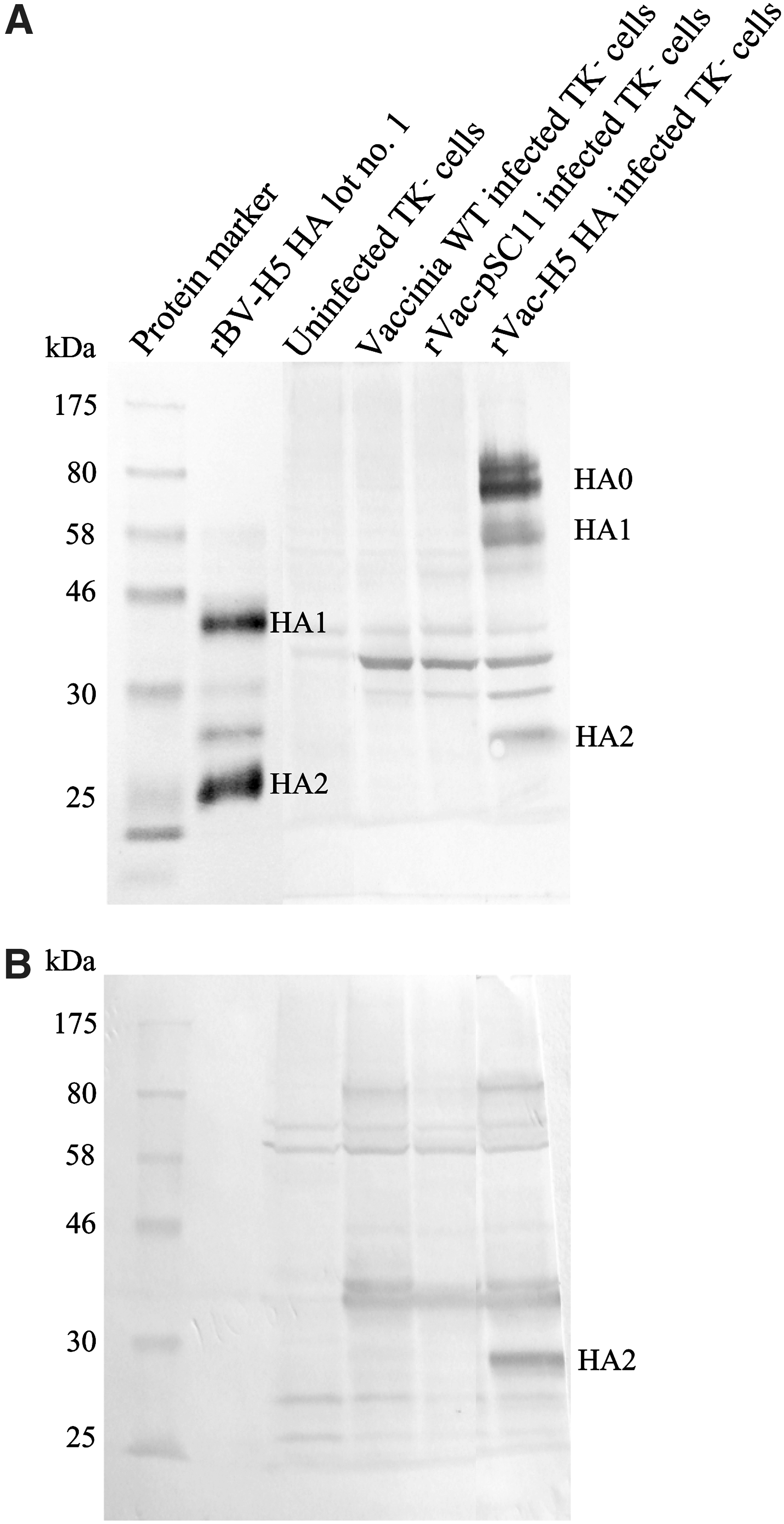
Recombinant baculovirus H5 HA protein (rBV-H5 HA) of known molecular weights (72 kDa for HA0, 45 kDa for HA1, and 25 kDa for HA2 domains as indicated in the product brochure) was used to characterize specificity of a goat antiserum and mouse monoclonal antibody against H5 HA by WB assay. Collective results from three lots of rBV-H5 HA demonstrated that the goat antiserum was reactive against HA0, HA1, and HA2 domains, whereas mouse monoclonal antibody was reactive only to HA0 and HA1 domains (Fig. 1).
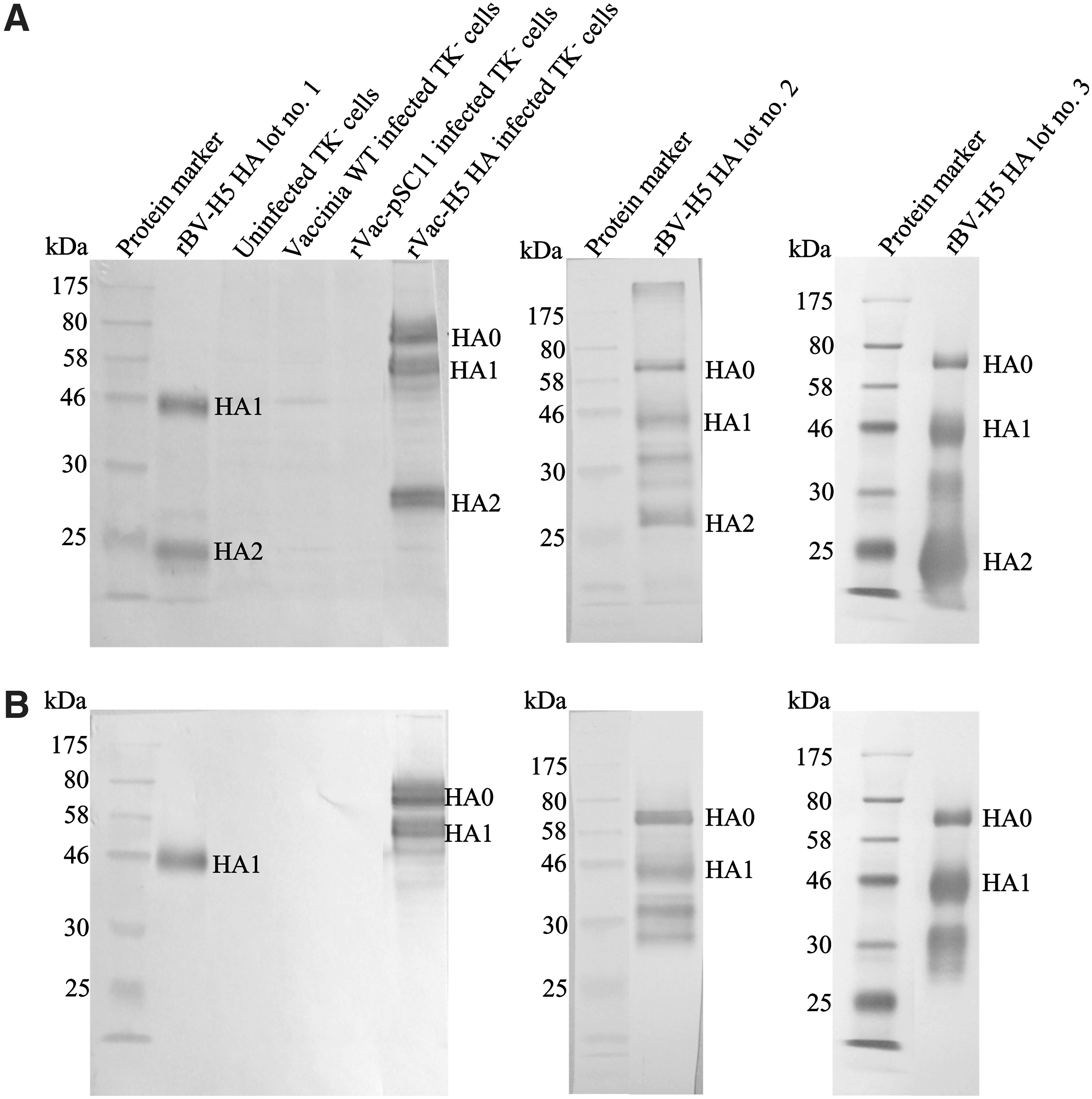
Specificity of a goat antiserum
The goat antiserum and mouse monoclonal antibody were further used to characterize our rVac-H5 HA. Lysates of TK- cells infected with rVac-H5 HA virus were analyzed on their mobility in SDS-PAGE and WB assay in parallel with those of TK- cells infected with rVac-pSC11 as the negative control. Three bands of proteins at MW of approximately 75, 55, and 27 kDa corresponding to the uncleaved HA0 precursor and its HA1 and HA2 cleavage products, respectively, were observed as analyzed with goat anti-H5 HA antiserum (Fig. 1A). On the other hand, 2 bands corresponding to HA0 and HA1 polypeptides were observed when anti-H5 HA monoclonal antibody was employed (Fig. 1B). Notably, the MWs of HA1 and HA2 derived from the recombinant baculovirus virus were clearly lower than those derived from the recombinant vaccinia virus. The discrepancy in MW between our system and the baculovirus system may be due to differences in the glycosylation pattern of HA produced in insect cells compared to vertebrate cells, as discussed below.
Expression and localization of rVac-H5 HA protein expressed in the infected TK- cells
Localization of rVac-H5 HA in the infected TK- cells was determined by indirect immunofluorescence assay using a goat anti-H5 HA antiserum and mouse anti-H5 HA monoclonal antibody. The rVac-pSC11 virus infected TK- cells were used as the negative control. The result showed that the fluorescent rVac-H5 HA protein localized both in the cytoplasm and on the cytoplasmic membrane of the infected cells; no fluorescent signal was observed in the rVac-pSC11 infected TK- cell control as visualized under a confocal fluorescence microscope (Fig. 2). It was noted that our recombinant H5 HA could be recognized by antibodies raised against wild-type HA derived from strain VN1203, which belongs to the same clade.
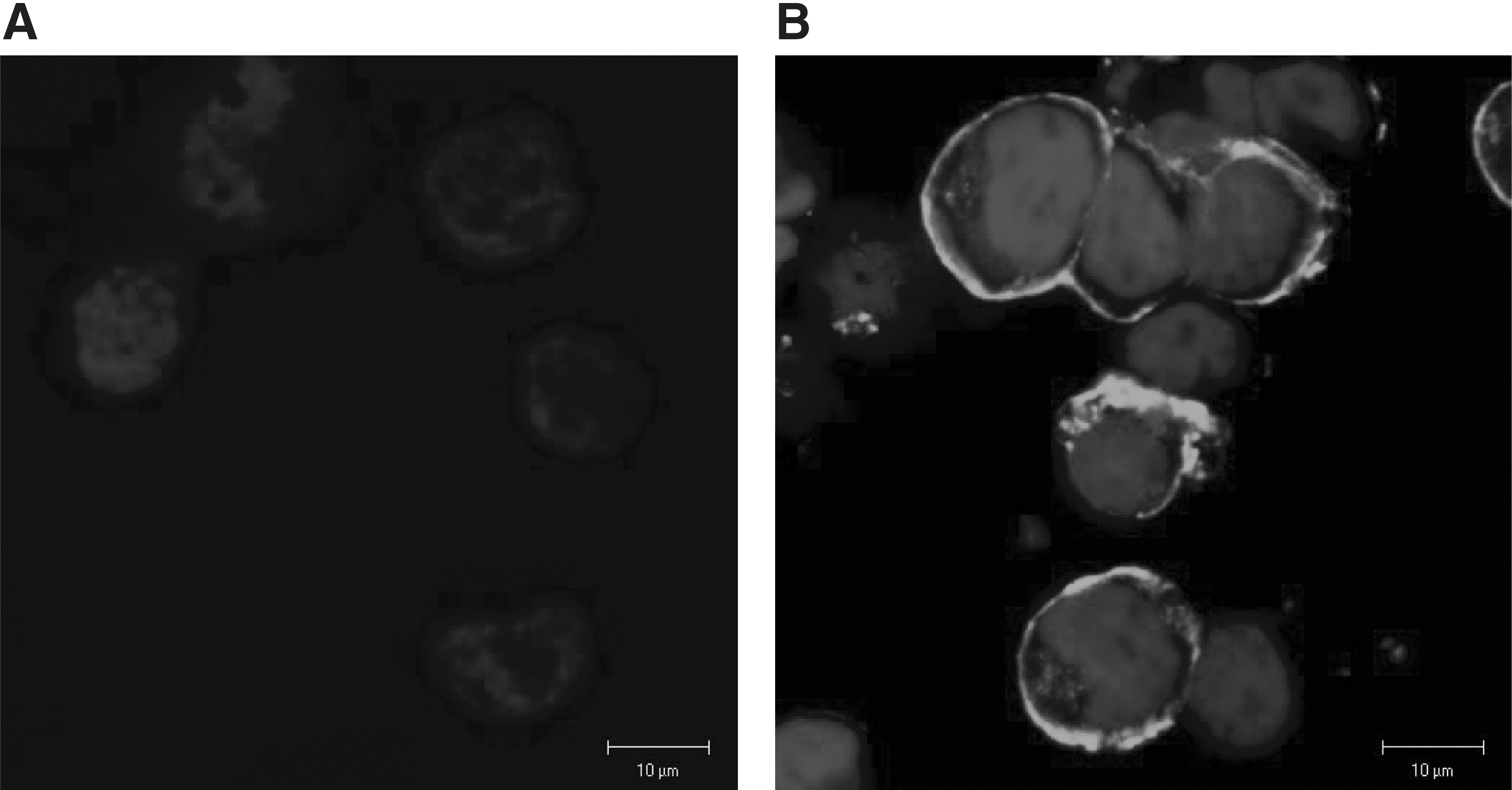
Expression and localization of H5 HA protein in TK- cells infected with recombinant vaccinia virus (rVac-H5 HA) as demonstrated by IFA using goat anti-H5 HA antiserum: TK- cells infected with rVac-pSC11 virus as the negative control
Comparison between rVac-H5 HA and wild-type H5 HA
The recombinant H5 HA protein expressed in TK- cells infected with rVac-H5 HA virus was compared with the wild-type HA protein synthesized in MDCK cells infected with KAN-1 virus. The results showed that HA proteins produced in both virus-cell systems were similar in size and number of the cleavage products (Fig. 3).
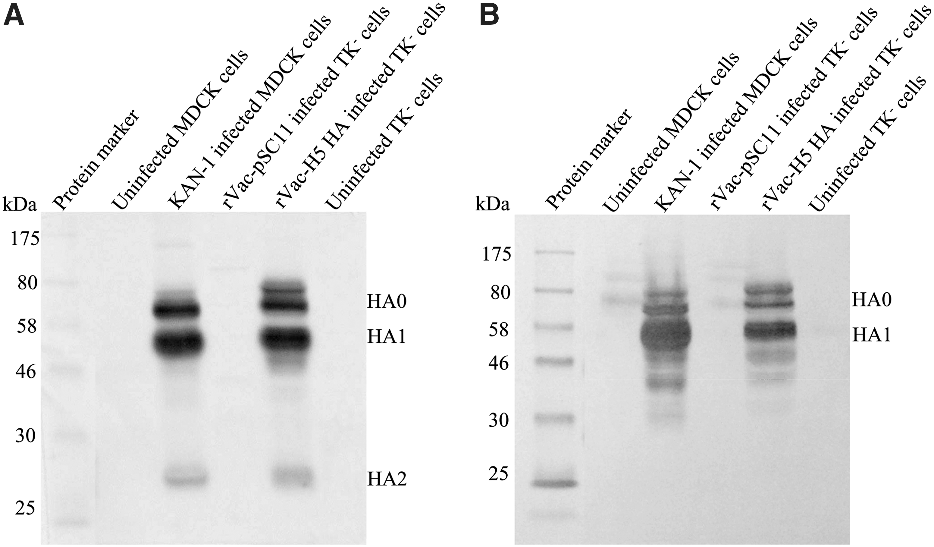
Comparison on the Western blot banding patterns between the recombinant H5 HA protein produced in TK- cells infected with rVac-H5 HA virus and the wild-type H5 HA produced in MDCK cells infected with H5N1 KAN-1 virus. Three bands of the same MWs for HA0, HA1, and HA2 domains are demonstrated by goat anti-H5 antiserum
Immunogenicity and antigenicity of rVac-H5 HA
The immunogenicity of our rVac-H5 HA in the induction of antibody response was investigated in a mouse model. Serum samples collected from BALB/c mice immunized with rVac-H5 HA virus (gift from Dr. Molvibha Vongsakul) were reactive with rVac-H5 HA protein and demonstrated reactivity against HA0, HA1 and HA2 domains (Fig. 4), while serum samples from non-immunized mice, or the mice immunized with rVac-pSC11 virus were nonreactive by WB assay (data not shown). Furthermore, the mouse anti-H5 HA antiserum recognized the HA0, HA1, and HA2 domains present either in the recombinant H5 HA expressed in the baculovirus-insect cell system or in the wild-type H5 HA synthesized in MDCK cells infected KAN-1 virus.
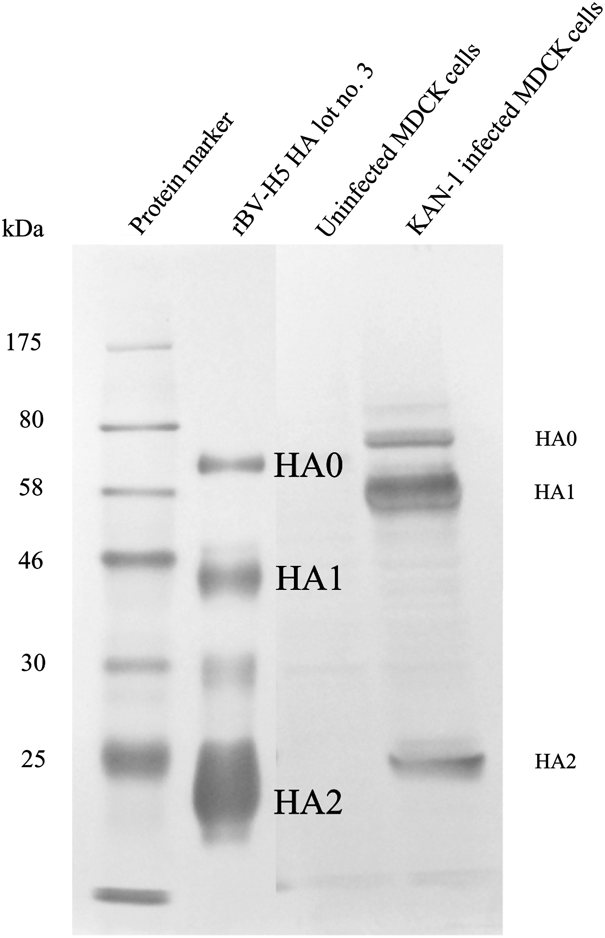
Immunogenicity of rVac-H5 HA protein in the induction of antibody response in BALB/c mice. Pooled sera from mice immunized with lysates of TK- cells infected with rVac-H5 HA virus are used to stain the recombinant H5 HA produced in the baculovirus-insect cell system (rBV-H5 HA lot no.3) and the wild-type H5 HA produced in the KAN-1 virus infected MDCK cells by WB assay. The result shows that three forms of HA proteins, HA0, HA1, and HA2 proteins, are expressed and immunogenic in the mouse model.
Reactivity of human sera to rVac-H5 HA protein
Human sera from various groups of subjects were assayed for the presence of anti-H5 antibody by WB assay using crude cell lysates containing rVac-H5 HA and the recombinant H5 HA expressed in insect cells (rBV-H5 HA lot nos. 1 and 3) as the test antigens. The result showed that sera from all three H5N1 survivors contained antibodies to HA0, HA1, and HA2 domains, while the H5N1-uninfected subjects contained antibody to HA2 domain only (Fig. 5 and Table 1). We further investigated these sera for presence of NT antibody against various influenza virus subtypes. All 3 H5N1 survivors contained high titers of anti-H5N1 NT antibody, whereas none of the H5N1-uninfected subjects including 8 H3N2-infected patients and 9 healthy individuals did so. Interestingly, these H5N1-uninfected subjects had NT antibody to seasonal H1N1 and H3N2 viruses. Therefore, it is plausible that the heterosubtypic antibodies cross-reacted with the H5 HA2 domain in the WB assay. Nevertheless, the frequency of antibody against the HA2 domain in H5N1-uninfected subjects varied according to sources of the recombinant protein antigen used. The recombinant H5 HA antigen from BEI Resources yielded the strongest reactivity with antibody to HA2, whereas our rVac-H5 HA virus expressed a lower amount of HA2 protein as demonstrated by a weaker reaction with antibody to HA2 antigen.
WB assay for antibody to H5 HA in human sera using the recombinant H5 HA expressed by baculovirus vector (rBV-H5 HA lot no. 1) and vaccinia virus vector as the test antigens. The results demonstrate three reactive bands of antibodies directed to HA0, HA1, and HA2 domains in H5N1 survivor
Protein Sciences lot no. 45-05034RA-2 and bBEI Resources lot no. 59137402. ND, not done.
Discussion
In the present study, the rVac-H5 HA virus was constructed and its immunogenicity was characterized in comparison to wild-type H5 HA produced in MDCK cells infected with KAN-1 virus and to recombinant H5 HA produced in a baculovirus-insect cell system using a goat antiserum and a mouse monoclonal antibody against H5 HA protein. The MW of approximately 75, 55, and 27 kDa of HA0, HA1, and HA2 as expressed by our rVac-H5 HA virus were comparable to those derived from the KAN-1 virus infected MDCK cells. The results demonstrated that the expressed rVac-H5 HA protein was localized in the cytoplasm and on the cytoplasmic membrane of the TK- cells infected with rVac-H5 HA virus; and this expression was stable after serial propagation of the recombinant virus in TK- cells (data not shown). This is the first documentation of H5 HA expression in TK- cells which is a human cell line in origin, however, our recombinant virus had been previously shown to infect and express the H5 HA protein in U937 cells, a human monocyte cell line (41).
The TK- cells infected with our rVac-H5 HA virus expressed three forms of HA: the HA0 polyprotein precursor and its HA1 and HA2 cleavage products, suggesting post-translational proteolytic cleavage of the precursor protein. All these forms of HA were immunogenic, as sera from mice immunized with crude lysates of rVac-H5 HA virus infected cells revealed three bands of the appropriate size for HA0, HA1, and HA2 against the recombinant H5 HA expressed in insect cells or wild-type H5 HA produced in MDCK cells infected with KAN-1 virus in a WB assay. Additionally, we have also investigated for the presence of specific neutralizing antibody in sera from BALB/c mice immunized with our rVac-H5 HA virus by the CPE-based NT assay using the highly pathogenic H5N1 (KAN-1) parental virus as the test virus. We found that the mouse sera contained the neutralizing antibody at titers between 80 and 640.
The proteolyic cleavage of HA0 precursor into HA1 and HA2 domains is required for the viral infectivity (9,14). The HA protein of HPAI viruses which contain multiple basic amino acids at cleavage site will be cleaved intracellularly by ubiquitous subtilisin-like proteases, while the HA that contains single arginine at the cleavage site (as found in nonpathogenic viruses) is cleaved extracellularly by trypsin-like proteases (9,14). Our rVac-H5 HA virus carries the full length HA coding sequence with multiple basic amino acids PQRERRRKKRG at cleavage site (GenBank accession number AY555150). This explains why the expressed HA was found in the cleaved forms (HA1 and HA2) together with residue of the uncleaved form (HA0). Other investigators who work on the expression of HA of influenza viruses that contain a monobasic amino acid at the cleavage site had incorporated trypsin into the virus growth medium in order to achieve post-translational proteolytic cleavage of the HA product (42).
The MW of approximately 75, 55, and 27 kDa for HA0, HA1, and HA2, respectively, as expressed by our rVac-H5 HA virus were comparable not only to the wild-type H5 HA derived from the KAN-1 virus infected MDCK cells, but also to that reported by other investigators using DF-1 cell line infected with modified vaccinia Ankara containing H5 HA from the VN1203 virus (18). However, the MW of these domains were higher than those of the recombinant H5 HA expressed by the recombinant baculovirus (i.e., 72, 45, and 25 kDa for HA0, HA1, and HA2), respectively, as shown in this study and also indicated in the product brochure. However, the MW of 70 kDa for HA0 and 50 kDa for HA1 were previously reported by the other group of investigators (43). Several published data demonstrated that the glycosylation of HA in insect cells was significantly retarded and less efficient than in vertebrate cells; the reduction in carbohydrate contents and truncated oligosaccharides was also observed (45,46). Moreover, the HA produced in different types of insect cells (Spodoptera frugiperda cells and Estigmene acrea cells) was different in glycosylation patterns (47).
The MW of deglycosylated forms of HA0, HA1, and HA2 is 64.5, 37.6, and 25.1 kDa, respectively, according to our prediction by using two web-based tools: Compute pI/Mw tool (
Our rVac-H5 HA virus at the concentration of 6×10⁶ pfu/50 μl reaction did not agglutinate goose erythrocytes and the infected TK- cells did not exhibit a hemadsorption property (data not shown). This is in contrast to recombinant vaccinia viruses expressing the HA protein of H1N1 or H3N2 viruses, constructed by Itamura et al. (34), which had hemagglutination, hemadsorption, and cell fusion activities. We have found out that the H5 HA sequence of the recombinant vaccinia virus in this study is different from its parental HA sequence originally reported by our group for the wild type KAN-1 virus (GenBank accession number AY555150) by two positions (R139G and K218E, H5 numbering). These two positions were responsible for recognizing the sialic acid receptor present on the host cell membrane according to those previously reported for the HPAI H5N1 virus (48 –50). It might be possible that the inability to recognize the cell receptor caused the loss of hemagglutinating activity of our recombinant virus. There are 5 antigenic sites on HA1 domains for seasonal and 2009 pandemic A(H1N1) (sites Sa, Sb, Ca1, Ca2, Cb) and H3N2 viruses (sites A–E), which are not related to the receptor binding site (51,52). The antigenicity-associated sites corresponding to the sites A–E have been reported for H5N1 virus (52). This might be an explanation why our recombinant virus could induce the production of neutralizing antibody in BALB/c mice despite its inability to agglutinate the goose red blood cells.
Our previous work confirmed the presence of antibody directed against different H5 HA domains by WB assay using recombinant VN1203 HA expressed in a baculovirus-insect cell system (Protein Sciences) as the test antigen. HA1 and HA2 specific antibodies were detected in all of 4 H5N1 survivors and lasted for years (53). Unfortunately, that lot of recombinant HA did not contain HA0 as characterized by the reference antiserum (53), while the second and the third lots in this study did. Our WB assay demonstrated that H5N1-uninfected subjects contained the antibodies directed to the HA2 domain, but not the HA0 or HA1 domains. However, the number of individuals with HA2 antibodies varied according to the lot of the recombinant proteins used. These H5N1-uninfected subjects had no NT antibody to H5N1 virus, while all had NT antibody to seasonal H1N1 and H3N2 viruses. We can conclude that the subjects who have no experiences of H5N1 infection contained cross-reactive antibodies against H5 HA2 as the result of previous or recent infection with seasonal influenza viruses. Therefore, our study can strengthen the WHO criteria for serodiagnosis of HPAI H5N1 in that the true H5N1 positive case should contain antibodies which directed against both the HA1 domain and the HA2 domain in a WB assay.
In terms of vaccine development for preparedness against pandemic influenza, it is difficult to predict which subtype of vaccine should be prepared in advance. Collective information suggests that HA2 peptide might be a good candidate (54). While the antibodies to HA1 is strain specific, the antibodies direct against HA2 are conserved and can cross-neutralize various influenza subtypes, including H1, H2, H5, H6, H18, H9, H11, H12, H13, and H16 (16,55,56). In addition, it has been shown that B lymphocytes from healthy individuals without experience of H5N1 HPAI could generate antibodies to H5 HA2 domain that exhibited neutralizing activity across influenza subtypes including the HPAI H5N1 virus (55). However, those works have generated the clone of monoclonal antibodies with cross reactive neutralizing activity against influenza virus subtypes by phage display technique. These monoclonal antibodies should be derived from the selected clones and assayed in purified and concentrated form to exhibit the neutralizing activity. On the other hand, our assay for neutralizing antibody against H5N1 virus in the non-H5N1 subjects employed the native sera. Even though those native sera could bind H5 HA2 protein as demonstrated by WB assay, it is likely that those sera contained undetectable levels of the neutralizing antibody but higher levels of binding antibody against the non-neutralizing epitope. An additional advantage is a potential robust booster effect in vaccines, most of whom have experienced natural influenza virus infection before. Thus, a high level of anti-HA2 antibodies that broadly neutralize across heterologous influenza subtypes might be expected. However, the antigenicity and stability of the protein produced is of concern, and especially, the host cell species for HA2 protein production.
Footnotes
Acknowledgments
This study was supported by the Thailand Research Fund for Senior Research Scholar, the National Center for Genetic Engineering and Biotechnology (BIOTEC), Thailand, and the Office of the Higher Education Commission and Mahidol University under the National Research Universities Initiative. The authors would like to thank all participants who enrolled in the study and Dr. Andrew Broadbent for editing manuscript. We used the laser scanning confocal microscope at the Division of Medical Molecular Biology, Department of Research and Development, Faculty of Medicine Siriraj Hospital, Mahidol University.
Author Disclosure Statement
No competing financial interests exist.