
Abstract
To understand the mechanistic basis for the reported outcomes of influenza A virus (IAV) infection during pregnancy, the effects of mouse adapted and pandemic (pdm) IAV infection in human choriocarcinoma cells were examined. Both viruses were able to infect and replicate in human placental cells, with pdm IAV being more apoptotic. A strong induction of innate signaling molecules, type I interferon and pro-inflammatory cytokine production, were associated with pdm IAV infection of human placental cells, with implications for adverse immediate and late outcomes during pregnancy.
I
Systemic distribution of the virus and pathophysiology of influenza in pregnant women remains to be explored, as the precise etiology of poor maternal and fetal outcomes in a proportion of pregnant women is unknown. Various studies in pregnant mice have shown that the increased severity and immunopathology of the 2009 pdm virus in pregnancy was probably due to severe pulmonary disease caused by elevated cellular recruitment and the effect of pro-inflammatory cytokines and chemokines (6,25). The poor fetal outcome post-influenza in pregnant women could also be attributed to an influx of peripheral cytokines that cross the placenta, or virus replication and cytokine production in the placenta itself (9,28). However, the effect(s) of seasonal and pdm influenza on the maternal–fetal interphase has never been explored. We hypothesized that increased mortality and morbidity in pregnant women due to seasonal and pdm influenza was a result of virus replication and consequent inflammatory cytokine production in the placental membranes. Therefore, we sought to compare the outcome of infection with a mouse adapted (A/PR/08/34, hereafter referred to as PR/08) and pdm (A/Ca/04/09, hereafter referred to as Ca/04) IAV on human placental choriocarcinoma cells (JEG-3).
In order to examine the susceptibility of JEG-3 cells toward different influenza viruses, JEG-3 cells were infected with either PR/08 or Ca/04 viruses at 0.01 multiplicity of infection (MOI) in the presence of 5% normal allantoic fluid and incubated at 37°C. Virus inoculum was removed after 1 h. At 24 h post infection (PI), immunofluorescence assay (IFA) was performed using swine anti-Ca/04 polyclonal sera and fluorescein isothiocyanate (FITC)-conjugated anti-swine secondary antibody. Cells were observed to be susceptible to both viruses (Fig. 1a), though increased fluorescence intensity was observed in PR/08 infected cells. We also compared the replication kinetics of PR/08 or Ca/04 viruses in JEG-3 cells. At different time points, supernatants were collected from infected (with either PR/08 or Ca/04 viruses at 0.01 MOI) JEG-3 cells and viral titers were determined using plaque assay. We observed cell rounding at around the same time with both viruses, but a slightly more pronounced cell death was observed in Ca/04 infected cells at 72 h PI (Fig.1b). Both viruses were observed to replicate at low levels reaching peak titers at 48 h PI. However, the PR/08 virus replicated to significantly higher titers (p=0.0017, 0.0199, and 0.0024, respectively) at all time points (Fig.1c). These results suggest that both IAVs can infect and replicate in placental cells.
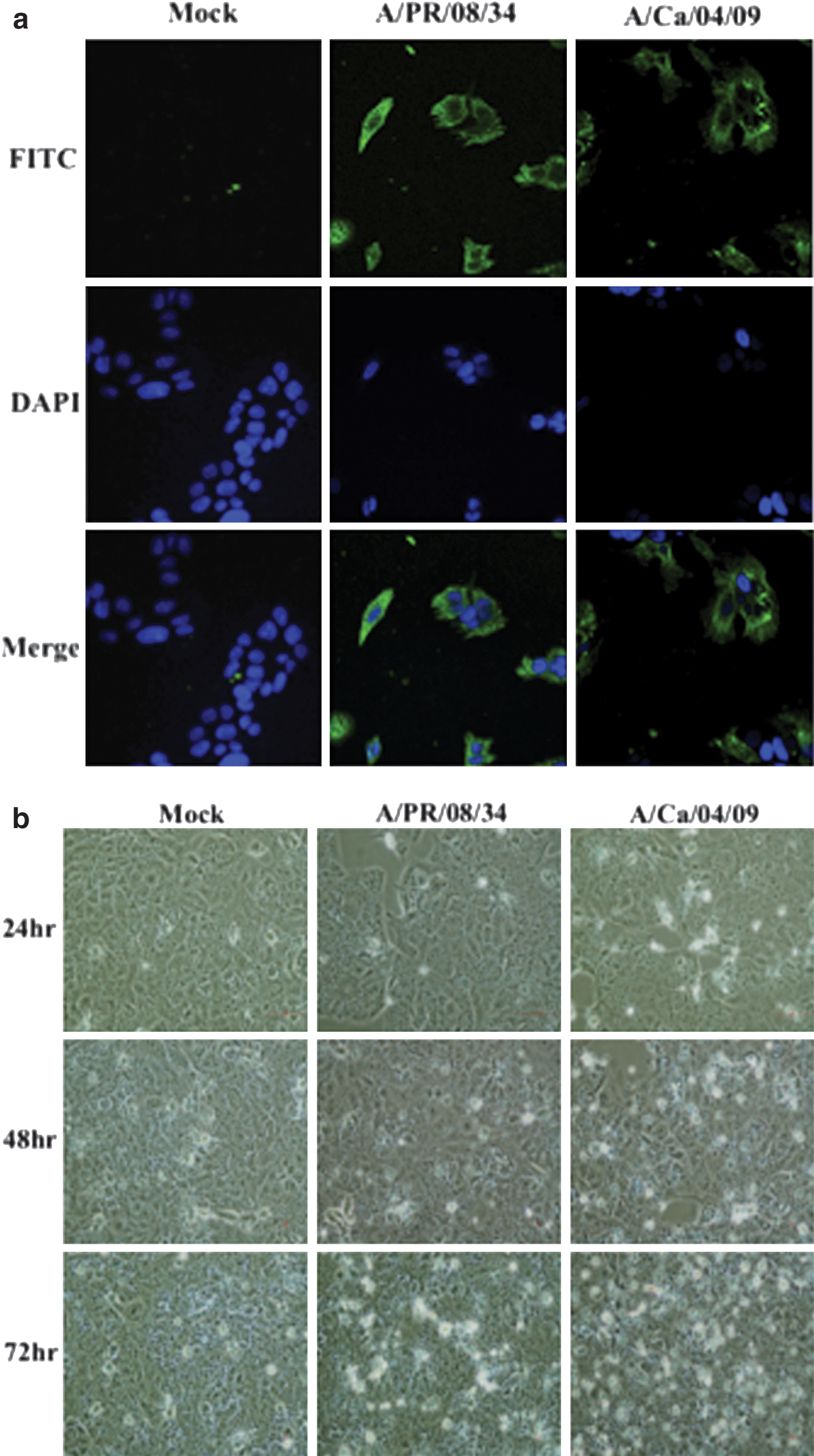
Infection and replication kinetics of PR/08 and Ca/04 viruses in JEG-3 cells.
These results led us to compare the apoptotic potential of the two viruses as increased cytopathic effect was observed in pdm virus-infected JEG-3 cells. Cells infected with 1 MOI of either PR/08 or Ca/04 virus were stained with annexin V antibody and propidium iodide at 12 h PI to determine early apoptosis and analyzed by flow cytometry. 1%–2% of cells were found to be annexin V+ in PR/08-infected cells, whereas ∼9% of cells were annexin V+ in Ca/04-infected cells (p=0.0028) (Fig. 1d). The percentage of annexin V+, propidium iodide+cells was slightly higher (22%–24%) in Ca/04-infected cells (statistically nonsignificant) as compared to both mock infected and PR/08-infected cells. These results indicate that pdm IAV induces a slightly more pronounced apoptosis in choriocarcinoma cells.
The innate immune system plays a crucial and central role in combating influenza virus infection. Cells of the placental membrane, including trophoblasts, uterine epithelial cells, and chorionic villi are capable of recognizing various pathogens associated molecular patterns through a variety of pattern-recognition receptors such as toll-like receptors (16,21,36). Therefore, we sought to measure the expression of key molecules of innate immunity upon viral infection of human chorionic carcinoma cells.
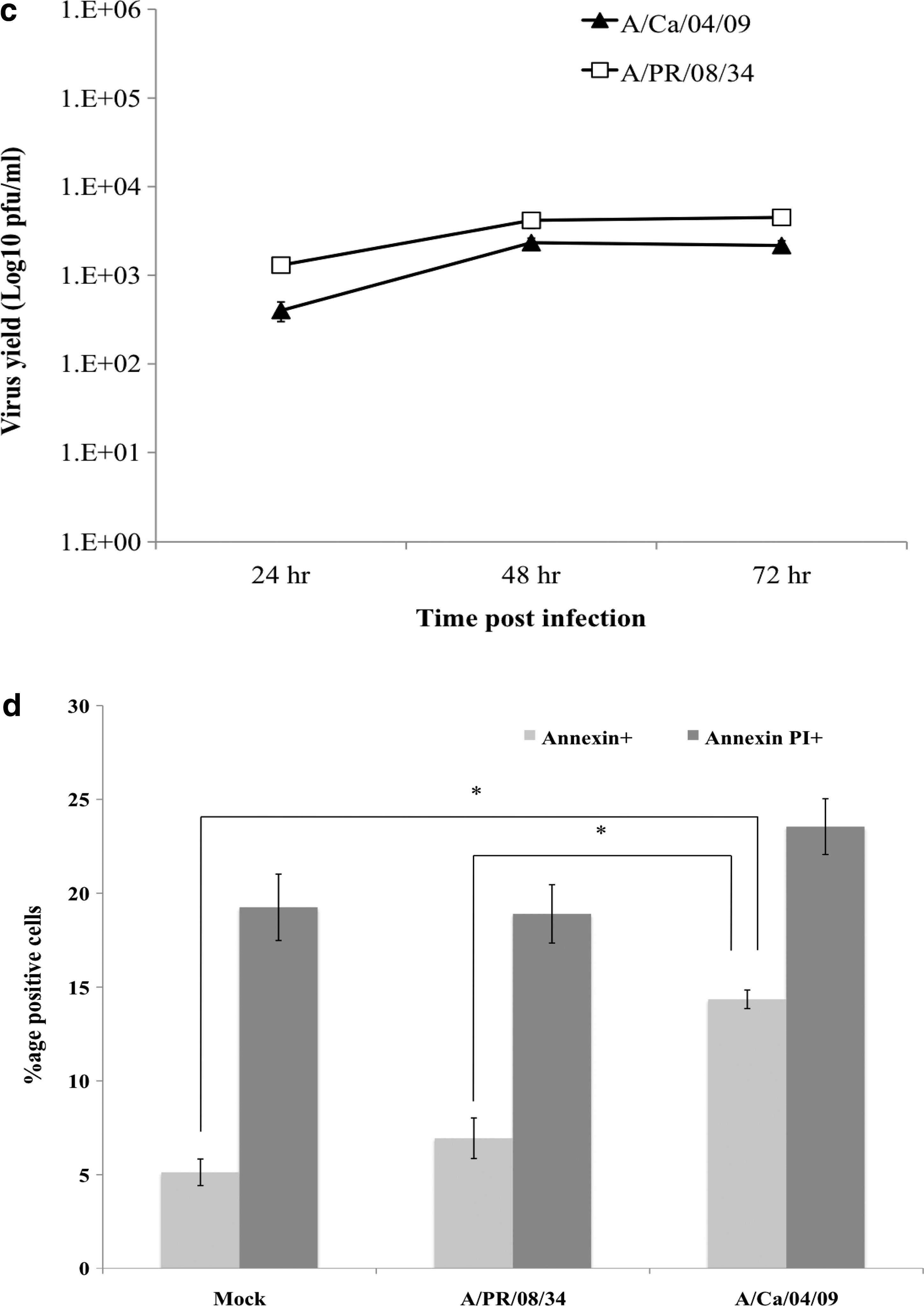
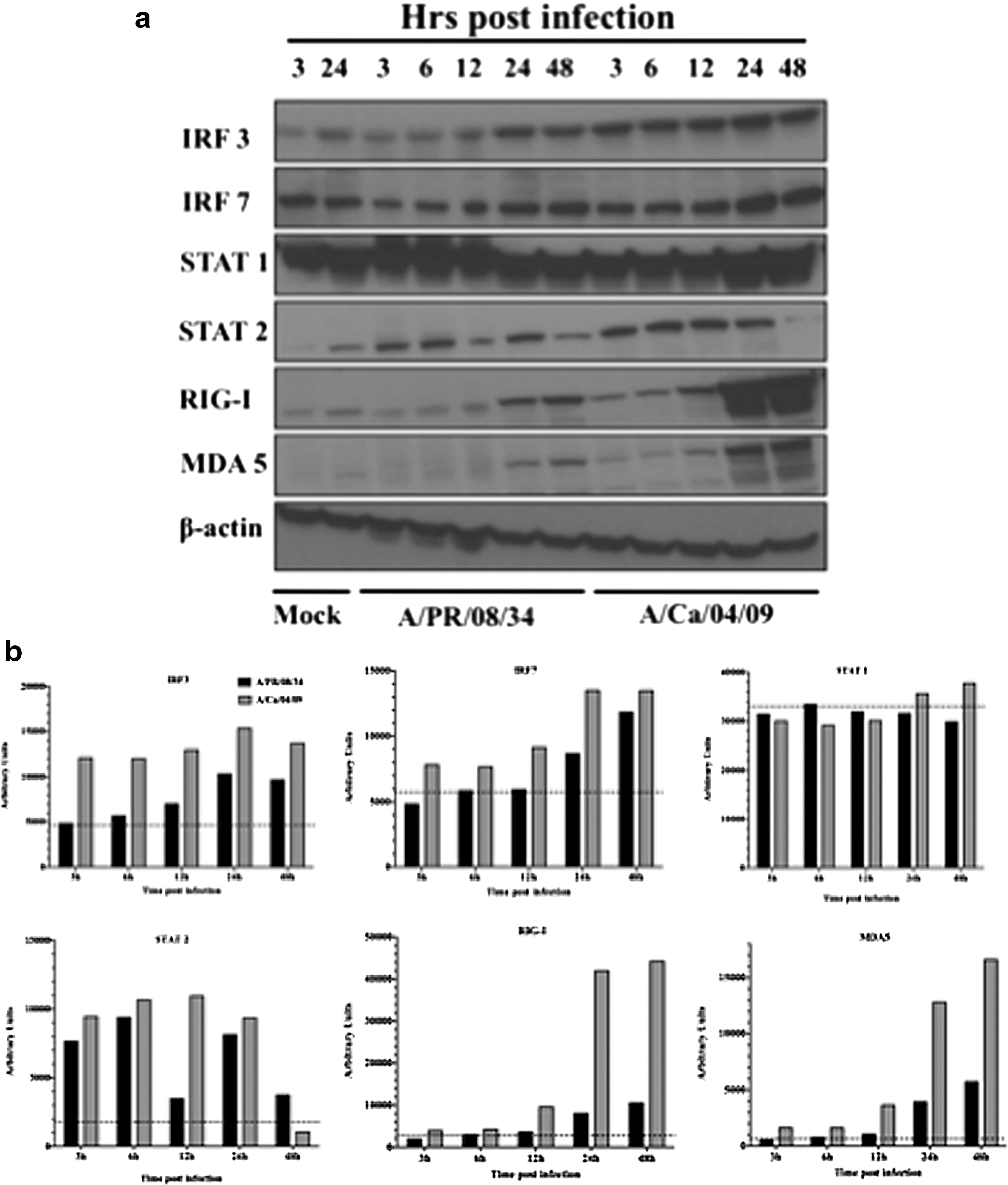
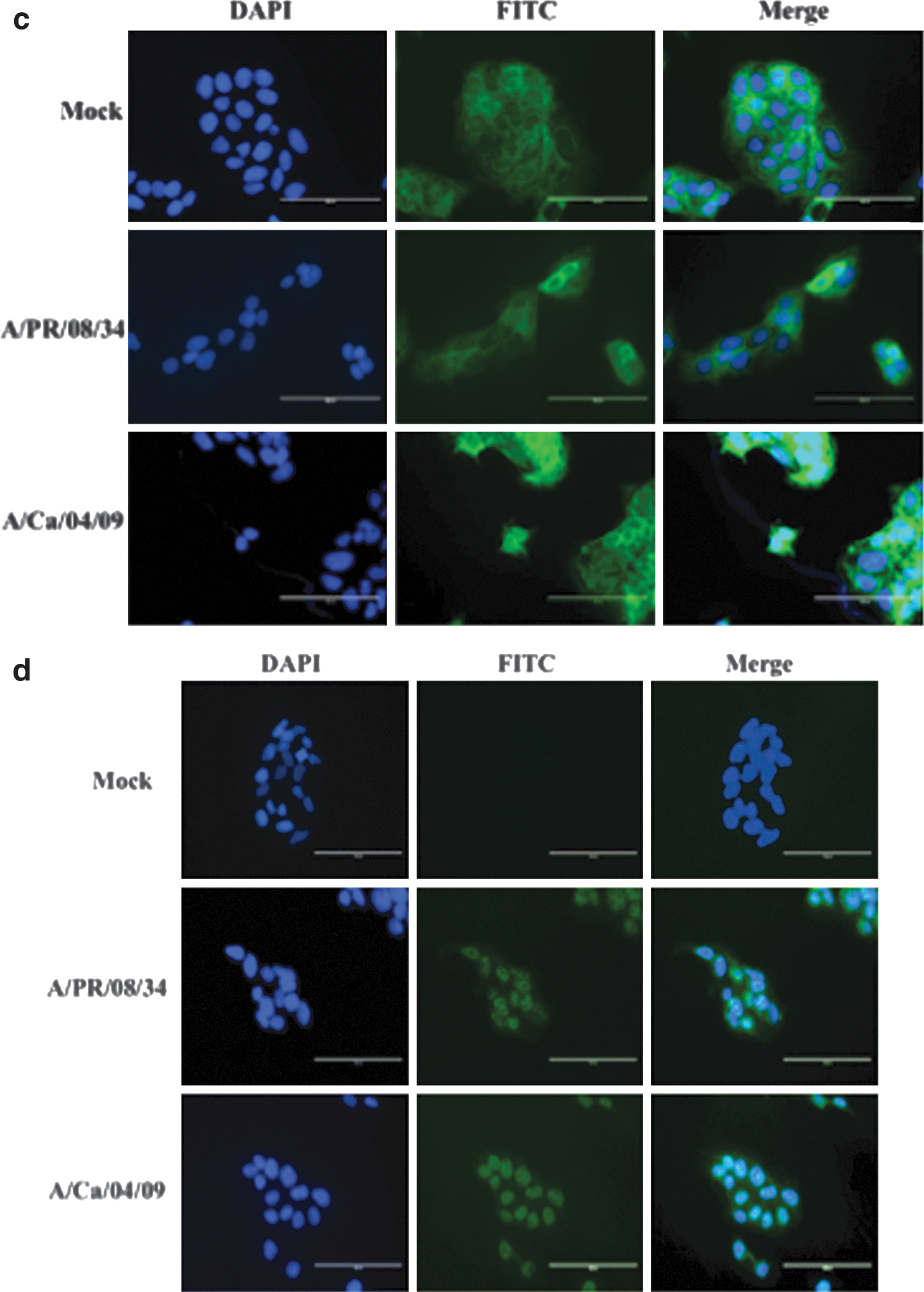
Expression levels of RIG-I, MDA-5, IRF3, IRF7, STAT1, and STAT2 were analyzed by immunoblotting of JEG-3 cell lysates infected with 0.01 MOI of each virus (Fig. 2a and 2b) using antibodies against β-actin, RIG-I, MDA-5, IRF3, IRF7, STAT1, and STAT2 (Santa Cruz Biotechnology, Santa Cruz, CA). Both viruses activated RIG-I as early as 3 h PI, with peak expression at 48 h PI, but the levels were much higher in Ca/04-infected cells at all time points tested. MDA5, another sensor of exogenous dsRNA in the cytoplasm, followed the same activation pattern in Ca/04-infected cells, whereas detectable levels were observed only after 24 h PI in PR/08-infected cells. Higher expression of IRF3 was observed in Ca/04-infected cells as early as 3 h PI. In both PR/08- and Ca/04-infected cells, expression levels of IRF3 peaked at 24 h PI. Similarly, expression of IRF7 peaked at 24 h PI and was higher in Ca/04-infected cells. Nuclear localization of IRF3 and IRF7 upon infection was confirmed by IFA. Higher expression and diffuse nuclear and cytoplasmic localization of IRF3 was observed in Ca/04-infected cells at 12 h PI (Fig. 2c). With IRF7, nuclear localization was evident upon infection with both viruses (Fig. 2d). Both viruses induced higher levels of STAT1 at all time points, while the expression of STAT2 was fractionally higher in Ca/04-infected cells. STAT2 expression was observed to decrease at 48 h PI with both viruses (Fig. 2a and 2b).
Ca/04 virus induced higher expression of innate immune signaling molecules in JEG-3 cells.
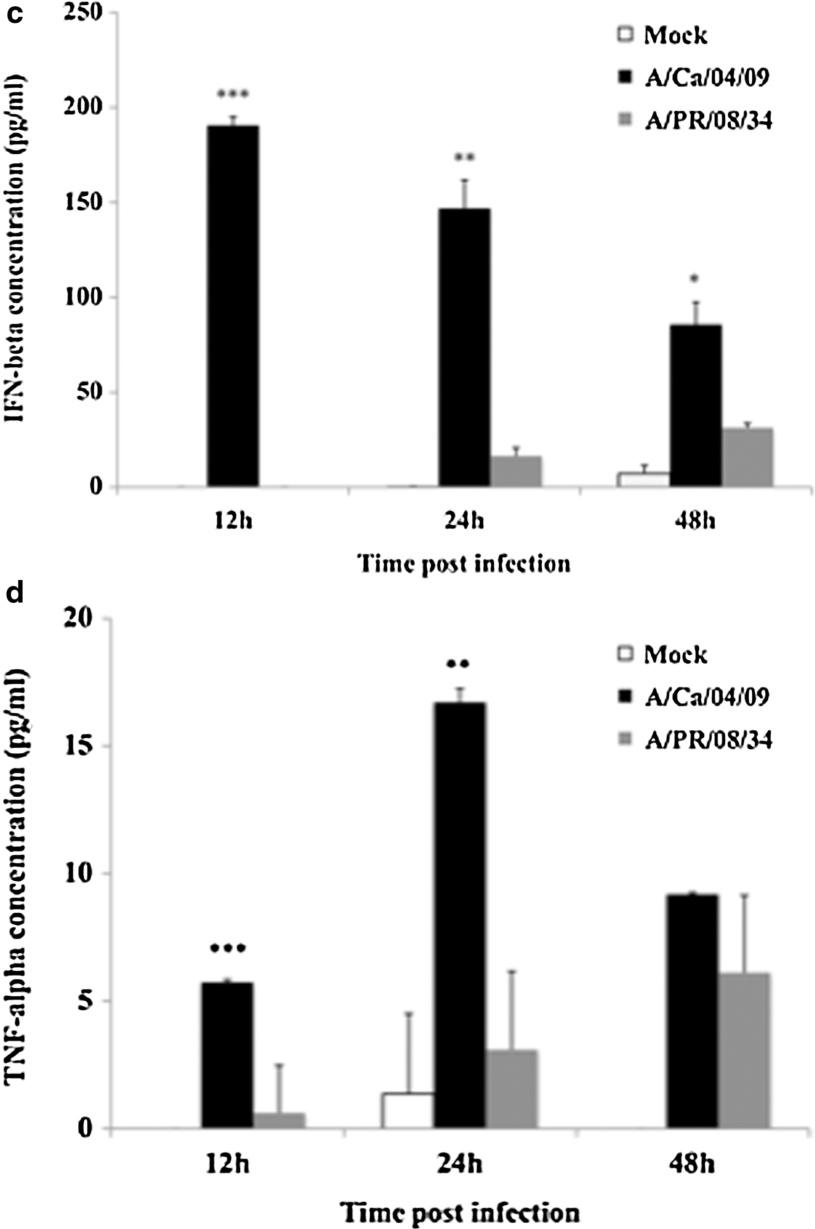
Signaling through RIG-like helicases leads to subsequent expression of type I IFN, pro-inflammatory cytokines, and chemokines. JEG-3 cells infected with 0.01 MOI of PR/08 or Ca/04 were examined for the differential mRNA expression of IL-6, RANTES, IP-10, MIP-1α, IFN-α, IFN-β, and TNF-α by real-time RT-PCR (primers available upon request) at 24 h PI. Fold change values were normalized to 18s RNA and calculated using the 2−ΔΔCT formula. IFN-β and its downstream chemokines RANTES, IP-10, and pro-inflammatory cytokines of the NFkB pathway, IL-6, and TNF-α were upregulated in Ca/04-infected cells compared to PR/08-infected cells (Fig. 3a). Protein levels of a few representative cytokines showing high mRNA expression levels were analyzed by cytokine-specific ELISA (eBioscience, CA). A significant increase in the expression levels of IL-6, IFN-β, and TNF-α in the supernatants of Ca/04-infected cells was evident (Fig. 3b–d).

Pdm influenza A virus induces stronger type I IFN and pro-inflammatory response in JEG-3 cells.
Though extrapulmonary infection and viremia in influenza is less common, it has been reported in influenza-infected patients (23,26,34,40,43). Studies have also shown transplacental transmission and fatal neonatal infection with seasonal H1N1, H3N2, avian influenza H5N1, and the 2009 pdm IAV (1,13,17,22,43). The ability to infect and replicate in JEG-3 cells suggest that IAV, given access, can infect and grow in placental tissue, leading to cell death and cytokine production. Together, these observations imply that transplacental transmission of influenza virus can result from placental infection, leading to preterm labor and abortion in viremic patients. Inflammatory cytokines and chemokines have been shown to contribute to the pathogenesis associated with IAV infection (2,35,41). An involvement between seepage of dysregulated cytokines into the bloodstream in pdm influenza infection and preterm labor and abortions in mice was observed (6,25). Maternal induction of pro-inflammatory cytokines may mediate the neurodevelopmental effects of maternal infections on the developing fetus (28). IRF3 and p38 MAPK have been indicted as major players in the induction of a stronger pro-inflammatory cytokine response in H5N1 influenza virus infected macrophages (18). We observed early and higher expressions of RIG like helicases and IRFs in Ca/04-infected cells, but did not observe a difference in the levels of p38 MAPK in PR/08- and Ca/04-infected cells (data not shown). Further, considerably higher levels of the key pro-inflammatory cytokines IL-6 and TNF-α were observed at both mRNA and protein levels in Ca/04-infected cells. A potential link between maternal influenza infection and fetal growth such as small for gestational age, prematurity issues, and neurological defects, including schizophrenia, has been reported in recent years (15,33,37,39,44). Maternal pro-inflammatory cytokines IL-6, IL-8, and TNF-α have been shown to play a major role in mediating the effects of infection on schizophrenia risk (3,5,28,39). The mechanisms of these adverse effects on fetal–placental units have not been defined. Our results demonstrate that pdm IAV infection may potentially result in virus replication at the maternal–fetal interface. Also, strain- specific innate immune responses to influenza, in immune and other cells may lead to dysregulated inflammatory milieu. The predominant pro-inflammatory cytokine environment and the apoptotic activity of the replicating virus in the placental membranes may lead to adverse immediate and late outcomes during pregnancy. Further studies are required to delineate the mechanisms that govern fetal and maternal responses to influenza virus infection during pregnancy.
Footnotes
Acknowledgments
The authors would like to thank Dr. Terrence M. Tumpey, Centers for Disease Control and Prevention, for providing swine A/Ca/04/09 virus and antiserum, and Ms. Melissa Makris, Virginia-Maryland Regional College of Veterinary Medicine, for help with flow cytometry.
Author Disclosure Statement
The authors have no competing financial interests.