
Abstract
Hepatitis C virus (HCV) is a dreadful viral disease, responsible for 170 million cases worldwide, of which most are from Asia and Africa and approximately 10 million people are from Pakistan. Currently, the pegylated interferon alpha (PEG-INF-α) has been approved as the standard of care in combination with ribavirin and Boceprevir/Telaprevir. Many studies regarding gene expression analysis of liver biopsy samples of patients with chronic HCV infection have been carried out previously. However, there are very few reports of expression analysis carried out using blood samples of HCV patients. Therefore, in this study, gene expression of human immune responsive genes (MMP-9, OAS1) and fibrogenic responsive gene (KRT19) was done in the peripheral blood mononuclear cells (PBMCs) of chronic HCV infected patients having differences in viral titers. Blood samples were collected from different hospitals in Pakistan. RNA was extracted and cDNA was synthesized according to the protocol prescribed by the Enzynomics™ M-MLV Reverse Transcriptase® Kit. The synthesized cDNA was amplified through polymerase chain reaction (PCR) using specific primers of immune responsive genes. The results were further evaluated using real-time PCR. There was a significant increase in the expression of the immune responsive genes (MMP-9, OAS1, CXCL6, CXCR3, ApoA1, and MYC) of HCV genotype 3a patients compared to controls. Similarly, the expression of the fibrosis genes was upregulated in HCV genotype 3a patients compared to controls. The information gained through this study is helpful to identify a noninvasive marker to determine liver fibrosis, and may also give useful information to understand HCV pathogenesis and develop better therapeutic regimens.
Introduction
H
HCV is a small positive-stranded enveloped virus belonging to the Flaviviradae family with a RNA genome of 9.6 Kb. The genome of HCV encodes a polyprotein precursor, which is cleaved into 10 viral proteins in order of NH(2)-Core, E1, E2, p7, NS2, NS3, NS4A, NS4B, NS5A, and NS5B-COOH. The three structural proteins (C, E1, and E2) have a role in viral assembly, and six nonstructural proteins (NS2, NS3, NS4A, NS4B, NS5A, and NS5B) are involved in viral replication and other cellular functions (11,19). HCV has six major genotypes, along with multiple subtypes throughout the world due to high mutation rate and lack of proofreading activity of RNA-dependent RNA polymerese (20). Currently, because of strain variation, no vaccination has been developed to prevent this infection. The combination therapy of pegylated interferon and ribavirin has been historically accepted as the standard care until 2011 (7). This therapy developed sustained virological response (SVR) in 40–50% patients of genotype 1, but recently a new treatment has been approaved by the Food and Drug Administration, which is the combination of pegylated interferon alpha (IFN-α) along with ribavirin and Boceprevir/Telaprevir (8).
In recent years, a link has been established between variations in immune response genes and the outcome of acute HCV infection (23). Thus, an initial innate immune response is an important host defense against the virus, but viral clearance is also dependent upon an effective adaptive immune response (21). However, there is no clear explanation about the variability in spontaneous clearance of HCV infection (23). Fibrogenesis is also linked with immune damage, and cytokines play an important role. Furthermore, it appears feasible to develop drugs against the fibrogenic effector cells, and it is being anticipated that, in upcoming years, an individualized and effective treatment may be developed, together with the evolution of serological markers of hepatic fibrogenesis (18).
Many studies regarding gene expression analysis of liver biopsy samples of patients with chronic HCV infection have been carried out previously. However, there are very few reports of expression analysis carried out using blood samples of HCV patients. Therefore, in this study, gene expression of human immune responsive genes was done in the peripheral blood of chronically infected HCV patients with differences in viral titers. This study was designed to investigate the effect of HCV infection on the genes involved in immune pathways. The information gained through the study may provide significant information that can help in understanding the HCV infection and may also help to identify noninvasive markers to determine liver fibrosis. Ultimately, this knowledge could be helpful in developing novel and better therapeutic regimens.
Methodology
Blood sample collection
Blood samples from HCV-infected patients were collected from the Jinnah Hospital, Lahore, and D.H.Q. Hospital, Faisalabad. Informed consent for blood sample collection was obtained from the participating subjects. The subjects of the current study were both male and female; the children and pregnant women were excluded. All patients were HBsA negative with no history of other diseases such as diabeties, high blood pressure, or kidney disease. A total of five blood samples from healthy individuals were taken as a control for the study. Briefly, 3 cc of blood was collected from each patient in a sterile tube containing 300 μL of 0.5 M ethylene diamine tetra acetate (EDTA) to prevent clotting and to slow down RNA degradation. This blood was immediately processed for total RNA isolation. HCV RNA quantification and genotyping tests were performed at the Department of Bioinformatics and Biotechnology, GC, University, Faisalabad. Only those patients with quantitative and genotype data were included in the current study.
RNA isolation, cDNA synthesis, and PCR amplification
All the steps of RNA isolation from whole blood were carried out in the type IIB Biosafety hood (Beckman Coulter). RNA from collected blood samples was extracted using a Trizol (Invitrogen). The quantity and the quality of the RNA was determined using a NanoDrop ND-1000 spectrophotometer. Extracted RNA was reverse transcribed into complementary DNA (cDNA) using a M-MLV Reverse Transcriptase® Kit (Enzynomics). A set of primers for immune and fibrosis genes were designed for polymerase chain reaction (PCR) amplification genes from cDNA of HCV 3a infected patients. Sequences were retrieved from the National Center for Biotechnology Information (NCBI), and primers were designed using Primer3 software (
Statistical analysis
All statistical analysis was done using GraphPad Prism software. Data are presented as mean±standard deviation (SD). Numerical data were analyzed using one way analysis of variance (ANOVA). A p-value of<0.05 was considered statistically significant.
Results
Blood sample collection
Blood samples of HCV patients were collected from the Jinnah Hospital, Lahore, and D.H.Q. Hospital, Faisalabad. A total of 16 patients were included in the study, of which 10 were male and 6 were female. Data for the patients and controls are given in Table 2.
HCV, hepatitis C virus.
Expression study
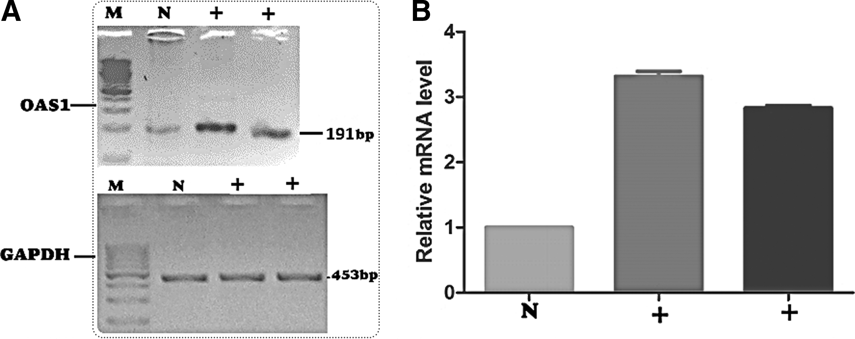
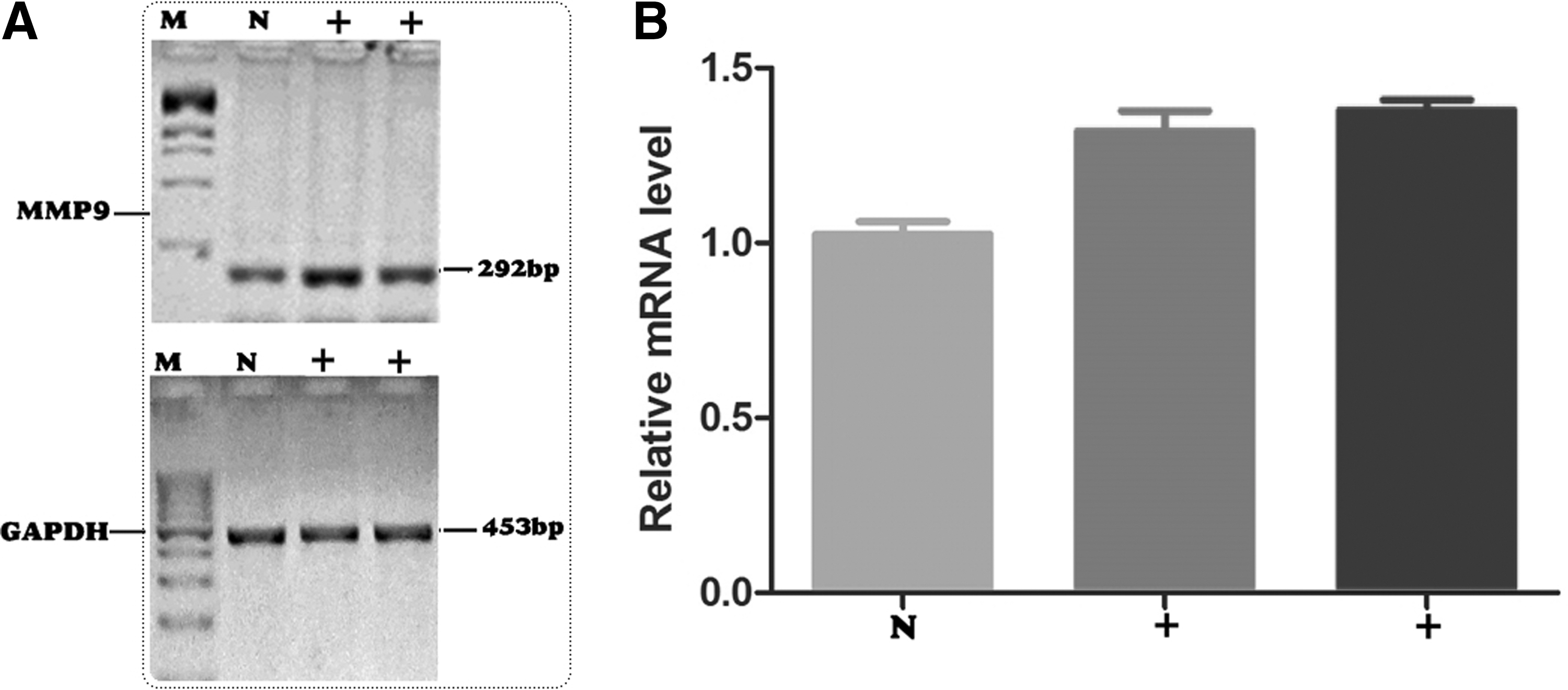
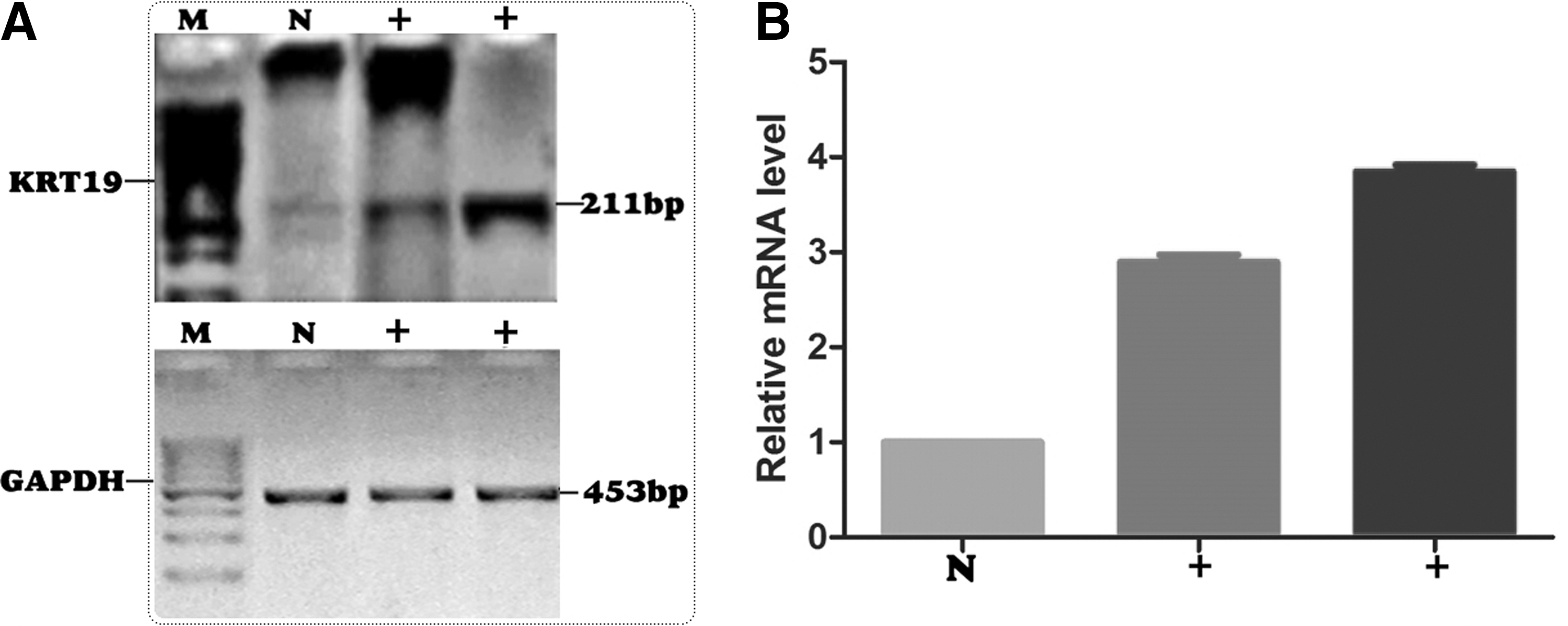
Expression analysis was carried out by PCR using gene-specific primers, and the results were checked on 2% agarose gel. Further gene expression analysis was carried out using real-time PCR to check the difference of expression change in infected individuals and controls. GAPDH was used as the internal control.
Expression of OAS1, MMP9, and KRT19 was checked on agarose gel, and it was found that expression was increased in the blood of HCV 3a genotype patients compared to controls. To investigate the expression change further, expression was checked through real-time PCR, and a one-way ANOVA was implemented to check the significant p-value using GraphPad Prism software. A significant increase in expression of OAS1 (Fig. 1), MMP9 (Fig. 2), and KRT19 (Fig. 3) genes was observed in HCV genotype 3a patients compared to controls. A p-value of<0.05 was regarded as significant.
Discussion
Liver cirrhosis is caused by chronic liver diseases, which can lead to the loss of liver function. Excessive accumulation of extracellular matrix (ECM) leads to fibrosis. Fibrogenesis can be stimulated by hypoxia, viruses, toxins, and bile stasis. Mechanical stress or cytokine activation can lead to excess synthesis of ECM, which in turn causes fibrogenesis. Fibrosis is a process of removing excess ECM through proteolytic activities in the acute phase of liver diseases. It has been reported that HCV affects genes that are involved in immune and fibrosis pathways. Thus, it is important to study the effect of HCV infection on these immune response genes. The genes involved in immune regulation, inflammation, and tissue regeneration are considered as important for fibrogenesis regulation (18).
Matrix metalloproteases (MMPs) are zinc-dependent endopeptidases that are the key proteases involved in the ECM degradation and are also known as matrixins. These MMPs can degrade a broad range of extracellular molecules, and are classified into six groups: collagenases (MMP-1, -8 and -13), gelatinases (MMP-2 and MMP-9), matrilysin (MMP-7 and MMP-26), stromelysins (MMP-3, -10 and -11), membrane type (MT) MMPs (MMP-14, -15, -16, -17, -24 and -25), and various other MMPs, including MMP-12, -19, -20, -21, -23, -27, and -28. These MMPs are involved in cell migration, cell proliferation, angiogenesis, differentiation, apoptosis, and host defenses. Previous studies prove that MMP-1, -2, -3, -8, -9, -12, -13, and -14, which are expressed in the liver, are considered the most important genes (4,12). The circulating MMP-2/TIMP-1 ratio has been correlated to the histological degree of fibrosis in hepatitis C (14). According to an expression study, MMP-1 and -2 expressions increase in the late stages of fibrosis and cirrhosis. During recovery, the MMP-3 expression of macrophages greatly increases in the unresolved fibrous septa (10). In the present study, the expression level of the MMP-9 gene was analyzed in the blood of HCV-infected patients, and the results show a significant increase in mRNA expression of MMP-9 while the GAPDH level remained constant.
There are three major genes of the 2′-5′-oligoadenylate synthetase (OAS) family: OAS1-3, OAS-Like (OASL), and an inactive gene. The IFN is responsible for the active transcription of these genes (15). According to Bran et al., IL-28A and IL-29 induced mRNA expression of the 2′,5′-OAS has been abolished by overexpression of SOCS-1 (6), but there has been no systematic expression studies of these genes during viral infections (15). Therefore, in our study, we analyzed the host OAS1 gene expression in the blood of HCV-infected patients.The results of our study showed a significant increase in mRNA expression of OAS1 while the GAPDH level remained constant. KRT19 belongs to the keratin family and is responsible for the structural integrity of epithelial cell. KRT19 is involved in the cytoskeleton, and it is associated with he liver damage in liver diseases (3). Previously, KRT19 expression has been studied in neuroblastoma, and a decrease in expression was observed (16). In the present study, the expression level of the KRT19 gene was analyzed in the blood of HCV-infected patients. The results of our study show a significant increase in mRNA expression of KRT19 while the GAPDH level remained constant. Taking into account the above information, in this comparative study, we also investigated the expression of the host genes MMP-9, OAS1, and KRT19, which play an essential role in immune and fibrosis pathways. Our results show enhanced mRNA expression of these genes in the blood of HCV genotype 3a patients compared to controls. These findings reveal that the expression of immune and fibrosis-related host genes are significantly high in HCV-infected genotype 3a patients compared to controls.
Conclusion
Conclusively, these results suggest that the enhanced expression of immune response genes OAS1 and MMP-9 and fibrosis gene KRT19 in HCV-infected patients is responsible for an increased level of HCV infection. In addition, controlling the expression level of immune response and fibrosis genes might be an important therapeutic target for the treatment of HCV infection.
Footnotes
Author Disclosure Statement
No competing financial interests exist.