
Abstract
Few reports have shown the relationship between the distribution of human immunodeficiency virus type 1 (HIV-1) proviral DNA in CD4 subsets during acute HIV-1 infection and HIV disease progression. In this study, we enrolled two groups with distinct differences in disease progression. The CD4 counts of one group fell below 200 cells/μL within 2 years (rapid progressors), whereas the other group maintained CD4 counts above 500 cells/μL (slow progressors). We collected blood samples during Fiebig stage III–IV of the two groups, and sorted CD4+ naïve, central memory, and effector memory lymphocytes. Real-time polymerase chain reaction was used to quantify HIV-1 DNA of the subsets. We found that HIV-1 DNA content was higher in memory T-cells than in naïve cells in both groups, and a higher HIV DNA content was found in naïve CD4+ T-cells during acute HIV-1 infection in rapid progressors. This suggests that higher HIV DNA in naïve CD4+ T-cells may associated with rapid progression.
Introduction
H
Patients and Methods
Forty patients who were recently infected with HIV-1 were recruited from an HIV-1-negative high-risk men who have sex with men (MSM) cohort, and screened every 2 months for HIV-1 infection at Beijing Youan Hospital (10,11). According to the results of standard clinical laboratory tests, all 40 patients were at Fiebig stage III–IV (7,8) when they first tested HIV-1 positive. Among the 40 patients, 20 patients progressed rapidly, and their CD4 counts fell below 200 cells/μL within 2 years (CD4 low group), whereas the other 20 patients maintained CD4 counts above 500 cells/μL (CD4 high group).
This study was reviewed and approved by the Beijing You'an Hospital Research Ethics Committee.
During the first visit, we collected blood samples from the HIV-1 positive group and sorted CD4+ naïve lymphocytes (CD3+ CD4+ CD45RA+ CCR7+), central memory lymphocytes (CD3+ CD4+ CD45RA− CCR7+), and effector memory (CD3+ CD4+ CD45RA− CCR7−) lymphocytes with an average purity >98% (Fig. 1A). Total DNA was extracted from each cell subset using the QIAamp DNA Blood Mini Kit (Qiagen). Real-time polymerase chain reaction was used to quantify HIV-1 DNA based on the protocol used in previous studies (1,4,19).
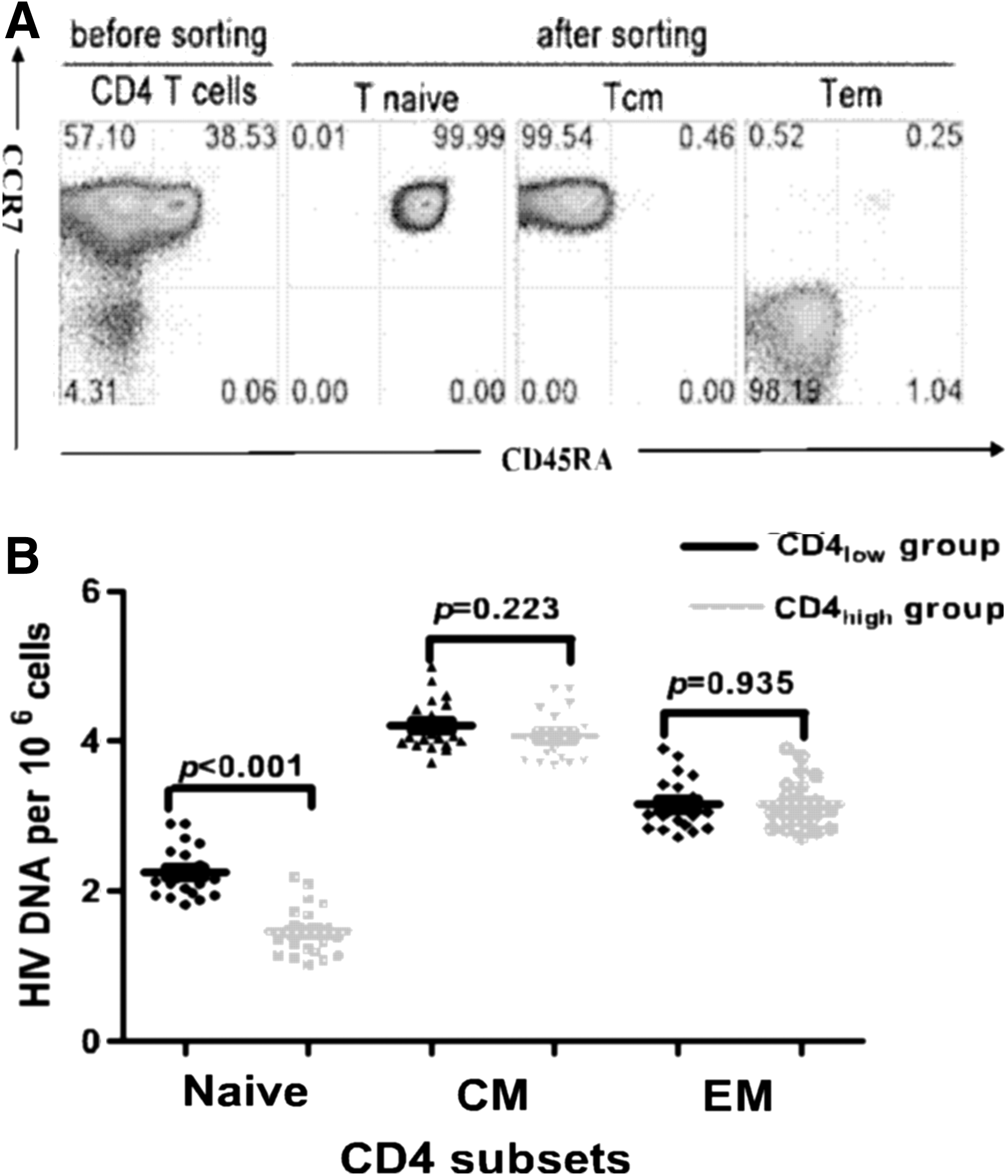
Comparison of the viral DNA content in the CD4 subsets between the CD4high and CD4 low groups. An example is shown for the flow cytometric sorting strategy for CD4+ T-lymphocytes subsets
Results
We found that in both CD4high and CD4low groups, memory CD4+ T-cells and naïve cells were involved in viral dissemination, and the viral DNA content was higher in the memory T-cells (average 4,199 copies per million CD4+ memory cells) than in the naïve cells (average 75 copies per million CD4+ naïve cells; Fig. 1B).
We compared the levels of HIV-1 DNA in each cell subset between the two groups and found that naïve CD4+ T-cells had a higher HIV DNA content in the CD4low group than in the CD4high group (average 160 and 28 copies per million CD4+ naïve cells respectively; p<0.001; Fig. 1B). No statistical difference was found in the HIV DNA content in the central memory (average 15,206 and 10,142 copies per million CD4+ central memory cells respectively; p=0.223) and effector memory (average 1,369 and 1,456 copies per million CD4+ effector memory cells respectively; p=0.935) subsets between the CD4low and CD4high groups (Fig. 1B).
Discussion
In this study, among patients who had acute HIV infection at Fiebig stage III–IV, we found a higher level of HIV DNA in naïve CD4+ T-cells in the CD4low group than in the CD4high group. This finding suggests that infection and the loss of naïve CD4+ T-cells during acute HIV infection may be related to rapid disease progression. A nonmutually exclusive hypothesis could explain this result. CD45RA− CD4+ T-cells can be infected by both nonsyncitium-inducing and syncitium-inducing variants, whereas CD45RA+ CD4 T-cells are exclusively infected by the syncitium-inducing virus (3,12,15). Co-receptor use is highly correlated with the ability to induce syncytia in the MT2 T-cell line: nonsyncytium-inducing variants use CCR5 but not CXCR4. Syncytium-inducing variants, which emerge in about 50% of infected individuals preceding an accelerated CD4 cell decline and progressive clinical course of infection, use CXCR4 (2,17). We previously reported that the primary CXCR4 co-receptor used in acute HIV infection caused rapid disease progression in the AE subtype in Chinese patients who were infected with HIV through sexual transmission (14). CXCR4 co-receptor usage of primary HIV-1 may be one of the reason for a higher level of HIV DNA in naïve CD4+ T-cells in the CD4low group. Further study of the relationship between HIV infected naïve cells during acute HIV-1 infection and HIV reservoir is needed. In the present study, our finding of higher HIV DNA in CD4+ naïve cells during acute HIV-1 infection in rapid progressors suggests that higher HIV DNA in naïve CD4+ T-cells in acute HIV-1 infection may associated with rapid progression.
Footnotes
Acknowledgments
This study was supported in part by the National Natural Science Foundation of China (81101250, 81371803), the National 12th Five-Year Major Projects of China (2012ZX10001-003, 2012ZX10001-006), National Special Research Program for Important Infectious Diseases of China (2013ZX10001004), and Development and Application Research of Beijing AIDS Clinical Data and Sample Repository 4, (D131100005313005).
Author Disclosure Statement
No competing financial interests exist.