
Abstract
The antimicrobial peptide cathelicidin is critical in killing pathogens by innate immune cells, including Mycobacterium tuberculosis and Candida albicans. These pathogens often cause infections in opioid users, a risk that is greatly increased with concurrent human immunodeficiency virus (HIV) infection. Therefore, we examined the association between opioid use and cathelicidin in HIV-infected subjects from Bandung, Indonesia. The following three groups of HIV-infected individuals were included: (i) Active drug users: used heroin in the last 30 days; (ii) Methadone clients: received methadone maintenance therapy in the last 30 days; and (iii) Controls: never used opioids or did not use opioids in the year preceding inclusion. In addition to interviews, blood samples were taken to examine the RNA expression of cathelicidin. We found that the RNA expression of cathelicidin was significantly decreased (p=0.007) in heroin users, compared with controls. Opioids are associated with immunosuppression, and cathelicidin could be an important factor in this association. However, more research is needed to examine the direct effects of decreased cathelicidin levels.
Introduction
I
Chronic opioid use is associated with diminished vitamin D, which is at least partly due to the overall behavior of addicted individuals (13). In addition, HIV infection is known to cause nutritional deficiencies, such as a diminished vitamin D (10,20). Vitamin D deficiency has been linked to a wide range of diseases (28), and it is able to modulate the innate immune response by upregulation of antimicrobial peptides (AMPs) (30). Cathelicidins are a large family of AMPs and the only human cathelicidin LL-37/hCAP-18 is regulated by the biologically active form of vitamin D: (1,25(OH)2D3) (7,30). Cathelicidin targets inflammatory pathways, and it forms an important link between the innate and adaptive immune system. It can modulate the innate immunity, activate different cell types, induce chemotaxis, and support cytokine secretion (17,32).
Previous studies have shown a strong relationship between the expression of cathelicidin and the occurrence of infections. In cathelicidin knockout mice, infections of cornea, gastrointestinal tract, and skin are frequently seen (11,12,18). In addition, humans who suffer from cathelicidin deficiency are more susceptible to periodontal diseases (21), respiratory tract infections (14,15), and candidiasis (8). Interestingly, these infections are very common among drug users, especially among those infected with HIV. Therefore, we hypothesize that the expression of cathelicidin is downregulated in opioid users, which we examined in HIV-infected individuals in Bandung, Indonesia.
Materials and Methods
Study setting and subjects
For this study, we included HIV-infected adults (>16 years) from different settings in West-Java, Indonesia: an HIV clinic, a methadone clinic, and the community. The clinics were established as a part of a 5-year program (2006–2011) called “IMPACT,” aimed at improving prevention, control, and treatment of HIV among injecting drug users in West-Java (31). To contact individuals in the community, we collaborated closely with Rumah Cemara, a local NGO focusing on increasing the quality of life for drug users. This study was approved by the Health Research Ethics Committee at the Faculty of Medicine of Padjadjaran University/Dr. Hasan Sadikin General Hospital in Bandung, Indonesia.
All individuals included were antiretroviral therapy (ART) naïve and had no signs of opportunistic infections, such as tuberculosis or oral thrush. We included the following three groups of HIV-infected individuals: (i) Active drug users: individuals who used heroin in the last 30 days; (ii) Methadone clients: individuals who received methadone maintenance treatment (MMT) in the last 30 days; and (iii) Controls: individuals without a history of drug use or individuals who did not use opioids in the year preceding inclusion. All participants were informed about the study and provided a written consent. Data on demographic factors, history of drug use, co-morbidity, self-reported tuberculosis treatment, and history of ART were collected through an interview with standardized questionnaires. In addition, blood was collected in EDTA tubes and PAXgene Blood RNA tubes (PreAnalytiX; Qiagen) for full blood counts and CD4 cell count, respectively, quantitative real-time polymerize chain reaction (qPCR) to detect the RNA expression of cathelicidin.
RNA expression of cathelicidin
PAXgene Blood RNA tubes were placed in a −20°C freezer until RNA extraction. RNA was isolated using the PAXgene Blood RNA Kit (PreAnalytiX; Qiagen) according to the manufacturer's instructions. RNA (0.2 μg) was reverse transcribed using the iScript cDNA Synthesis Kit (Biorad) following the manufacturer's instructions. The cDNA was stored at −20°C until qPCR.
The qPCR was used to determine the RNA expression of cathelicidin. The results were normalized using the housekeeping gene GAPDH, and quality control was performed by measuring multiple melting curve points. We used the following primers systemized by Biolegend: cathelicidin forward, TGCCCAGGTCCTCAGCTAC, and reverse, GTGACTGCTGTGTCGTCCT. Each reaction contained 5 μL SYBR Green PCR Master Mix (Applied Biosystems), 2.92 μL H2O, 0.04 μL of 100 μM forward and 100 μM reverse primers, and 2 μL cDNA sample. Amplification of the genes was performed for 10 min at 95°C, followed by 40 cycles at 95°C for 15 sec, annealed at 60°C for 20 sec, and extended at 90°C for 15 sec. Data were analyzed using the StepOnePlus Q-PCR (Applied Biosystems). Relative changes were analyzed using the 2(−Delta Delta C(T)) method, and measurements were repeated if the reaction efficiencies were >95%.
Statistical analysis
The Kruskal–Wallis and Mann–Whitney-U analyses were used to compare differences between groups. For this study, we used an alpha of 5%; for Mann–Whitney-U analyses, this results in a p-value of 0.016 after Bonferonni adjustment. All statistical analysis was performed using the Statistical Product and Services Solutions package version 18.0 and GraphPad Prism version 5.0.
Results
In total, we included 89 HIV-infected individuals, of whom 26 were active heroin users, 25 individuals received MMT, and 38 had never used opioids or used opioids more than 1 year ago (Table 1). The majority was male and had an average CD4 cells count of 343 cells/μL (interquartile range [IQR]: 192–496). We found no significant differences between the three groups with regard to gender, ART, and CD4 cells (Table 1). However, the group receiving MMT had a significantly lower hemoglobin level (p=0.011) and nonusers were significantly younger (p=0.001) (Table 1).
Heroin users: individuals who used heroin in the last 30 days; MMT: individuals who received MMT in the last 30 days; and Controls: individuals who never used opioids or did not use opioids in the year preceding inclusion.
p-Values calculated by comparing groups with Kruskal–Wallis.
ART, antiretroviral therapy; Hb, hemoglobin levels; IQR, interquartile range; MMT, methadone maintenance treatment; n, number of people.
We did not find a difference in the production of GAPDH between heroin users, MMT clients, and nonactive users (Kruskal–Wallis: p=0.303). The median relative RNA expression of cathelicidin was 0.030 (IQR: 0.018–0.077). We found a significant difference in the expression of cathelicidin between groups (Kruskal–Wallis: p=0.014). Figure 1 shows that the cathelicidin expression was significantly lower in heroin users compared with controls (p=0.007) and those receiving MMT (p=0.058). The median cathelicidin expression was 0.022 in heroin users (IQR: 0.011–0.040), compared with 0.031 in those receiving MMT (IQR: 0.18–0.087) and 0.040 in controls (IQR: 0.022–0.130) (Fig. 1).
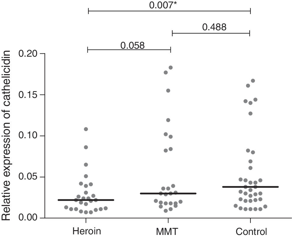
Gene expression of cathelicidin for human immunodeficiency virus (HIV)-infected individuals in those using heroin or methadone maintenance treatment (MMT) in the last 30 days, and individuals who never used opioids or did not use opioids in the year preceding inclusion (Controls). Data are shown as individual and median RNA expression. There was a significant difference in expression between groups (Kruskal–Wallis: p=0.014). After Bonferonni adjustment, a p-value below 0.016 represents a statistically significant difference with an alpha of 5%. *p<0.05.
Discussion
This study indicates that the whole blood cathelicidin expression in HIV-infected heroin users is downregulated compared with controls which may play a role in the increased susceptibility to certain opportunistic infections in these patients. To our knowledge, no previous studies have examined the association between opioids and cathelicidin.
Cathelicidin is important in the innate and adaptive immunity and for the clearance of pathogens, including infections frequently seen in drug users. Infections such as tuberculosis and candidiasis are more common among drug users, which cannot be completely explained by higher exposure to these pathogens (5,29). Over the past few decades, studies show that opioids increase the susceptibility to microbial infections in experimental animal models by modulating the immune response. Infections with herpes simplex virus and Candida albicans have proved lethal in opioid-treated animals (23,26). Cathelicidin severely affects the membrane morphology of C. albicans, which results in leakage of proteins, release of vital components, and subsequent cell death (3). Opioids might diminish this defense mechanism by decreasing cathelicidin and thus resulting in more (severe) candidiasis, as seen in the animal models. An important risk factor for these infections is, of course, HIV itself; however, the decreased expression of cathelicidin may play a role in the increased risk of opportunistic infections in injecting drug users.
LL-37-mediated inflammasome activation has also been described. Cathelicidin is able to activate the P2X7 receptor, resulting in an increase of IL-1β expression and release (4). If opioids decrease the expression of cathelicidin, this would lead to decreased clearance of gram-negative bacteria. Supporting this hypothesis, LPS-induced sepsis was indeed more common in opioid-exposed rats (19).
Cathelicidin enhances the resistance to mycobacterial infections by the activation of transcription of autophagy-related genes (33). Capturing and destroying of the immature phagosomes that harbor pathogens is an important host defense mechanism of human macrophages against Mycobacterium tuberculosis (9). Downregulation of cathelicidin would result in higher susceptibility to M. tuberculosis, as less accumulation of autophagosomes and decreased ability to fuse with lysosomes will lead to decreased clearance of intracellular pathogens. In line with our hypothesis, M. tuberculosis infection is more common among drug users (2).
Opioid receptors are expressed on immune cells, and signal transduction induced by opioids modulates the immune function of these cells (1). However, there has not yet been any link with the production of cathelicidin. However, cathelicidin production is linked to vitamin D, which is regulated by the biologically active form of vitamin D: (1,25(OH)2D3) (7,30). Possibly opiates either directly or indirectly affect cathelicidin via this pathway.
In this study, we focused on looking at the whole blood RNA expression of cathelicidin. Consequently, we were not able to determine the contribution of each cell type or conclude anything on the actual amount of released cathelicidin in the blood or at the level of mucous membranes. Therefore, more research is needed to explore the relationship between opioid use and the cathelicidin.
Conclusion
In conclusion, cathelicidin could be considered an important factor in the immunosuppression induced by opioids. This study provides unique evidence on decreased expression of cathelicidin in opioid users, but more research is essential for a better understanding of underlying mechanisms and direct effect of this result on risk of infection.
Footnotes
Acknowledgments
The authors would like to thank Dr. Bayu Wahyudi, Director of Hasan Sadikin General Hospital, and Prof. Tri Hanggono Achmad, Dean of the Medical Faculty, Universitas Padjadjaran, for encouraging and accommodating research at their institutions. Everyone working at the HIV clinic and the methadone clinic in the hospital is thanked for providing HIV care and collecting the data used for this study. In particular, the authors would like to thank Yusandi Sastra Atmaja, Nuni Haeruni, Dwi Febni Ratnaningsih, and Diah Wulandari for their support during patient inclusion as well as Suharyani Soedarmo, Fitri Utami, and Nurul Setia Rahayu for their assistance in the laboratory. Great appreciation is due to all Rumah Cemara staff, especially Dehan Mulyana and Hendra Ferdian, not only for helping the authors with this project, but also for showing them the work they do and all the care they provide. The authors also wish to thank Arif Rusman for taking them all over Bandung to include subjects.
Author Disclosure Statement
All authors report no competing financial interests.