




Abstract
Filoviruses are causative agents of hemorrhagic fever, and to date no effective vaccine or therapeutic has been approved to combat infection. Filovirus glycoprotein (GP) is the critical immunogenic component of filovirus vaccines, eliciting high levels of antibody after successful vaccination. Previous work has shown that protection against both Ebola virus (EBOV) and Marburg virus (MARV) can be achieved by vaccinating with a mixture of virus-like particles (VLPs) expressing either EBOV GP or MARV GP. In this study, the potential for eliciting effective immune responses against EBOV, Sudan virus, and MARV with a single GP construct was tested. Trimeric hybrid GPs were produced that expressed the sequence of Marburg GP2 in conjunction with a hybrid GP1 composed EBOV and Sudan virus GP sequences. VLPs expressing these constructs, along with EBOV VP40, provided comparable protection against MARV challenge, resulting in 75 or 100% protection. Protection from EBOV challenge differed depending upon the hybrid used, however, with one conferring 75% protection and one conferring no protection. By comparing the overall antibody titers and the neutralizing antibody titers specific for each virus, it is shown that higher antibody responses were elicited by the C terminal region of GP1 than by the N terminal region, and this correlated with protection. These data collectively suggest that GP2 and the C terminal region of GP1 are highly immunogenic, and they advance progress toward the development of a pan-filovirus vaccine.
Introduction
F
While the filovirus VLP can express multiple viral proteins, in its simplest form it is made up of the matrix protein (VP40) and GP. VLP containing these two proteins have proven efficacy in mouse, guinea pig, and nonhuman primate (NHP) models of filovirus infection (29,44,45,47,50,53). While VP40 is required for particle formation, it was shown that the inclusion of GP in the VLP vaccine and on other vaccine platforms is critical for protection from challenge (5,44).
The Ebola virus (EBOV) GP gene encodes two distinct proteins: sGP, which is a secreted form of GP and shares the first 295 amino acids of the N-terminal region of GP1, and the envelope GP, which is expressed on the surface of virions (24,40,41). Approximately 80% of transcripts encode for sGP as opposed to full length GP (24,41), and evidence suggests that sGP may serve as an immune decoy for antibody responses against surface-expressed GP (32,40). Both Marburg virus (MARV) and EBOV GP are expressed as trimers and are made up of two distinct subunits: GP1 and GP2. A furin-cleavage site in GP0 separates GP1 and GP2, and cleavage occurs in the host cell during viral assembly (41,48). GP1 contains the receptor-binding domain (RBD), while GP2 contains the transmembrane domain and is critical for membrane fusion within the phagosome.
The C-terminal region of GP1 contains the highly glycosylated mucin-like domain (MLD), which has been shown to be involved in initial virus attachment to cells but is not required for virus fusion (20,55). The N-terminal region of GP1 appears to be critical for host cell binding and includes the RBD, which is conserved between EBOV and MARV (4,10,21,27). Multiple glycosylation sites throughout GP create a glycan cap. The glycan cap mostly consists of N-linked glycosylation sites, while the MLD contains additional N- and O-linked glycosylation sites (25,26). The surface glycosylation has a role in cell attachment, but may also protect potential viral epitopes from detection (6,9,25,54) and prevent detection of host cell proteins, such as major histocompatibility class I, on infected cells' surfaces (13,39). GP lacking the MLD elicited higher neutralizing antibody titers than full length GP, suggesting that the MLD may block development of potentially important immune responses (28).
Considering the unpredictable nature of filovirus disease outbreaks, the development of a single filovirus vaccine with efficacy against multiple filovirus variants is of great interest. The genus Marburgvirus contains two viruses: MARV and Ravn virus (RAVV). The genus Ebolavirus contains five viruses: Sudan virus (SUDV), EBOV, Bundibugyo virus (BDBV), Reston virus (RESTV), and Taï Forest virus (TAFV) (22). The first hurdle in development of a pan-filovirus vaccine is to achieve cross-protection among variants of a virus belonging to a particular genus. It has previously been shown that a VLP expressing MARV-Musoke can provide protection against infection with MARV-Musoke, RAVV, or MARV-Ci67 strains in NHPs (45). Additionally, however, there are efforts to produce a vaccine that confers protection from infection with filoviruses belonging to multiple genera and to emerging viruses (16). A mixture of VLPs expressing EBOV VP40 and GP with VLPs expressing MARV VP40 and GP resulted in protection from infection with both viruses in the guinea pig model, suggesting that there is no inhibitory effect of vaccinating with both agents (44). This finding was supported in studies utilizing multiple vaccine platforms. A complex adenovirus-based vaccine, which expressed the viral GP of five filoviruses and variants (EBOV, SUDV, MARV-Musoke, MARV-Ci67, and RAVV) as well as nucleoprotein (NP) from EBOV and MARV-Musoke, protected NHP from an initial infection with either EBOV or MARV-Musoke; animals were also protected from a secondary infection with MARV-Ci67 or SUDV, respectively (47). Similar results obtained using a DNA vaccine platform and an rVSV-based vaccine platform suggest that mixing antigens from multiple filoviruses or variants does not result in immunological interference and can yield protection from infection with multiple agents (12,14,15,31,42).
While the data suggest that protection against multiple filovirus species can be achieved by co-expressing GP from each virus of interest, cross-protection does not seem to be achievable using a single viral protein. In this study, trimeric hybrid GP constructs were used to inform on the most relevant immunogenic regions of GP. GP constructs expressing different N-terminal GP1, C-terminal GP1, and GP2 domains with regions derived from EBOV, SUDV, and MARV GP were developed, and the ability of these constructs to express GP and to be expressed on the VLP platform was evaluated. The immunogenicity of these constructs were then tested in the guinea pig model of filovirus infection. The C-terminal region of GP1 was found to be more immunogenic than the N-terminal region of GP1, and GP2 and the C-terminal region of GP1 were sufficient for partial or complete protection from filovirus challenge.
Methods
Trimeric hybrid glycoprotein development
Trimeric hybrids containing one segment each of the EBOV (Kikwit 1995, Genbank AY354458.1), SUDV-Boniface (Genbank U28134.1), and MARV-Musoke (Genbank AY430365.1) GP proteins were designed. Drawing on the known structure of a mutant form of EBOV GP in complex with an antibody, the sequence of GP1 was subdivided into two subdomains with relatively independent structural features (25). These are shown as GP1-N and GP1-C in Figure 1. The structure of the MLD included as part of GP1-C is unknown. The third gene segment encoded GP2, which is naturally cleaved from GP1 by a furin protease. The boundary points for the Sudan and Marburg genes were chosen on the basis of sequence alignment with Ebola. Genes for the six permutations containing one segment from each viral species were constructed by complete gene synthesis (DNA 2.0; Supplementary Fig. S1; Supplementary Data are available online at
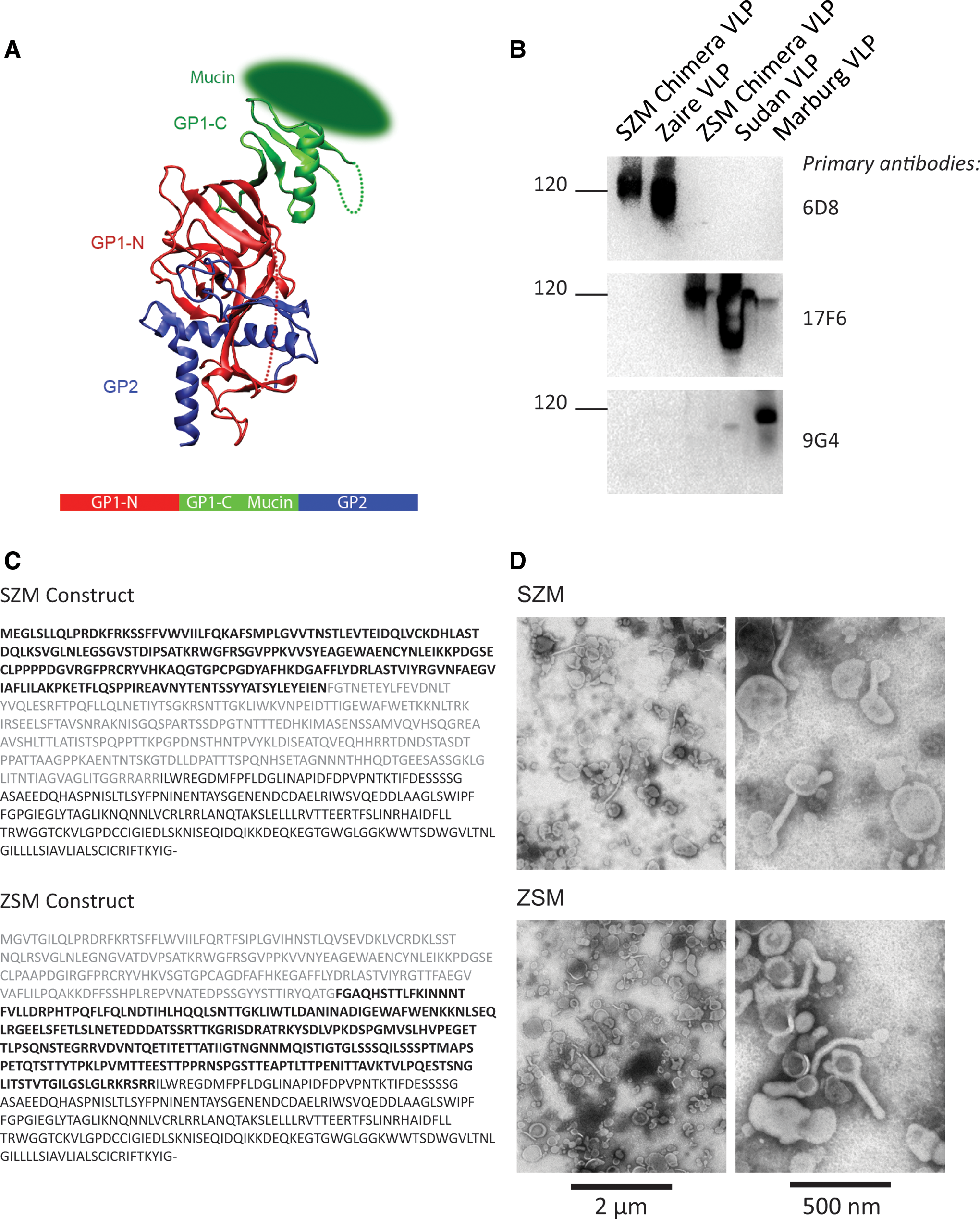
Production of virus-like particles (VLPs) containing trimeric hybrid glycoprotein (GP).
Protein quantification and Western blot
Total protein in VLP preparations was determined using the bicinchoninic acid assay (BCA; Thermo Scientific). For Western blot analysis, samples were boiled in SDS for 5 min at 95°C and loaded onto a 4–20% Tris-Glycine gel along with various concentrations of recombinant GP or VLPs, which served as controls. The iBlot system was used for transfer to nitrocellulose. Blots were incubated for 4 h with blocking buffer (PBS-T and milk), and then were incubated overnight with primary antibodies. After washing, blots incubated for 1 h with anti-mouse IgG HRP were washed, and then the Novex ECL substrate was used for detection (Life Technologies). The 6D8 (specific for Ebola GP amino acids 389–405) (54) and 17F6 (specific for Sudan GP amino acids 353–361; US patent application 20120164153) antibodies were used to detect epitopes in Ebola and Sudan GP, respectively, and the AE11 (specific for Ebola VP40; precise epitope unknown; thought to target C-terminal 16 amino acids of VP40 (36)) antibody was used to detect VP40. The anti-MARV GP antibody 9G4 was used to demonstrate no cross-reactivity with this antibody. GP content was approximately quantified using a dose curve of recombinant GP from 15 μg to 0.94 μg. Specificity of each construct for predicted anti-GP antibodies was tested by loading 1 μg of total protein of ZSM, SZM, EBOV, SUDV, and MARV VLPs. The binding of primary antibodies 6D8, 17F6, and 9G4 were tested individually. After exposure with each primary antibody, the blot was stripped using boiling water, blocked for 4 h, and then incubated overnight with the next primary antibody.
Electron microscopy
Samples were adsorbed to formvar/carbon-coated copper supports (SPI, Inc.) for electron microscopy and contrasted with phosphotungistic acid or uranyl salts. Samples were examined at 80 kV in a transmission electron microscope and digitally imaged.
Ethics statement
Research was conducted under an IACUC approved protocol in compliance with the Animal Welfare Act, PHS Policy, and other Federal statutes and regulations relating to animals and experiments involving animals. The facility where this research was conducted, USAMRIID, is accredited by the Association for Assessment and Accreditation of Laboratory Animal Care International, and it adheres to principles stated in the 8th Edition of the Guide for the Care and Use of Laboratory Animals, National Research Council, 2011.
Animals, vaccines, and challenge
Female Hartley guinea pigs (300–400 g) were purchased from Charles River Laboratories. Animals were monitored at least once daily, and their status was evaluated according to an intervention score sheet approved by USAMRIID IACUC. Monitoring increased to two to three times daily if the animals were given a score of three or four. Euthanization was by CO2 inhalation. Animals were euthanized if they reached a score of five or more; otherwise animals were euthanized on day 14 of the study.
VLPs were produced by transfecting 293T cells with expression vectors for viral GPs and VP40 (Ebola VP40, Genbank AAQ55047.1), essentially as previously described (46). PolyICLC (Hiltonol) was provided by Oncovir, Inc. VLP were diluted in sterile saline and combined with polyICLC prior to administration to animals. The vaccine consisted of 300 μg of VLP based on total protein and 10 μg of polyICLC (Hiltonol) diluted in sterile saline to a total volume of 200 μL per animal. Control groups included animals vaccinated with adjuvant alone. Challenge with 1,000 pfu guinea pig-adapted-MARV (gp-MARV) or guinea pig-adapted-EBOV (gp-EBOV) occurred 4 weeks after the second vaccination and was administered via the intraperitoneal route. Two weeks after the final vaccination, blood was collected from anesthetized guinea pigs via the cranial vena cava for a total blood volume of 500 μL.
Anti-GP antibody titers
Antibody titers were determined using an enzyme-linked immunosorbent assay (ELISA) with recombinant GP as the antigen. The recombinant proteins used as ELISA antigen were expressed via transient transfection of HEK293E cells. Plasmids were specific for Ebola GP (Kikwit), Sudan GP (Boniface), and Marburg GP (Ci67) lacking the transmembrane domain, which allowed for secretion from the transfected cell, and including a His-tag, which is used for isolation. Two μg/mL of recombinant Ebola, Sudan, or Marburg GP were plated in a flat-bottom 96-well plate overnight. Plates were incubated with blocking buffer (5% milk, 0.05% Tween in PBS) for 2 h, and then serum samples were added to plates. The protocol used half log dilutions starting at a 1:100 dilution. After 2 h, plates were washed with PBS+0.05% Tween, and goat anti-guinea pig IgG-HRP secondary antibody was added at a 0.6 μg/mL. One hour later, plates were washed and exposed using Sure Blue TMB 1-component substrate and stop solution (KPL), and the absorbance at 450 nm was recorded. Serum from unvaccinated animals was used to establish background, and titers were defined as the serum dilution resulting in an absorbance >0.2, where background was universally <0.2. Serum from animals previously determined to contain anti-GP antibody was included on each plate to serve as an internal positive control (average and standard deviation of positive control at 1:1,000 dilution across nine plates per antigen: Ebola, 1.912, 0.25; Sudan 3.19, 0.13; Marburg, 0.542, 0.06).
Pseudovirion neutralization assay
The pseudovirion neutralization assay (PsVNA) used to detect neutralizing antibodies in sera was essentially described previously, with key differences being the use of filovirus pseudovirions (23). Briefly, heat-inactivated guinea pig sera was first diluted 1:10, followed by fivefold serial dilutions that were mixed with equal volume of Eagle's minimum essential medium (EMEM) with Earle's salts and 10% fetal bovine sera containing 4,000 fluorescent focus units (FFU) of either EBOV, SUDV, or MARV pseudovirions and 10% guinea pig complement (Cedarlane). This mixture was incubated overnight at 4°C. Following this incubation, 50 μL was inoculated onto Vero cell monolayers in a clear bottom, black-walled 96-well plate (Corning) in triplicate. Plates were incubated at 37°C for 18–24 h. The media were discarded, and cells were lysed according to the luciferase kit protocol (Promega #E2820). A Tecan M200 Pro was used to acquire luciferase data. The values were graphed using GraphPad Prism software (version 6) and used to calculate the percent neutralization using cells alone and pseudovirions alone as the minimum and maximum signals respectively. The curves generated were interpolated to obtain PsVNA 50% and 80% neutralization titers.
Plaque reduction neutralization test
To determine the levels of plaque-neutralizing antibodies, guinea pig serum was initially diluted 1:100 in EMEM with 10% guinea pig complement. Subsequent twofold dilutions were made in the EMEM containing complement, and diluted sera was mixed 1:1 with ∼100 pfu of EBOV and incubated at 37°C for 1 h. As a result, the final dilutions of the serum samples in the neutralizing assay ranged from 1:200 to 1:6,400. The diluted samples containing virus were plated on confluent Vero E6 cells in duplicate, in six-well plates. After a 1 h adsorption, 2 mL of overlay media (2× EBME with 1% agarose) was added, and the plates were incubated for 7 days. On day 7, overlay media with 4% neutral red solution was added. Plaques were read on day 8. The percent of plaque reduction was calculated by comparing the number of pfu present in each sample to the pfu obtained with virus alone, similar to that described previously (18).
Statistics
The one-tailed Student's t-test was used to compare antibody titers. Survival studies were evaluated using Fisher's exact test. A p-value of<0.05 was considered statistically significant.
Results
Development of trimeric hybrid GP
Constructs were developed to express GP as a trimeric hybrid of three filoviruses: EBOV, SUDV, and MARV. Six different constructs were produced, expressing each combination of EBOV, SUDV, and MARV GP in the three regions: the N-terminal region of GP1 prior to the MLD and including the receptor binding domain (RBD); the C-terminal portion of GP1 was the second region, which included the MLD and most of the glycan shield region; and GP2 (Fig. 1A and Supplementary Fig. S1).
After transfection of the constructs into 293T cells and isolation of VLPs by sucrose gradient purification, the presence of GP and the relative concentration of the protein by Western blot were evaluated. The primary antibodies 5B4 (SUDV), 6D8 (EBOV), and 17F6 (SUDV) were used to quantify five of the constructs, which expressed the known linear epitopes for these antibodies. Serum from vaccinated animals was used as the primary antibody for the sixth construct, as no monoclonal antibodies were available that targeted this construct. Interestingly, only the two constructs in which MARV was represented by the third region (GP2) were successfully transcribed and translated into protein. The remaining four constructs did not result in protein expression as detected either in the VLPs or in transfection of the construct alone (data not shown). This is likely due to misfolding caused by incompatibility of the protein domains.
“SZM” is used to designate the construct in which the N-terminal region of GP1 expressed SUDV GP, while the C-terminal region expressed EBOV; “ZSM” is used to designate the construct in which the N-terminal region of GP1 expressed EBOV GP, while the C-terminal region expressed SUDV. Due to the length disparity between MARV and EBOV GP2, these constructs were 746 amino acids in length, rather than the standard 676 amino acids of EBOV GP and 681 amino acids of MARV. This is reflected in the size of the proteins as seen by Western blot (Fig. 1B). The sequences of the two constructs that resulted in protein production are shown in Figure 1C. Electron microscopy (EM) was used to confirm that transfection of the constructs resulted in VLPs with structure consistent with filovirus morphology. Figure 1D includes EM images of both the SZM and ZSM constructs. Both filamentous and globular particles were present, as has been observed previously.
VLPs expressing trimeric hybrid GP provide comparable protection from MARV challenge but distinct protection from EBOV challenge in Hartley guinea pigs
To test the immunogenicity and efficacy of the constructs, Hartley guinea pigs were vaccinated with either the trimeric hybrid VLPs SZM or ZSM, or with VLPs expressing MARV GP or EBOV GP. All vaccinations included 10 μg of polyICLC (Hiltonol); control animals received only adjuvant. Three weeks after the initial vaccination, animals were vaccinated a second time. Four weeks after the second vaccination, animals were challenged with 1,000 pfu of gp-EBOV or gp-MARV. There is no SUDV small animal challenge model, and so efficacy against SUDV challenge could not be examined.
The EBOV GP-expressing VLPs protected 100% of animals challenged with EBOV, and the MARV GP-expressing VLPs protected 100% of animals challenged with MARV. The VLPs expressing the trimeric hybrid GPs, however, had different effects on survival. The SZM trimeric hybrid VLP, in which SUDV GP was expressed in the N-terminal region of GP1 and EBOV GP was expressed in the C-terminal region of the GP1, protected 75% of the vaccinated animals, regardless of whether challenge was with gp-EBOV or gp-MARV. The ZSM trimeric hybrid VLP, in contrast, protected 100% of the animals challenged with gp-MARV but had no efficacy upon gp-EBOV challenge (Fig. 2).

VLPs expressing trimeric hybrid GP provide partial protection from challenge with either EBOV or MARV. Hartley guinea pigs were vaccinated twice with VLPs expressing EBOV VP40 and either EBOV, SUDV, or MARV GP, or one of the trimeric hybrid GPs, SZM or ZSM. The adjuvant polyICLC was included with vaccination. Animals were then challenged with either guinea pig (gp)-adapted MARV virus
The C-terminal region of GP1 elicits higher anti-GP antibody titers than the N-terminal region of GP1
Considering the distinct protection afforded by vaccination with the two constructs, the antibody response of vaccinated animals was compared against EBOV GP, SUDV GP, and MARV GP. Serum collected 2 weeks after the final vaccination was tested using an ELISA, with EBOV, SUDV, or MARV recombinant GP as the antigen. As shown in Figure 3A, the viral protein that was present in the C-terminal region of GP1 elicited a stronger antibody response than did the protein represented by the N-terminal region. Additionally, the titers obtained by vaccination with the C-terminal region of GP were comparable to the titers present in animals that were vaccinated with VLPs expressing full length EBOV or SUDV GP. As was anticipated, the anti-MARV GP responses present in animals vaccinated with the trimeric hybrid constructs were comparable; the same region of MARV GP, GP2, was present in both constructs. Of note, however, the antibody responses elicited only by this GP2 region were similar quantitatively to the levels elicited by vaccination with VLP expressing full length MARV GP (Fig. 3A).

Antibody titers of vaccinated animals. Serum was collected from vaccinated animals 2 weeks after the vaccine boost, and the serum was tested for anti-EBOV, SUDV, and MARV glycoprotein IgG antibody responses
Together the IgG titers suggested that vaccination with only a portion of GP, specifically GP2 or the C-terminal region of GP1, was sufficient to elicit a robust antibody response from vaccinated animals. The functionality of these antibodies was then examined. To evaluate the quantity of neutralizing antibody present in the vaccinated animals, a pseudovirion neutralization assay (PsVNA) was used. It was found that the neutralizing antibody profile paralleled the overall anti-GP IgG titers. Neutralizing antibody specific for EBOV or SUDV GP was higher in animals vaccinated with VLP expressing either full-length GP from that viral species or expressing the C-terminal portion of GP1 from that species (Fig. 3B). MARV neutralizing antibody titers were comparable between animals vaccinated with the two trimeric hybrid GP VLPs. A previously published neutralization assay for EBOV, the plaque reduction neutralization test (PRNT), was used to confirm the results observed with the PsVNA, and these data are presented in Supplementary Figure S2. The PRNT was run with a lower limit of detection of 1:200, and values were assigned based on the dilution of serum that neutralized a percentage of the plaques as compared to a control well treated with no serum. The PsVNA titers for each serum sample are interpolated from titration curves performed in triplicate. The lowest dilution tested in the PsVNA is 1:20, so titers <20 are considered below the limit of detection. Thus, while the data from both assays depicted the same trends in the neutralization titers, the absolute titers were not identical.
Discussion
The development of a single vaccine that confers protection against multiple filoviruses would be a significant advance in public health in parts of Africa where filovirus outbreaks are endemic, and it would also provide a level of protection against the threat of a bioattack using a filovirus. Efforts to date have demonstrated that there is no negative cross-reactivity associated with vaccination with multiple filovirus GP molecules, suggesting that such a goal is within reach. This study attempted to develop a single GP that could provide protection from multiple filovirus species, and this tool was used to explore the immunogenicity of different regions of the filovirus GP.
The presence of anti-GP IgG is one of the few correlates associated with protective vaccination from filovirus challenge (43). Recent studies have demonstrated that transfer of polyclonal convalescent antibody or a combination of antibodies to naïve animals post-infection can partially or completely protect NHP from either MARV or EBOV challenge (11,30,35,37,38). Administration of antibody has also been tested in humans infected during past filovirus outbreaks and during the ongoing outbreak in West Africa (33). A comprehensive understanding of what constitutes an effective antibody response to filoviruses is still elusive, however (56). Neutralization was once thought to be a potential correlate, but both neutralizing and poorly neutralizing antibodies have protective efficacy in vivo (11,34,54). The potential impact of the glycan shield on antibody–virus interaction, as well as the presence of secreted and soluble decoy molecules, complicates the understanding of the role of antibody in protection from viral challenge (8,24,40).
In this study, the N terminal region of GP1 elicited lower levels of neutralizing antibody than the C terminal region. Animals vaccinated with the SZM construct had lower anti-SUDV neutralizing antibody and overall IgG titers than did animals vaccinated with the ZSM construct, and the converse was observed for anti-EBOV virus antibody levels. The development of antibody specific for the N-terminal region of GP may be impacted by the masking effect of the glycan shield. Access to the RBD in the N-terminal region of GP1 is impeded by the glycan shield, as evidenced by the fact that cathepsin cleavage, which results in removal of the majority of the glycan shield and MLD, has been demonstrated to expose the RBD of GP1 (19).
The C-terminal region of GP1 is the most variable region of GP among filovirus species. In the current system, the C terminal region included the MLD and many of the residues that contribute to the glycan cap. It was anticipated, therefore, that this region would be largely shielded by glycosylation and would elicit lower levels of antibody than other GP regions. However, a previous study had found that deletion of the MLD, in part or in full, reduced the efficacy of a DNA-based EBOV vaccine (9). In that study, multiple glycosylation mutants of GP were produced, and removal of the MLD reduced vaccine efficacy and correlated with lower neutralizing antibody titers (9). In keeping with these findings, it was found that the C-terminal region of GP1 elicited higher antibody titers and conferred better protection on guinea pigs challenged with gp-EBOV than did the N-terminal region of GP1. Interestingly, evaluation of antibody responses in human EBOV infection survivors showed that the majority of anti-GP antibody responses targeted the C-terminal region of GP1, particularly the MLD, supporting the potential importance of this immunogenic region (2).
GP2 includes the region critical for fusion with the target cell membrane. It is also thought to be largely masked by the glycan shield and therefore a difficult target for neutralizing antibodies. The current data suggest, however, that the GP2 region of MARV is capable of conferring protection from MARV challenge. Inclusion of only the GP2 region of MARV GP resulted in antibody titers comparable to those elicited by full length MARV GP, and nearly all animals vaccinated with VLP expressing the hybrid GPs survived challenge. These data are particularly striking considering the minimal cross-protection between EBOV and MARV infection, which emphasizes that responses to GP2 alone were likely responsible for the protection against MARV challenge.
Crystal structures suggest that masking mediated by the glycan shield is not specific to the C-terminal region of GP1, but that in fact the majority of the GP is masked by the glycan shield (26). Several small regions are thought to remain exposed, including the region where GP1 meets GP2 (24). A recent study looking at the immunogenicity of this region showed that amino acid sequences from the GP1/GP2 junction can elicit neutralizing antibody (49), and it has been hypothesized that antibodies targeting this bridging region prevent the conformational changes necessary for GP fusion with the cell membrane (as reviewed in (24)). Interestingly, both the first SUDV neutralizing antibody and KZ52, the EBOV neutralizing antibody isolated from a human survivor, bind the GP1-GP2 junction (7,24). Antibody targeting this region of the constructs in the current study may have played a role in protection; further exploration of this region and the function of antibody targeting the GP1/GP2 interface would be of value.
The data from the present study add to the body of knowledge about the precise mechanism through which anti-GP immunity protects from filovirus infection, and they highlight the importance of GP2 and the C terminal region of GP1 in conferring protection. Future directions will test efficacy in the NHP model of filovirus infection. Additionally, the ability of such hybrid constructs to confer protection from filovirus variants and species not specifically represented in the vaccine construct will be evaluated.
Footnotes
Acknowledgments
PolyICLC (Hiltonol) was kindly provided by Dr. A. Salazar, Onocovir, Inc. This study was supported with funding from the Department of Defense DTRA project number CB2630. Opinions, interpretations, conclusions, and recommendations are those of the authors and are not necessarily endorsed by the U.S. Army.
Author Disclosure Statement
No competing financial interests exist.
References
Supplementary Material
Please find the following supplemental material available below.
For Open Access articles published under a Creative Commons License, all supplemental material carries the same license as the article it is associated with.
For non-Open Access articles published, all supplemental material carries a non-exclusive license, and permission requests for re-use of supplemental material or any part of supplemental material shall be sent directly to the copyright owner as specified in the copyright notice associated with the article.
