
Abstract
Background:
Hepatitis B virus (HBV) surface antigen (HBsAg) induces a vigorous neutralizing antibody response, which causes effective protection against HBV infection. Little is known about the profile of variable region genes of immunoglobuline heavy (VH) and light (VL) chains rearranged in anti-HBs antibodies, and also the possible association of this profile with specificity pattern of these antibodies to mutant forms of HBsAg.
Aims:
The present study determined the nucleotide sequence of VH and VL genes of mouse monoclonal antibodies (MAbs) generated against HBsAg.
Methods:
Hybridoma clones secreting anti-HBsAg MAbs were developed from hyperimmunized Balb/c mice. VH and VL gene sequences of all MAbs were determined by amplifying the genes using a panel of VH and VL family specific primers by reverse transcription polymerase chain reaction. The reactivity pattern of anti-HBs MAbs with different mutant forms of HBsAg was evaluated by enzyme-linked immunosorbent assay, and then the profile of antigen specificity and its association to VH/VL family expression was analyzed.
Results:
Twenty-three murine hybridomas producing anti-HBs MAbs were generated. Nucleotide sequence analysis revealed that heavy chains of these MAbs were encoded by IGHV genes from the HV1 (52%), HV6 (22%), HV5 (17%), and HV3 (9%) families in combination with IGHJ2 (57%), HJ1 (26%), and HJ4 (17%). Besides, 56% of MAbs used IGHD1 genes in their VDJ rearrangements. Concerning the IGKV gene, 26% and 22% of clones used KV4 and KV10 gene families, while the rest of the clones used KV8, KV6, KV1, KV12, and KV14 gene families. Besides, the IGKJ2 gene was the most represented KJ gene (43%). No association was found between the specificity pattern of MAbs to mutant forms of HBsAg with their preferential V, D, and J genes usage for most of MAbs.
Conclusion:
The data suggest that heavy chains of anti-HBs MAbs preferentially use genes derived from the IGHV1, IGHV6, IGHJ2, and IGHD1 families. In contrast to heavy chains, which predominantly use four families of IGHV genes, light chains use more diverse IGKV gene families.
Introduction
H
HBV surface antigen (HBsAg) can induce a strong immunogenic antibody response (21). Consistent with the major role antibodies play in defense against invading pathogens, antibodies to HBsAg have been shown to cause effective protection against HBV infection.
Production of anti-HBs antibodies is the major indicator of clinical features such as monitoring of seroconversion, vaccination, and immunoprophylaxis after liver transplantation (5). Most of these neutralizing antibodies are directed against the main antigenic determinant (“a” determinant), which is located between amino acids 124 and 147 within the major hydrophilic loop of HBsAg (21). Consequently, there is increasing concern about the emergence of mutant viruses with amino acid substitutions in the “a” loop. These kinds of mutations have been identified in chronic carriers and among fully vaccinated subjects and during prophylaxis of recurrent HBV infection with high-dose hepatitis B immunoglobulin (HBIG) in liver-transplant recipients (2,5,24).
The antigen recognition site of an antibody is composed of a paired immunoglobulin (Ig) heavy chain (IGH) and immunoglobulin light chain (IGL) variable domains. Both the IGH variable domain (VH) and IGL variable domain (VL) are made of conserved framework sequences that alternate with three hypervariable regions, the complementary-determining regions (CDRs), which are responsible for antigen recognition (9). Antigen-specific antibody response results in the production of a large number of individual B cell clones with diverse specificities (1,28).
This diversity is created by unique lymphocyte molecular mechanisms, including random rearrangement of variable (V), diversity (D), and joining (J) genes in the IGH locus and also V and J genes in either of the kappa (IGK) or lambda (IGλ) IGL locus. Additional diversity at the junctions of the V, D, and J genes would be created by insertion and deletion of nucleotides. The V, D, and J genes in IGH and IGK chains are called IGHV, IGHD, IGHJ, IGKV, and IGKJ genes, respectively (1,28).
Somatic hypermutation and receptor editing finally result in the formation of a repertoire of high-affinity antibodies (6). This process theoretically yields an almost unlimited number of antibody specificities. However, evidence from murine and human systems suggests that these processes may not occur randomly, leading to utilization of limited diversity in structure of antibodies and changing Ig repertoire during development (1).
Little is known about the profile of Ig VH and VL gene usage in production of anti-HBs antibodies. The present study determined the nucleotide sequence of IGHV, IGHD, IGHJ, IGKV, and IGKJ of 23 MAbs generated against this biologically significant antigen.
In order to assess if there is any correlation between antigen binding specificity of these MAbs with their VH and VL nucleotide sequence, the fine epitope specificity of these MAbs was analyzed using a panel of HBsAg with single mutations within the “a” determinant.
Materials and Methods
Production of anti-HBs monoclonal antibody
Murine hybridomas producing anti-HBs MAbs were generated, as previously described (8,25,26). Briefly, female Balb/c mice were immunized with HBsAg, Adw2 subtype vaccine (Heberbiovac, Heberbiotec, Cuba), emulsified in Freund's complete adjuvant (CFA; Sigma-Aldrich, St. Louis, MO) for the first and incomplete Freund's adjuvant (IFA; Sigma-Aldrich) for five additional booster immunizations, until the serum titer of anti-HBs antibody reached a plateau. Then spleen cells were harvested and fused to the mouse myeloma cell line SP2/0 (National Cell Bank of Iran, Pasteur Institute, Tehran, Iran) using PEG 1500 solution (Sigma-Aldrich). Hybridoma cell selection and cloning was carried out in RPMI-1640 medium containing 20% heat-inactivated fetal calf serum (FCS), supplemented with HAT medium (hypoxanthine, aminopterine and thymidine; Sigma-Aldrich). Anti-HBs antibodies, secreted from selected hybridomas, were detected by binding to wild type HBsAg in an indirect enzyme-linked immunosorbent assay (ELISA) (27). Positive hybridomas were cloned by limiting dilution at least three times to obtain stable single clones.
Construction and expression of plasmids encoding recombinant wild and mutant forms of HBsAg
Ten mutant forms of HBsAg (mtHBsAg) and wild type (wt) Adw were constructed by primer-specific site-directed mutagenesis and SEOing polymerase chain reaction (PCR) method, followed by cloning into the mammalian cell expression vector pCMV6-neo plasmid (Origene, Rockville, MD), as described elsewhere (27).
CHO cells (National Cell Bank of Iran, Pasteur Institute) were cultured in 12-well plates in RPMI-1640 medium supplemented with 10% FCS, under the condition of 5% CO2, 37°C. pCMV6-neo vector, which contains wt and mtHBsAg genes, were transfected in CHO cells using JetPEI reagent (PolyPlus-Transfection Co., Illkirch, France). Two days after transfection, the culture medium was harvested, and secretion of wt and mtHBsAg was evaluated with a commercial ELISA kit (BioELISA HBsAg, Biokit, Barcelona, Spain), which employs polyclonal antibodies for both capture and detection layers and recognizes mtHBsAg with high specificity and sensitivity. Besides, different concentrations of a commercial recombinant HBsAg (Heberbiovac) was used as standard to determine the quantity of wt and mtHBsAg in the supernatant of transfected CHO cells.
RNA isolation and cDNA synthesis
Hybridoma cells were collected, washed with phosphate-buffered saline (PBS), and counted. Total cellular RNA was isolated from five to six million hybridoma cells using RNA-Bee (Tel-test, Inc. Friendswood, TX) based on the manufacturer's instructions.
First-strand cDNA was synthesized from total RNA (first unfolded at 65°C for 5 min) using reverse transcriptase (RT) enzyme (Fermentas Life Sciences, Ontario, Canada), random hexamer primers (0.4 μg) and dNTPs (20 mM; Roche, Mannheim, Germany). The mixture was incubated at 42°C for 60 min followed by 10 min at 70°C for inactivation of RT enzyme.
In order to evaluate the quality of cDNA preparation, PCR was performed for β-actin as a universal housekeeping gene using the primers: forward GTG GGG CGC CCC AGG CAC CA, reverse CTC CTT AAT GTC ACG CAC GAT TTC.
For the reaction, the mixture was prepared containing 1 μL of cDNA from each hybridoma, one unit of Taq DNA Polymerase (CinnaGen Co, Tehran, Iran), 0.2 mM deoxynucleotides (Roche), and 1.2 mM magnesium chloride (25 μL total volume). The thermal cycling conditions used for this pair of primers were: 94°C for 2 min, 27 cycles of 94°C for 30 sec, 60°C for 30 sec, and 72°C for 30 sec, followed by a final period at 72°C for 3 min. PCR products were analyzed using horizontal electrophoresis in 1% agarose gel.
Determination of VH and VL gene sequence by PCR
Two PCR reactions were run for each hybridoma cDNA: one for heavy chain amplification, and the other for light chain.
Primer sequences that were used to amplify heavy and light chains are shown in Tables 1 and 2. PCR reactions were performed using pfu DNA polymerase (Fermentas Life Sciences), in 40 cycles, initiated by 5 min at 94°C followed by 94°C for 45 sec and 60°C for 30 sec then 72°C for 120 sec and 10 min at 72°C for the final extension.
PCR products were finally visualized by electrophoresis on 1% agarose gel containing ethidium bromide. The VH and VL Ig chains were sequenced, and the amino acid sequences deduced.
The identification of IGHV, IGHD, IGHJ, IGKV, and IGKJ sequences was performed by sequence comparison using the international immunogenetics database-IMGT (
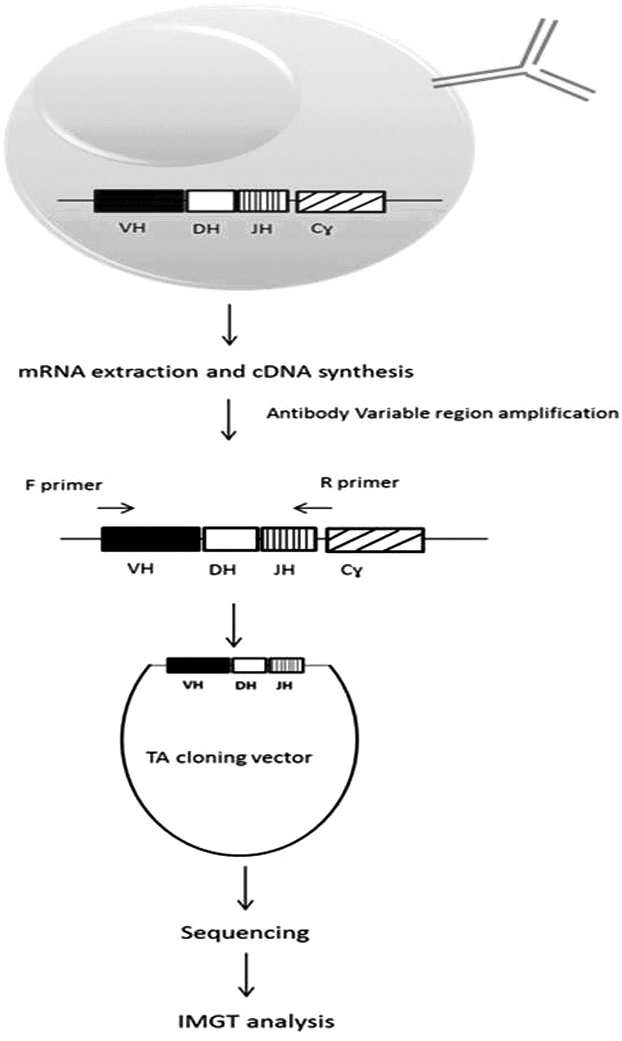
Schematic representation of the IGHV and IGLV cloning and analysis. Total cellular RNA was isolated from hybridoma cells, and cDNA was synthesized. The VH and VL regions of heavy and light chains were amplified with specific primers, cloned in TA vector, and sequenced eventually. The identification of IGHV, IGHD, IGHJ, IGKV, and IGKJ sequences was performed by sequence comparison using the IMGT.
Reactivity pattern of anti-HBs MAbs with mutant forms of HBsAg by ELISA
Different concentrations of purified MAbs were added in 50 μL volumes on the solid phase of a 96-well ELISA plate (Maxisorp, Nunc, Denmark) at 4°C overnight, as previously described (8). Briefly, after blocking with skim milk, supernatants of transfected CHO cells (wt and all mtHBsAg) were added to the wells at an adjusted concentration of 40 ng/mL for 1 h at 37°C. After washing, polyclonal biotin labeled sheep anti-HBs antibody conjugate (Pishtaz Teb, Tehran, Iran) was added followed by streptavidin horseradish peroxidase conjugate (Invitrogen, Carlsbad, CA). Subsequently, 3,3′,5,5′-Tetramethylbenzidine (TMB) substrate solution (Pishtaz Teb) was added, and the reaction was terminated by adding 1 M HCl, and optical density (OD) measured in an automated plate reader (Organon Teknika, Boxtel, Netherlands) at 450 nm.
Statistical analysis
A chi square test was performed to determine the significance of differences among frequencies of data between groups. p-Values of < 0.05 were considered to be significant.
Results
Characterization of anti-HBs MAbs
Twenty-three anti-HBs MAbs, mostly reported in previous works (8,25 –27), were employed in this study. All MAbs were purified from ascitic fluids by streptococcal protein G column. Purified MAbs were then characterized for isotype and HBsAg binding affinity (Table 3). All MAbs displayed IgG/K isotype with binding affinities ranging from 5 × 10⁷ to 1.8 × 10⁹ M/L.
ND, not determined.
Cloning and sequencing of the VH and VL genes
The variable region heavy (VH) and kappa chain (VK) genes rearranged in hybridoma clones producing the anti-HBs MAbs were amplified from cDNA and cloned into the TA cloning vector. The cloned VH and VK genes were then sequenced, and their nucleotide homologies to their germline counterparts were determined (Table 3).
Sequence analysis and family assignment of the VL genes
The light chain expressed by all MAbs belonged to the kappa isotype. With the exception of the 5G7 IGKV gene, which showed 81.7% nucleotide homology, other MAbs showed homology of ≥92% with their counterpart germline IGKV genes. 2G2, S7A, and 2F2 IGKJ genes also showed 71% and 84% nucleotide homology with their counterpart germline IGKJ genes, while the rest of MAbs displayed 91.9% or higher homology with germline IGKJ genes (Table 3).
The IGKV gene families (subgroup) as well as IGKJ genes were not equally distributed among anti-HBs MAbs. With regard to the IGKV gene, 26% of clones used KV4 gene family (subgroup), while 22%, 17.4%, 13%, 13%, 4.3%, and 4.3% of clones used KV10, KV8, KV6, KV1, KV12, and KV14, respectively (Tables 3 and 4).
The data represented as % of IGKV germline (7,14,20) and cDNA (16) in the literature belong to Mus musculus. IGKV percentages have been calculated among functional IGKV. The whole numbers of IGKV variable cDNA for all families were determined, and then the percentages of each family cDNA were calculated separately.
The IGKJ2 gene was the most represented KJ gene (43%), followed by KJ5 (35%), KJ1 (13%), and KJ4 (9%; Table 5).
Sequence analysis and family assignment of the VH genes
The amino acid sequences deduced from the IGHV sequences of anti-HBs MAbs demonstrated that 52% of anti-HBs MAbs use members of the IGHV1 family, while 22%, 17%, and 9% use HV6, HV5, and HV3 families, respectively (Table 6). The IGHJ2 gene encoded 57% of MAbs joining region, while HJ1 and HJ4 represented 26% and 17% of the IGHJ genes rearranged in anti-HBs MAbs, respectively.
The data represented as % of IGHV genes in IGHV germline (12,18) and cDNA (16) belong to Mus musculus and calculated based on IMGT database. IGHV percentages have been calculated among functional IGHV. The whole numbers of IGHV variable genes (or cDNA) for all 15 families were determined, and then the percentages of each family genes (or cDNA) were calculated separately.
Meanwhile, 50% of the hybridoma clones expressing IGHV1 gene family use IGHJ1 gene, while 33% use HJ2, and only 17% of them use HJ4 gene in their VDJ rearrangements. However, MAbs that use IGHV5 and HV6 gene families preferentially use just the HJ2 gene. The only D gene rearranged in the latter group (antibodies that use IGHV6 family) belonged to the IGHD1 subgroup.
Fifty-six percent of MAbs used IGHD1 genes in their VDJ rearrangements, while 22%, 11%, 5.%5, and 5.5% of them used HD2, HD4, HD3, and HD5, respectively. Besides, D–D fusion was not noted for any clone, and IGHD gene of five hybridomas could not be determined (Table 3).
The overall homologies of the IGHV regions of anti-HBs antibodies to their germline HV gene counterparts are between 88.5% and 98.2%. 4G4 and 3C9 MAbs show the lowest homologies (88.5%), while S4A has the highest homology (98.2%; Table 3).
2A7, 3A7, and 2C5 MAbs share similar or the same heavy and light chain sequences with mostly identical framework (FR) structure and few amino acid substitutions in CDR1 and CDR3 domains. The same pattern was detected for two other groups of MAbs 4A3, 6C1, and 2F4, as well as 5F9 and 6F9.
No significant correlation was found between the IGHV, IGHD, and IGHJ gene family usage and other aspects such as N-region addition and CDR3 features (data not shown).
Reactivity pattern of anti-HBs MAbs with mutant forms of HBsAg
Immunoreactivity of anti-HBs MAbs with 14 recombinant HBsAg mutants, including P120E, T123N, T126N, Q129L, Q129H, M133H, M133L, K141E, P142S, T143K, D144A, G145R, N146S, and C147S as well as wt HBsAg was assessed by ELISA. Supernatants of untransfected cells or cells transfected with pCMV-6 vector alone were used as negative control. All antibodies recognized wt HBsAg, but some MAbs either reacted weakly or failed to bind to some mutant antigens (Table 7). Based on these results, the MAbs could be classified into different groups with distinct reactivity profiles. The first group includes six MAbs (5F9, 3A7, 2C5, 6F9, 6E3, and 2A7) that failed to react with T123N and G145R. The second group with five MAbs (2F4, 5G7, 2G2, 4A3, and 2F2) did not react with T123N, T126N, and Q129L, while the third group including S15A and S11A MAbs failed to bind to 8/14 mtHBsAg. The remaining MAbs showed overlapping but different profiles with mtHBsAg.
Due to the insufficient amount of 1F1 and H12A, the reactivity pattern of these MAbs has not been included in this table.
Discussion
Development of neutralizing antibodies to HBsAg provides protection against HBV infection. However, limited information is available on the fine specificity and genetic origin of these neutralizing antibodies. The present study analyzed 23 anti-HBs MAbs generated by the hybridoma method. Establishment and characterization of some of these MAbs has already been reported (8,25 –27). Nucleotide sequence analysis revealed that heavy chains of these MAbs were encoded by IGHV genes from the HV1 (52%), HV6 (22%), HV5 (17%), and HV3 (9%) families in combination with IGHJ2 (57%), HJ1 (26%), HJ4 (17%), and IGHD1 (56%), HD2 (22%), HD4 (11%), HD3 (5.5%), and HD5 (5.5%; Tables 3 and 6). These data suggest that anti-HBs MAbs preferentially use VH genes derived from the IGHV1, IGHJ2, and IGHD1 families. In contrast to heavy chains, which predominantly use four families of IGHV genes, light chains use more diverse IGKV genes from the KV4 (26%), KV10 (22%), KV8 (17.4%), KV6 and KV1 (13%), KV12 (4.3%), and KV14 (4.3%) families (Tables 3 and 4).
Assessment of the preferential use of VH and/or VK genes or gene families in an antigen-specific antibody response is based on knowledge of the frequency of these genes in the germline configuration and also in circulating naive B cells from healthy subjects. Although in early life, a preferential rearrangement of the most VDJ genes has been reported in fetal and neonatal B cells both in humans and mice (3,23), predominancy of the rearranged genes in B cells is largely associated to their germline number in the chromosome (3). Strain-dependent variations that may not be due to the size of the VH gene families have also been reported (3).
Thus, the frequency of the VH and VK genes expressed in the panel of anti-HBs MAbs was compared with the frequency of both the germline and rearranged genes in mice (Mus musculus). We delineated the genes listed in the IMGT database which is regarded as the most comprehensive database for immunoglobulin genes. We calculated percentage of Mus musculus IGHV1-16 and also IGKV1-20 cDNA families usage frequency based on IMGT and compared them to those of families which were used in anti-HBs MAbs (Tables 4 and 6).
The mouse IGHV locus, which is located on chromosome 12, is composed of 218 functional IGHV genes. These genes are classified into 16 IGHV families, the most abundant family being HV1 (52.2%) followed by HV5 (12.4%) and HV2 (10%) families (Table 6) (12,18).
These three IGHV families are also more represented in the rearranged genes, as analyzed in cDNA transcripts from circulating B cells (Table 6), though the percent of IGHV genes in each family differs slightly from that of the germline state. Overall, the data regarding representation of IGHV gene families in the anti-HBs MAbs are similar to the normal representation of these genes, with some differences. Thus, while IGHV1 (52%) and HV5 (17%) are expressed at a similar level to the normal rearranged and non-rearranged (germline) IGHV genes, HV6 (22%) and HV3 (9%) are more represented, and HV2 gene family is totally absent in our anti-HBs MAb panel.
We found a statistically significant difference between only HV6 usage among our anti-HBs antibodies and normal rearranged IGHV genes (p value <0.0001).
The mouse IGHD genes are composed of 21 functional genes, which are classified into six families. There are only four functional IGHJ genes in the mouse genome (11,15). The majority of our anti-HBs MAbs employed the IGHD1 (56%) and IGHJ2 (57%) genes. The IGHD in five clones could not be clearly identified due to extensive CDR3 sequence diversification. As mentioned, 50% of the hybridoma clones expressing IGHV1 gene family use IGHJ1 gene, while 33% use HJ2 and only 17% of them use HJ4 gene in their VDJ rearrangements. Therefore, it can probably be assumed that IGHV1 containing transcripts preferentially use the joining region IGHJ2 and IGHJ1 rather than other IGHJ genes. We could not find a representative data about the frequency of rearranged IGHJ genes in mouse antibodies in order to compare them with those of IGHJ genes in our anti-HBs antibodies. Furthermore, there was no statistically significant correlation between any IgG subclasses and particular IGHV gene usage.
The IGKV genes in mouse (Mus musculus) are more diverse, with 98 functional genes organized into 20 families on chromosome 6 (14,22). The IGKV4 family is the mostly represented family (25.5%) of the mouse germline IGKV genes, followed by IGKV6 (12.2%), IGKV3 (9.2%), and IGKV1 and IGKV8 (8% each). IGKV1, 3, 4, 6, 8, 12, and 5 families together contain more than 70% of the total number of germline genes (Table 4). The same gene families are more represented in the cDNA rearranged sequences of mouse VK genes with one major exception, which is related to the IGKV1 family (Table 4), showing more representation in the rearranged as compared with the germline IGKV genes. Comparison of the present results with the frequency of rearranged mouse IGKV families indicates a similar pattern with three major exceptions regarding the IGKV10, 8, and 12 families. Only the frequency of IGKV8 and IGKV10 usage between the anti-HBs MAbs and that of the mouse IGKV was significantly different (p = 0.03 and p = 0.0004, respectively).
As for the IGKJ genes, there are four functional IGKJ genes in the mouse genome (13). The majority of kappa light chains of our MAbs are encoded by either IGKJ2 (43%) or IGKJ5 (35%). The remaining MAbs (n = 5; 22%) were encoded by two other KJ genes, including IGKJ1 and IGKJ4 (Tables 3 and 5). Comparison of the present results with the frequency of rearranged mouse IGKJ genes indicates a similar pattern (Table 5) (17).
Furthermore, it seems that there is no preferential association between the patterns of IGKV-KJ pairing. For examples, IGKV4 (as the highest used IGKV family) in 50%, 33%, and 17% of MAbs pairs with IGKJ5, KJ1, and KJ2, respectively.
The critical role of the VH chain in antigen recognition has already been reported (19). In the present panel of MAbs, there are some antibodies such as 2G2 and 2F4 that share similar VK genes and different VH genes, but display similar reactivity pattern with mtHBsAg. As shown in Table 3, 4A3, 2F4, and 6C1 MAbs shared almost the same VK and VH genes (but their isotype is different), so we could expect the same pattern of reactivity with mutant forms of HBsAg. However, due to the fine changes of amino acids in FRs and CDRs, there is a slight difference between the mtHBsAg reactivity patterns of 6C1 with the other two MAbs (Table 7). Therefore, hybridomas that are most likely derived from the same parental clones, after affinity maturation and class switching, could lose their ability to recognize some of the mutant forms of HBsAg or even show stronger reactivity with other mutants. Another examples are that 3C9, 5G7, and 3A7 shared almost the same VH sequence (with slight differences), but the reactivity pattern of only 3C9 and 5G7 with mutant forms of HBsAg is almost the same. It might also be expected that MAbs that show a similar pattern of reactivity with mutant forms of HBsAg are likely to share the same or similar V, D, and J genes compared with those that react differently with mutant forms of HBsAg.
Although this assumption could be justified for some MAbs, many of such MAbs are encoded by different VH and/or VK genes. For example, 4 MAbs (2F4, 5G7, 2G2, and 2F2) displayed the same pattern of mtHBsAg reactivity, but only two (2F4 and 2G2) were encoded by the same VK gene. There are two other panels of MAbs showing distinct reactivity patterns, including 5F9, 3A7, 2C5, 6F9, 6E3, and 2A7 as well as S15A and S11A (Table 7), but none of these MAbs was encoded by the same VH and/or VK genes (Table 3).
Using the specific primers for 12 IGHV and 11 IGKV families (Tables 1 and 2) to amplify all families of IGHV and IGKV is one of the limitations of this study. However, we were aware that the usage frequency of IGHV13-16 and IGKV12-20 families in mouse antibodies is only 4.8 and 16.8% of the whole IGHV and IGKV families, respectively. Besides, with these sets of primers, the variable region of all hybridomas was amplified, and neither of those variable regions belonged to IGHV13-16 and IGKV12-20 families.
Human antibody libraries against HBsAg in one study revealed diversity in specificity of antigen binding in the VH and/or VK genes (29), whereas in another study, all the VH and VK genes were derived from a single VHIII and VKI gene family (10). The results of the latter study showed no apparent trend in the usage of J gene segments. All four investigated clones in the latter study showed a high degree of homology to a single germline V gene segment, which implied a restricted V gene segments utilization in the human immune response to HBsAg.
The present data permit a more comprehensive picture of the influences of various IGHV and IGKV genes in the generation of antibody response to HBsAg in mice.
On the sole basis of the amino acid sequences and different V, D, and J genes of these antibodies, the results do not support the concept of a simple correlation between the VH/VL gene usage and antigen-binding specificity toward different mutant forms of HBsAg.
It is clear from the discussion so far that a variety of IGKV and IGHV genes encode our anti-HBs MAbs, and they appear not to be restricted to a small group of VH and VK gene segments.
Footnotes
Acknowledgments
We would like to thank Dr. Véronique Giudicelli from IMGT, the international ImMunoGeneTics information system for scientific consultation. This study was supported by grants from Avicenna Research Institute and Iran National Science Foundation.
Author Disclosure Statement
No competing financial interests exist.