
Abstract
Immunoglobulin Y (IgY) antibodies were generated against canine parvovirus virus-like particles (CPV-VLPs) antigen using chickens. Anti-CPV-VLPs-IgY was extracted from hen egg yolk and used for developing enzyme-linked immunosorbent assay (ELISA) and immunochromatographic assay (ICA) for the detection of CPV in dog feces. The cutoff negative values for anti-CPV-VLPs-IgY were determined using negative fecal samples (already confirmed by polymerase chain reaction [PCR]). In both ELISA and ICA, there was no cross-reaction with other diarrheal pathogens. Thirty-four fecal samples were collected from dogs with diarrhea, of which 26.47% were confirmed as CPV-positive samples by PCR, while 29.41% and 32.35% of the samples were found to be positive by ELISA and ICA, respectively. The developed ELISA and ICA exhibited 97.06% and 94.12% conformity with PCR. Higher sensitivity and specificity were observed for IgY-based ELISA and ICA. Thus, they could be suitable for routine use in the diagnosis of CPV in dogs.
Introduction
C
In recent decades, the generation of antibodies in chicken egg yolk has been recognized as a promising alternative source for large amounts of specific antibodies with high affinity for immunodetection and immunodiagnostics. In addition, the lack of cross-reactivity between immunoglobulin Y (IgY) and IgG could aid in reducing unwanted reactions in assays using anti-IgG antibodies (2,6,8,17). The present study aimed to prepare IgY against CPV-VPLs and develop IgY based in enzyme-linked immunosorbent assay (ELISA) and immunochromatographic assay (ICA) in order to detect CPV.
Materials and Methods
Materials
The experimental protocol was reviewed and approved by the Ethics Committee of Northwest A&F University for the use of Laboratory Animals. CPV-VLPs protein was obtained from Key Laboratory of Animal Virology of Ministry of Agriculture, Lanzhou Veterinary Research Institute, China. Two brown Leghorn hens (29 weeks old) were procured from a local poultry farm and maintained in cages with food and water ad libitum.
Chicken immunization
Chickens were immunized intramuscularly with CPV-VLPs protein mixed with Freund's adjuvant (Sigma-Aldrich, St. Louis, MO) at different sites of breast muscles. In the first immunization, 250 μL (1 mg/mL) of CPV-VLPs protein was emulsified with an equal volume of complete Freund's complete adjuvant (FCA), and four booster immunizations were followed up using Freund's incomplete adjuvant (FIA) at 2 week intervals. Eggs were collected daily, and were marked and stored at 4°C until they were processed.
Extraction and characterization of IgY
IgY antibodies were isolated from the eggs using PEG-6000 as described before (12,18). The titer of specific IgY antibody against CPV-VLPs protein was determined as described in the authors' previous report (6). Western blotting was employed to determine the purity and specificity of anti-CPV-VLPs-IgY; the protein was separated using 12% SDS-PAGE and transferred onto a polyvinylidene fluoride membrane using a Mini-Protean system (Bio-Rad, Hercules, CA). The unreacted sites were blocked with 5% (w/v) nonfat milk powder in TBST (20 mM Tris-HCl, 150 mM NaCl, 0.05% Tween 20 [v/v], pH 8.0) buffer for 1 h and then incubated with anti-CPV-VLPs-IgY antibody (1:1,000) for 1 h at room temperature. After washing three times with TBST, HRP-conjugated goat anti-chicken antibody (Abcam, Cambridge, MA) diluted in TBST (1:5,000) was added and incubated for 1 h at room temperature. The membrane was washed three times with TBST, and the results were detected using the Pierce ECL Western blot substrate (Thermo Scientific, Waltham, MA) and analyzed by Tanon-410 automatically gel imaging system (Shanghai Tianneng Corporation, China).
Determination of cutoff value and preparation of ICA
Fecal samples were collected using sterile swabs from 10 dogs. All specimens were confirmed for CPV negativity by PCR. Briefly, fecal samples were obtained by rectal swabs, and were homogenized (10%, w/v) in phosphate-buffered saline (PBS; pH 7.2) containing streptomycin (100 μg/mL) and penicillin (100,000 IU/L). The supernatant was boiled as template. Primers were described in the previous report (6). The template was denatured at 94°C for 5 min, followed by 30 cycles of 95°C for 30 sec, 55°C for 30 sec, 72°C for 1 min, and a final extension at 72°C for 10 min. ELISA was performed as described in the authors' previous study (6). Microtiter plates (NUNC, Roskilde, Denmark) were coated with fecal samples (100 μL per well, and triplicates were maintained), 100 μL of CPV-VLPs antigen (5 μg/mL) as positive control, and CBS as negative control.
Colloidal gold was prepared using a method described previously (11). Briefly, an aqueous solution of chloroauric acid (100 mL of 0.01%HAuCl4•4H2O) was heated to boiling and, with rapid stirring, 2 mL of 1% sodium citrate solution was added. After an additional 10 min of continuous boiling, the colloid gold was gradually cooled with continuous stirring for an additional 15 min. The colloidal gold solution was then stored at 4°C in a dark-colored glass bottle. Murine monoclonal antibodies against CPV-VLPs (kindly provided by Leposen Ltd., Henan, China) was added to 10 mL of gold colloid solution and adjusted to pH 8.0. The mixture was stirred vigorously for 30 min, and 1.3 mL of 10% (w/v) bovine serum albumin (BSA) aqueous solution was added to block excess reactivity of the colloidal gold, followed by an additional 30 min stirring, and sprayed onto glass fiber pads and then dried. Anti-CPV-VLPs IgY was microsprayed onto nitrocellulose membranes at 1.0 μL/cm at a position that became the capture test line on completed strips. Polyclonal rabbit antimurine-IgG antibody was microsprayed onto the same nitrocellulose membranes at 1.0 μL/cm at a position that became the capture control line. The membrane was sliced into 25 mm long and 5 mm wide strips. Finally, membranes were dried.
Evaluation of ELISA and ICA
To determine the cross-reactivity of the test strip, positive clinical samples of canine coronavirus (CCV), canine distemper virus (CDV), enteropathogenic Escherichia coli, Salmonella enteritidis, Staphylococcus aureus, and Clostridium difficile were tested against the prepared strips. From the sample suspension, 100 μL (10 times diluted) was added into the test strip, and results were observed within 20 min. Fecal samples of 34 dogs with suspected diarrhea were collected from the rectum using sterile swabs, which was repeatedly washed in 1 mL of PBS (pH 7.4). That suspension was then centrifuged at 12,000 g for 10 min and supernatant was filtered using 0.22 μm microfiltration membrane and stored at −20°C until use. ELISA and ICA were used to confirm the samples. PCR was then conducted to evaluate the conformity of the results.
Results
Anti-CPV-VLPs IgY antibodies
IgY was isolated from egg yolk by the PEG-6000 method, and was confirmed by SDS-PAGE under reducing conditions (Fig. 1). This showed two bands of light and a heavy chain corresponding to 23 kDa and 67 kDa, respectively. Furthermore, some lower molecular weight bands around 40 kDa were also observed. The titers of specific IgY antibodies against CPV-VLPs protein in immune egg yolk were determined by indirect ELISA. A specific IgY titer (1:6,400) was increased after the first booster injection, reaching a peak (1:64,000) after the third booster injection, and was found to be stable at the highest titer for more than 1 month. It was then gradually decreased to 1:16,000 and remained at this level until the end of the experiment (Fig. 2). Binding activity of IgY antibodies to CPV-VLPs was further determined by Western blot. The isolated IgY bound to their specific protein (65 kDa) at a dilution of 1:1,000 (Fig. 3), thus showing that extracted IgY has good sensitivity to the antigen.
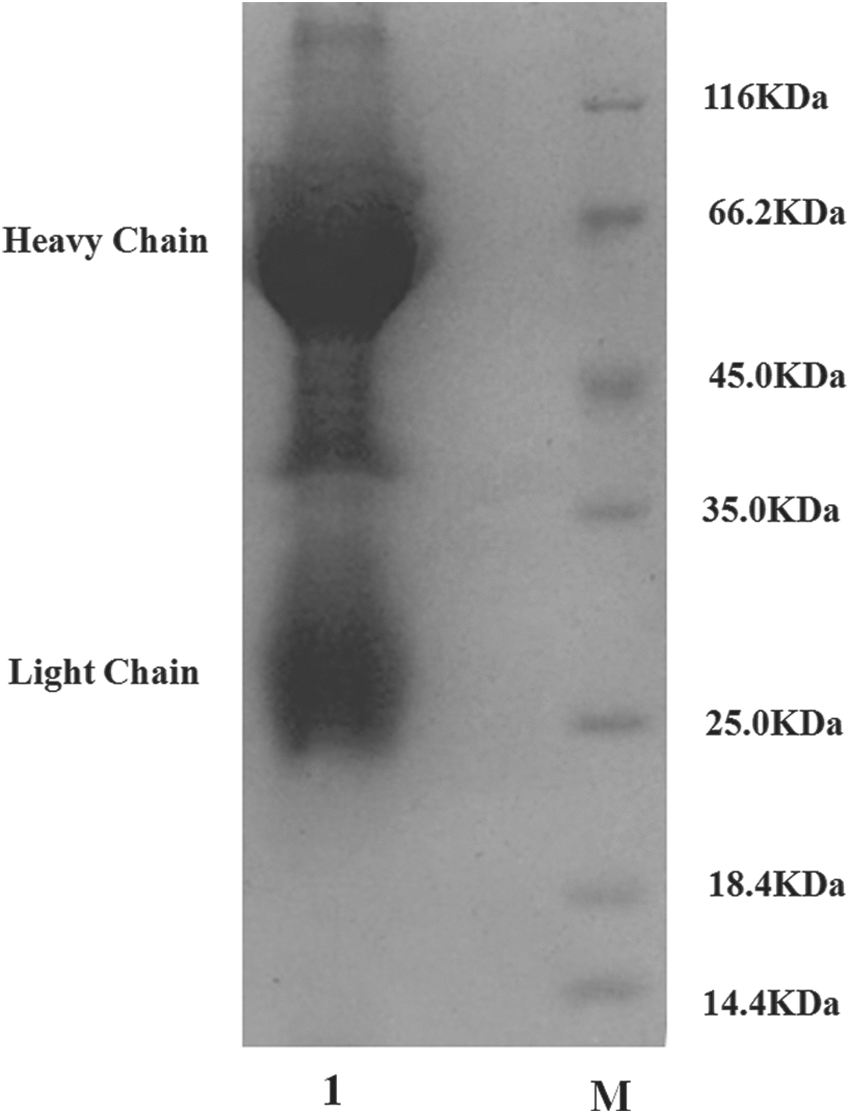
SDS-PAGE analysis of the isolated immunoglobulin Y (IgY) antibody by PEG-6000. Lane M, marker; Lane 1, IgY antibody.
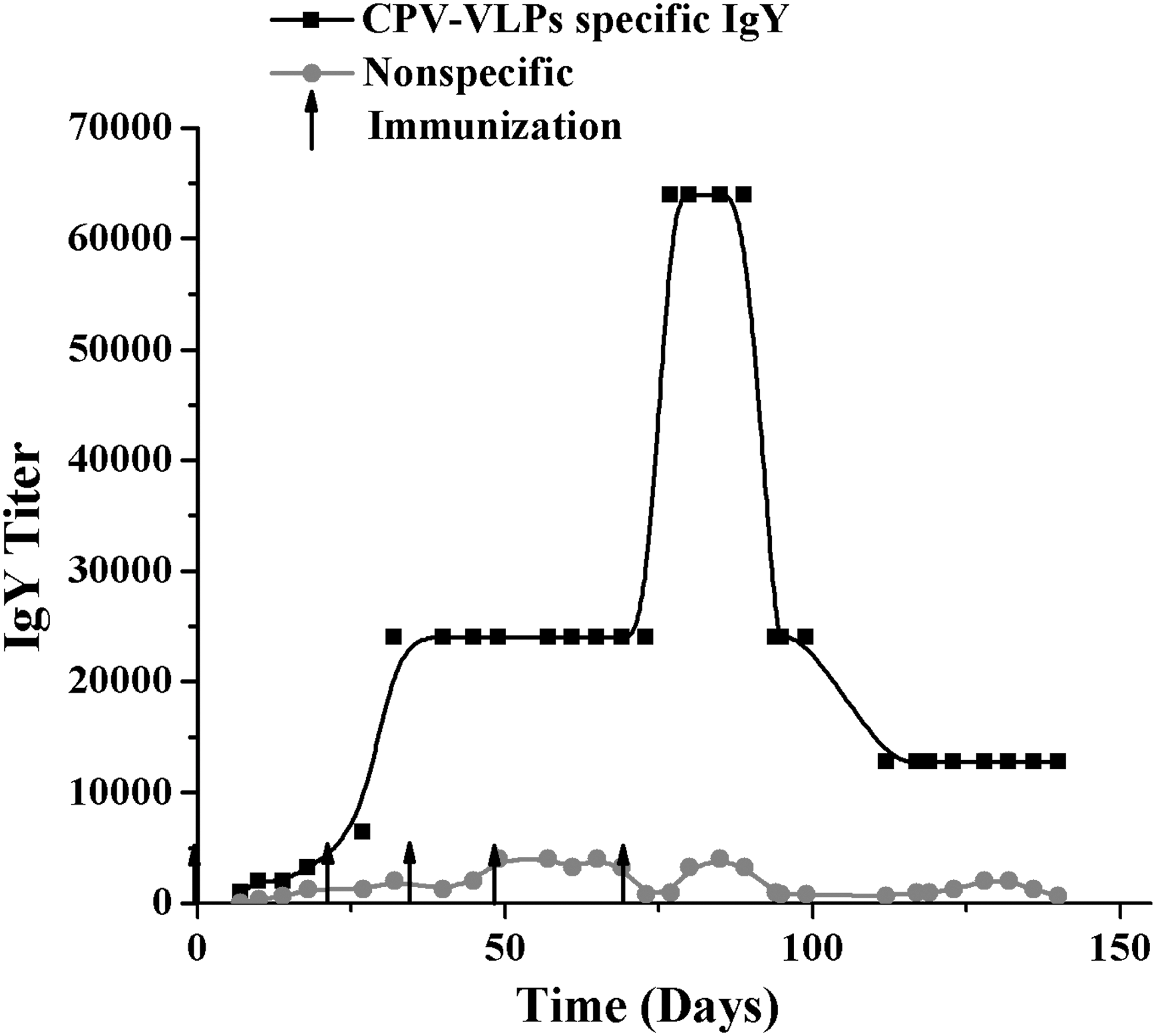
Development of IgY titer against canine parvovirus virus-like particles (CPV-VLPs) protein.
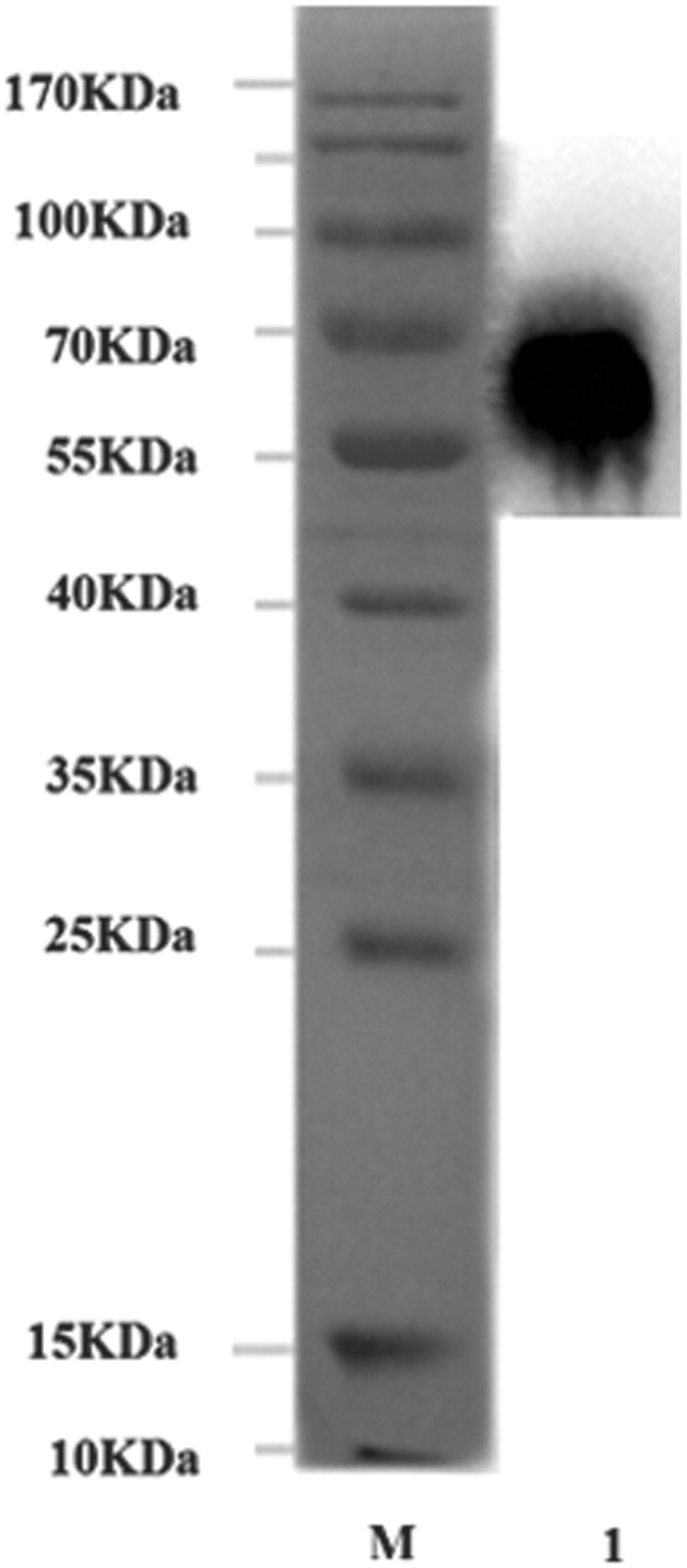
Western blot analysis of the binding activity of the anti-CPV-VLPs IgY. Lane M, marker; Lane 1, CPV-VLPs protein recognized by IgY.
Development of the cutoff values
Indirect ELISA was applied to evaluate the cutoff values of negative samples verified by PCR. The group of CPV-VLPs and positive sample almost showed the same values with a big gap from negative samples (Fig. 4). The average OD450 of negatives values was 0.2133, and the min–max negative values were 0.2044 and 0.2256, respectively, which were regarded as cutoff values of the developed method. Additionally, the value of the positive/negative (P/N) values was 2.03.
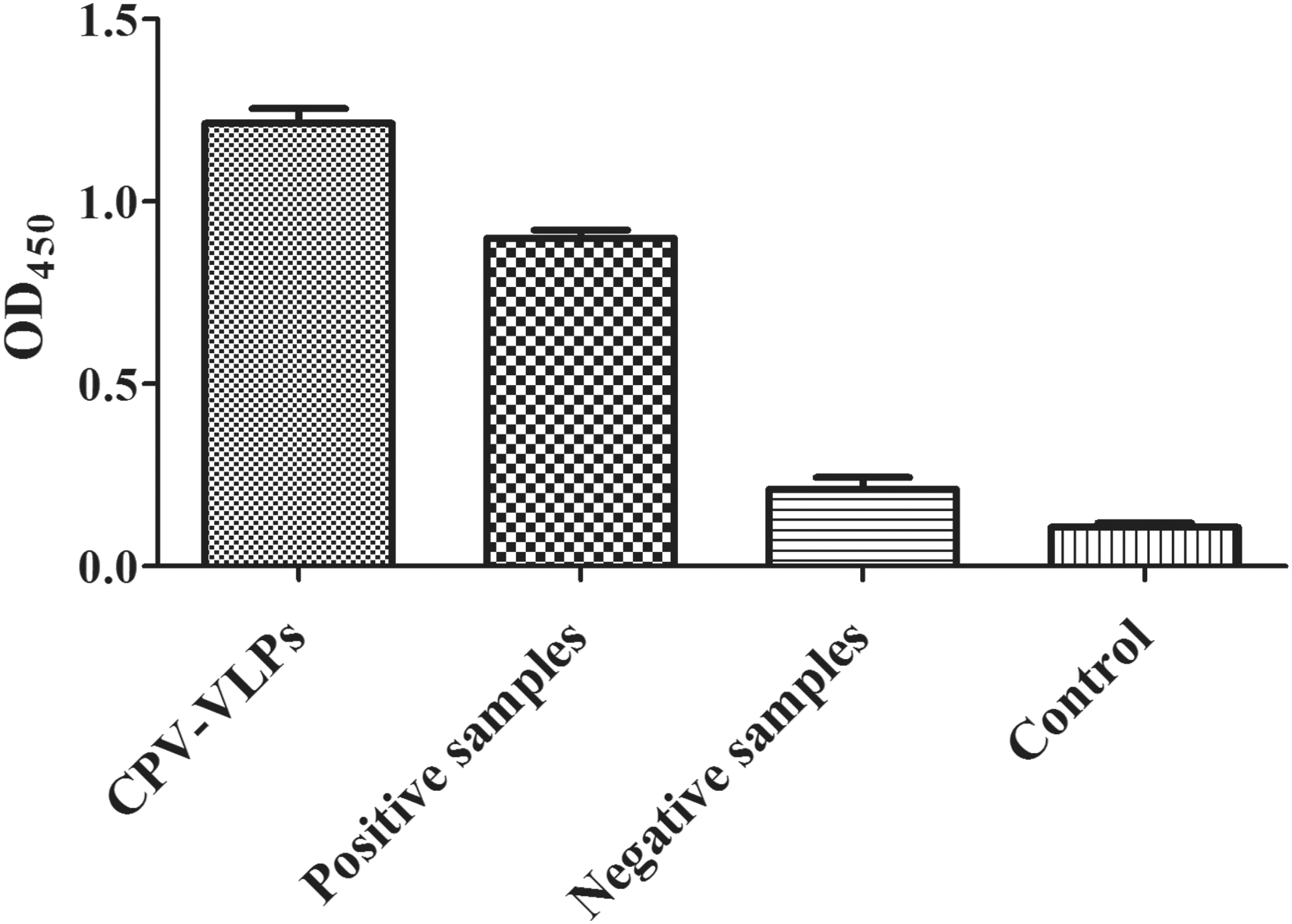
The OD450 values of different samples.
Specificity of ELISA and ICA
The obtained IgY antibody did not show any cross-reactivity with other diarrhea-causing pathogens (Table 1) while demonstrating strong reactivity with CPV-positive samples. The same results were observed with ICA (data not show). These observations suggest that the developed method based on anti-CPV-VLPs-IgY exhibited exquisite specificity (Table 1). The 34 suspected samples were tested in parallel with three different methods. The coincidence of ELISA and ICA with PCR was 97.06% and 94.12%, respectively (Fig. 5 and Table 2).
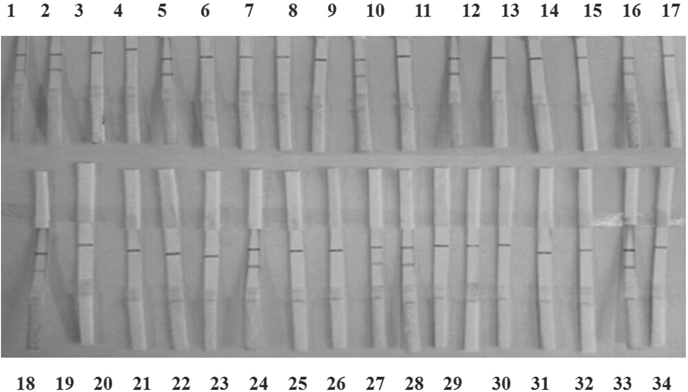
ICA for detection clinical test samples. Among 34 samples, samples 1, 2, 5, 10, 12, 16, 18, 23, 26, 27, and 33 showed positive results. ICA, immunochromatographic assay.
CPV-VLPs, canine parvovirus virus-like particles; IgY, immunoglobulin Y; ELISA, enzyme-linked immunosorbent assay; CCV, canine coronavirus; CDV, canine distemper virus; /, not clear; +, positive detecting result; –, negative detecting result; P/N, the ratio of positive and negative values.
ICA, immunochromatographic assay; PCR, polymerase chain reaction; +, positive detecting results; −, negative detecting results.
Discussion
As an alternative to prokaryotic and eukaryotic protein expressions, VLPs have been considered as a promising choice for antigen preparation and vaccine development. A high level of anti-CPV antibody titer and specificity was observed in hens immunized with CPV-VLPs compared with the chicken that received VP2 protein expressed in E. coli (6). Furthermore, the target IgY showed almost the same reaction values between CVP-VLPs and CPV clinical samples. Both ELISA and ICA did not show cross-reactivity with other diarrheal pathogens. This emphasizes the excellent specificity of anti-CPV-VLPs-IgY. In this study, IgY-based ELISA and ICA confirmed that 29.41% and 32.35% of the samples, respectively, were positive for CPV, whereas PCR revealed only 26.47%. The coincidence of ELISA and ICA with PCR was 97.06% and 94.12%, respectively. To date, several methods have been developed for CPV detection (Table 3). Previously reported methods for CPV detection are not cost-effective, especially for detection at the molecular level with high sensitivity and specificity (5). Because of the high specificity, the anti-CPV-VLPs-IgY-based ELISA and ICA developed in this study could be better choice for CPV immunodiagnostics. Noticeably, the ICA does not require any special equipment or expertise for CPV detection. This advantage could aid veterinarians to confirm CPV infection rapidly in pet clinics. Further investigations are necessary to prepare anti-CPV-VLPs-IgY with a high level of diagnostic efficiency.
LAMP, loop-mediated isothermal amplification; AC-ELISA, antigen-capture ELISA; IEM, immune-electron microscopy; –, standard detection of results; /, not standard detection of results; NG, not given; HA, hemagglutination; IC, immunochromatograph; SAT, slide agglutination test; VI, virus isolation.
Footnotes
Acknowledgments
This work was supported by Key Program for International S&T Cooperation Projects of Shaanxi Province (2012KW-20); Key Construction Program of International Cooperation Base in S&T (2015SD0018) Shaanxi Province, and Ministry of Education and State Administration of Foreign Experts Affairs “Overseas Teacher” project (No. MS2011XBNL057), China.
Author Disclosure Statement
No competing financial interests exist.