
Abstract
Our group has developed a subunit vaccine candidate against Dengue virus (DENV) based on two different viral regions, the domain III of the envelope protein and the capsid protein. The chimeric proteins for each serotype (DIIIC1-4), aggregated with the oligodeoxynucleotide 39 M, form the tetravalent formulation named Tetra DIIIC. Tetra DIIIC induces a protective immune response in mice when it is inoculated by intraperitoneal route. However, if children are the main targets for a DENV vaccine, then a needle-free route of administration should be attractive and advantageous. In this study, we evaluated for the first time, in vivo, a vaccine candidate against DENV based on recombinant proteins using the intranasal route. After three doses of Tetra DIIIC in mice, we measured the humoral immune response against the four DENV serotypes and the corresponding recombinant proteins. Moreover, the functionality of these antibodies was evaluated through a plaque reduction neutralization test. Finally, to assess the cellular immune response induced, we measured the IFN-γ-levels secreted by spleen cells after in vitro stimulation with DENV. The results presented in this study indicate that the intranasal immunization with Tetra DIIIC favors the generation of DENV-specific cell-mediated immunity. On the other hand, the immunization using intraperitoneal and intranasal routes, simultaneously, generate functional antibodies (anti-DIIIC and anti-DENV) and an in vitro response of IFN-γ secretion.
Introduction
D
It is estimated that 390 million DENV infections occur annually in the world, of which 96 million are apparent. Moreover, 3.6 billion people live in areas at risk of DENV transmission (3). Infection with any DENV serotype can cause a broad spectrum of clinical manifestations that goes from self-limiting illness to potentially lethal and/or severe dengue disease (14).
The only DENV vaccine that has been registered to date (Dengvaxia, developed by Sanofi Pasteur Company) had an efficacy of 56.5% and 60.8% in phase III clinical trials in Asia and Latin America, respectively (4,33). Prospective analyses of phase III trials suggest higher rates of hospital admissions accompanying breakthrough dengue infections in vaccinated seronegative individuals, notably among children younger than nine years old. This is probably due to nonprotective infection-enhancing antibodies raised by Dengvaxia (15). Therefore, the development of an effective vaccine inducing long-life immunity is still a need.
Our group has developed a subunit vaccine candidate against DENV based on two different viral regions [the domain III of the envelope (E) protein and the capsid protein (C)] to combine B cell and T cell epitopes. The chimeric protein for each serotype (DIIIC 1-4) forms aggregates of particles of around 50–55 nm, upon incubation with the oligodeoxynucleotide (ODN) 39 M (31). The ODN 39 M contains CpG motifs and stimulates antigen-specific immune responses in mice, monkeys, and humans (12). The tetravalent formulation (Tetra DIIIC) induces protection measured by the viral load in the mouse encephalitis model. Moreover, it was demonstrated that Tetra DIIIC induced neutralizing antibodies and memory cell-mediated immune response with IFN-γ secreting and cytotoxic capacity in nonhuman primates (30).
It is well established that DIII is not the target of neutralizing antibodies in humans upon virus infection (34,35). However, there are several reports that demonstrate the immunogenicity and protective capacity in monkeys of domain III as fusion protein and adjuvanted on Freund’ adjuvant or alum (2,16,30,32) suggesting that such vaccination strategy should not be discounted.
Children are the main targets for a DENV vaccine since they have the highest risk of severe disease. Therefore, a needle-free route of administration should be attractive and advantageous. Several studies have described the use of the intranasal route (IN) for evaluating vaccine candidates against viral infections (1,8,37). In general, this noninvasive needle-free immunization route favors the induction of strong immune responses and requires low amount of antigen (9). Antigens administered through this way are internalized by microfold cells, which are highly efficient in capturing aggregated antigens and transporting them to antigen-presenting cells in local lymphoid organs (24). Moreover, several authors describe strong cell-mediated immune responses in systemic compartments using this immunization route (10,36,37).
Tetra DIIIC has been evaluated using the intraperitoneal administration route in mice and intramuscular, subcutaneous, and intradermal routes in nonhuman primates (30). This is the first study, where it is evaluated, in vivo, a vaccine candidate against DENV based on recombinant proteins using the intranasal route. We evaluated in mice the immunogenicity of Tetra DIIIC administered by intranasal route and coadministered, simultaneously, by intranasal and intraperitoneal routes. Intranasal administrations did not include alum since it cannot be used in mucosal vaccines (22,26) possibly due to its toxicity and so far, there is not a mucosal adjuvant licensed for human use. After three doses we determined the humoral immune response against the four DENV serotypes and the corresponding recombinant proteins. Moreover, the functionality of these antibodies was evaluated through a plaque reduction neutralization test. Finally, to assess the cellular immune response induced, we measured the IFN-γ-levels secreted by spleen cells after in vitro stimulation with DENV.
Materials and Methods
Cells and viruses
African monkey kidney (VERO) cells were received from the National Institute for Biological Standards and Control and were used in the PRNT assay. Cells were grown at 37°C in Eagle's Minimum Essential Medium (MEM) supplemented with 10% heat-inactivated fetal bovine serum.
For the neutralization assays we used the strains Jamaica (DENV-1, AF42562), SB8553 (DENV-2, isolated in 1997 in Malaysia, FM986658), Nicaragua (primary DENV-3 isolate from the Nicaraguan 1994 epidemic, FJ882576), and Dominica 814669 (DENV-4 isolate, AF326573). A concentrated preparation of each virus was used to stimulate mouse splenocytes in vitro. A mock preparation was prepared from the supernatant of uninfected VERO cells following a similar procedure. We used the virus strains Hawaii (DENV-1), New Guinea C (DENV-2), H-87 (DENV-3), and H241 (DENV-4) (6) for antibody detection assay by ELISA as well as for positive control immunogens.
Animals and ethics statement
Female BALB/c (Bc, H-2d) mice (aged 6–8 weeks) were purchased from CENPALAB, and housed in appropriate animal care facilities during the experimental period. Mouse experiments were carried out in strict accordance with the recommendations of the Guide for the Care and Use of Laboratory Animals of the Center for Genetic Engineering and Biotechnology (CIGB), Cuba. The protocols were approved by the Committee on the Ethics of Animal Experiments of CIGB.
Recombinant proteins
The design, cloning, expression, and purification of the recombinant proteins DIIIC 1, DIIIC 2, DIIIC 3, and DIIIC 4 were previously described (31). All the recombinant proteins were purified in the presence of 7 M urea by a procedure consisting of ammonium sulfate precipitation, followed by a cation exchange chromatography, and an immobilized metal ion affinity chromatography. The purified proteins were subjected to an in vitro aggregation procedure as previously described with few modifications (12). It consists of 400 μg of the recombinant proteins incubated with 500 μg of ODN 39 M (5′-atc gac tct cga gcg ttc tcg ggg gac gat cgt cgg ggg-3′) in TE buffer (Tris 10 mM, EDTA 6 mM, pH 7.4). The mixture was incubated for 30 min at 30°C and finally stored at 4°C.
Mouse experiments
Five groups of 15 mice were immunized intraperitoneally on days 0, 15, and 45. Groups 1–3 were immunized with Tetra DIIIC (Table 1). The remaining two groups were used as controls. One of them received three doses of ODN 39 plus alum (Placebo) and the others received 105 FFU of DENV, each of a different serotype. All the formulations used by intraperitoneal route were adjuvanted with aluminum hydroxide (alum) (Alhydrogel) (Brenntag Biosector) at a final concentration of 1.44 mg/mL. The formulations used by intranasal route were not adjuvanted. One month after the last dose, all the animals were bled and splenectomized to study humoral and cellular immune responses.
IP, intraperitoneal; IN, intranasal; DIIIC, domain III-capsid protein; ODN, oligodeoxynucleotide; FFU; focus formation units.
Measurement of humoral immune responses
Detection of anti-DIIIC antibodies
Polystyrene 96-well plates (Costar) were each coated with 5 μg/mL of DIIIC protein during 2 h at 37°C in coating buffer (0.16% Na2CO3, 0.29% NaHCO3, pH 9.5). We used a mock purification run as a negative control. This mock run was from cells transformed with the modified pET28a plasmid without the genes of DIIIC protein (31). The plates were blocked with 5% skimmed milk in coating buffer for 1 h at 37°C. After three washes with PBS containing 0.05% Tween 20 (PBS-T), 100 μL per well of sera from each group were tested by serial dilutions in PBS-T, starting at 1:1,000. Anti-mouse IgG-peroxidase conjugate (Sigma) was added and the plates were incubated for 1 h at 37°C. After washing, substrate solution (0.04% O-phenylenediamine in 2% Na2HPO4, 1% citric acid buffer pH 5, and 30% H2O2) was added, the plates were kept for 15 min at 25°C, and the reaction was stopped through the addition of 12.5% H2SO4. Absorbance at 492 nm was read in a microplate reader (SensIdent Scan; Merck, Germany). The positive cutoff value was set at twice the mean absorbance value of the negative control sera.
Detection of anti-DENV IgG antibodies
An amplified sandwich ELISA system was used to detect anti-DENV IgG antibodies. We coated 96-well polystyrene plates (Costar) for 2 h at 37°C with 100 μL per well of a mixture of anti-DENV human immunoglobulins (IgG) (5 μg/mL) in coating buffer (0.16% Na2CO3 and 0.29% NaHCO3; pH 9.5). Then we blocked the plates with coating buffer containing 5% skimmed milk for 1 h at 37°C and after that we washed them three times with PBS containing 0.05% Tween 20 (PBS-T). The viral antigen solution (100 μL per well) and the negative control antigen solution were then added and the plates were incubated overnight at 4°C. After three washes with PBS-T, 100 μL per well of sera from each group were tested by serial dilutions in PBS-T, starting at 1:1,000. After incubating the plates for 1 h at 37°C and washing them as described above, 100 μL per well of anti-mouse IgG peroxidase conjugate (Amersham Pharmacia) diluted 1:35,000 were added and the plates were incubated for 1 h at 37°C. Then the plates were washed again and afterward we added 100 μL per well of 0.04% substrate O-phenylenediamine in buffer (2% Na2HPO4 and 1% citric acid; pH 5.0). The plates were kept for 30 min at room temperature and the reaction was stopped with 50 μL per well of 2.5 M H2SO4. Absorbance was read at 492 nm in a SensIdent Scan device (Merck, Helsinki, Finland). The positive cutoff value was set at twice the mean absorbance value of the negative control serum.
Plaque reduction neutralization test
Neutralizing antibody titers were measured by plaque reduction neutralization test (PRNT) in VERO cells as previously described (21). Neutralizing antibody titers were identified as the highest serum dilution that reduced the number of virus plaques by 50% (PRNT50). We used murine hyperimmune ascitic fluids specific to DENV serotypes, as positive controls.
Cell culture and viral stimulation
Spleen cells were obtained under aseptic conditions, eliminating erythrocytes by lysis in NH4Cl 0.83%. The cells were washed twice with PBS with 2% FBS (PAA Laboratories) and resuspended at 2 × 10⁶ cells/mL in RPMI 1640 medium (Sigma Aldrich) supplemented with 100 U/mL penicillin, 100 μg/mL streptomycin (Gibco), 2 mM glutamine (Gibco), 5 × 10⁻⁵ M 2-mercaptoethanol (Sigma), and 5% FBS. Finally, 2.5 × 10⁵ cells/well were cultured in 96-well round bottom plates and stimulated with DENV at different multiplicities of infection (MOI) (DENV-1, -2, and -4 were used at MOI = 0.5 and DENV-3 at MOI = 0.1). Concanavalin A (Sigma) was used as a positive control. Three wells were plated for each antigen in all experiments. After a 4-day incubation period, culture supernatants were collected and stored at −20°C
Cytokine detection in culture supernatants by ELISA
The culture supernatants of splenocytes, previously stimulated with each DENV serotype, were analyzed in duplicate to determine their IFN-γ concentration by ELISA, using monoclonal antibody (MAb) pairs (INF-γ; Mabtech, Inc.). The ELISA protocol recommended by the manufacturers was used with slight modifications. The lowest limit of detection for the cytokine was 10 pg/mL.
Statistical analysis
Prism version 5.00 for Windows (GraphPad Software) was used for statistical analysis. Data normality was verified using D'Agostino–Pearson test, and homogeneity of variance was checked using Bartlett's test. Nonparametric analyses of more than two groups were performed using the Kruskal–Wallis test and Dunn's Multiple Comparison test.
Results
The immunogenicity of the tetravalent formulation Tetra 20, based on 5 μg of each of the four DIIIC proteins (the domain III of the E protein fused to the N-terminal of the C protein for each dengue serotype) was evaluated in BALB/c mice. One group received Tetra 20 by the intraperitoneal route (IP), another by the intranasal route (IN), and a third one by both immunization routes, simultaneously (IP/IN). All the animals received three doses and 1 month afterward, we evaluated the humoral and cellular immune responses. The formulations used by intraperitoneal route were adjuvanted in alum, whereas those immunized by intranasal route did not include any adjuvant. Five more groups were included as controls; one received the placebo formulation (containing the same amount of the ODN 39 M included in the tetravalent formulation and adjuvanted in alum as well) and the remaining four received a viral preparation of a different serotype as positive controls.
Humoral immune response
The presence of a humoral immune response was determined by measuring anti-DIIIC IgG antibodies by ELISA. Animals immunized with Tetra 20 (IP) had a positive anti-DIIIC response with antibody titers having a statistically significant difference (p < 0.01) from those of animals receiving Tetra 20 (IN) (Fig. 1). Similar results were achieved in the group of animals immunized using both immunization routes, simultaneously. As expected, none of the animals immunized with the placebo exhibited detectable anti-DIIIC antibodies. Similar with previous results from an evaluation of a tetravalent formulation of DIIIC in mice (30), sera from animals immunized with virus, slightly recognized the homologous DIIIC proteins (Fig. 1).
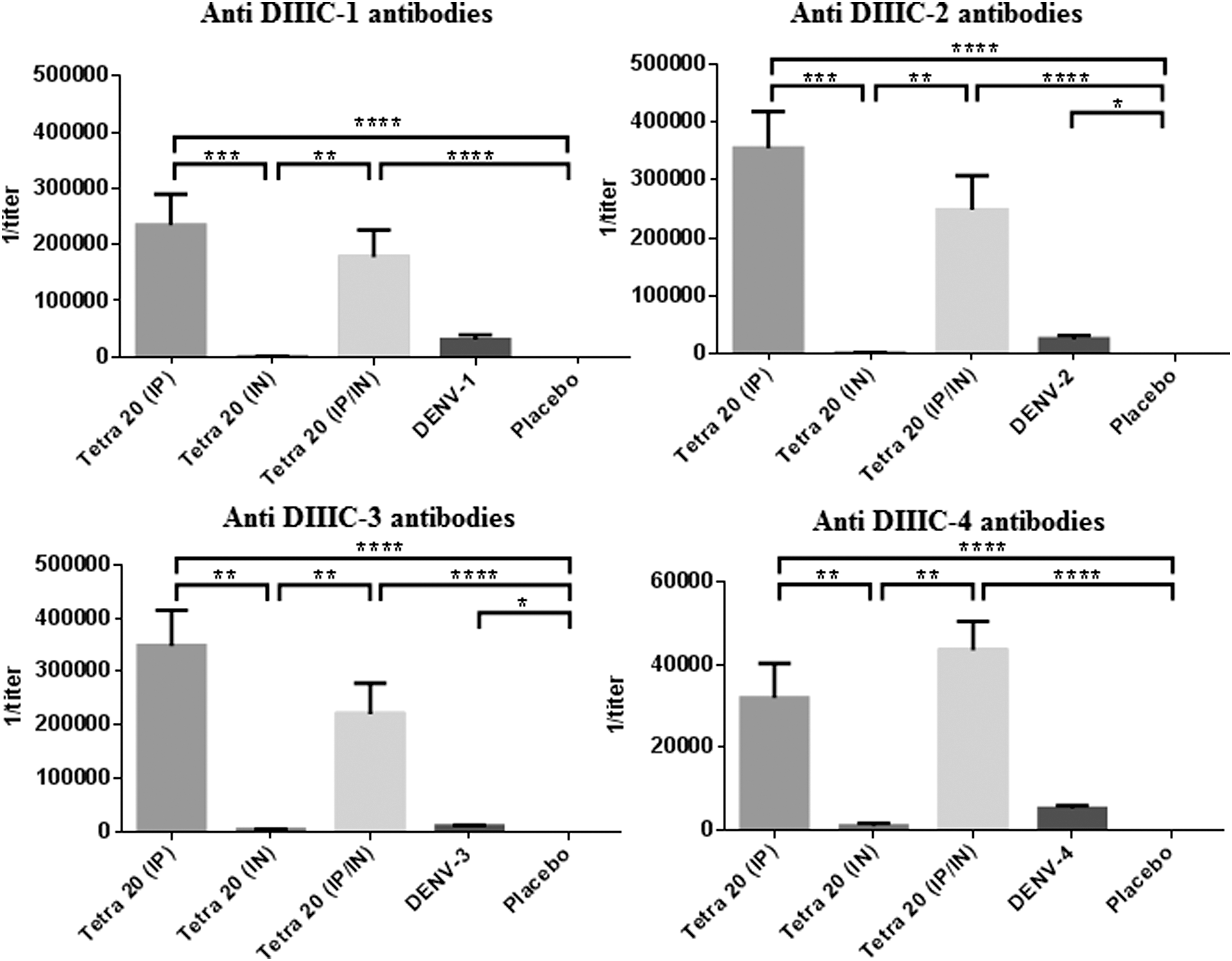
Anti-DIIICs antibody responses induced in mice. Three groups of BALB/c mice were immunized with a tetravalent formulation of recombinant DIIIC proteins by different immunization routes. A negative control group and four positive control groups were immunized with a placebo formulation and infective DENV-1–4, respectively. One month later, the animals were bled and anti-DIIIC IgG antibody titers were measured by ELISA. The charts plot mean with standard error media (n = 9). The statistical analysis was performed using Kruskal–Wallis and Dunn's multiple comparison tests (*p < 0.05; **p < 0.01; ***p < 0.001; ****p < 0.0001). The results are representative of two independent experiments. DENV, dengue virus; DIIIC, domain III-capsid protein; IP, intraperitoneal; IN, intranasal.
The functionality of these humoral immune responses was evaluated by means of two different assays: a capture ELISA using virus preparations as the antigen, and PRNT50. In accordance with the anti-DIIIC IgG response, animals immunized with Tetra 20 (IP) developed anti-DENV-1, -2, and -3 titers higher and having a statistically significant difference (p < 0.05) from those of animals receiving Tetra 20 (IN) (Fig. 2). Similar results were achieved in the group of animals immunized with Tetra 20 (IP/IN) for DENV-1 and -2. For DENV-3, all the animals immunized with Tetra 20 (IP/IN) had a positive antibody response taking into account as cutoff, the double of the mean titers in the Placebo group. Animals immunized with Tetra 20 (IP/IN) received only the half of the amount of antigen by IP route in comparison with the group Tetra 20 (IP). It is possible to think that comparable levels of antibody were due to a synergistic effect between IP and IN routes on humoral response. However, in a recent dose–response study using the intraperitoneal route in mice, we had evidences that 10 μg of Tetra DIIIC induces humoral immune response similar to 20 μg (data pending publication). In general, the highest antiviral response was reached in mice immunized with the homologous viral serotype.
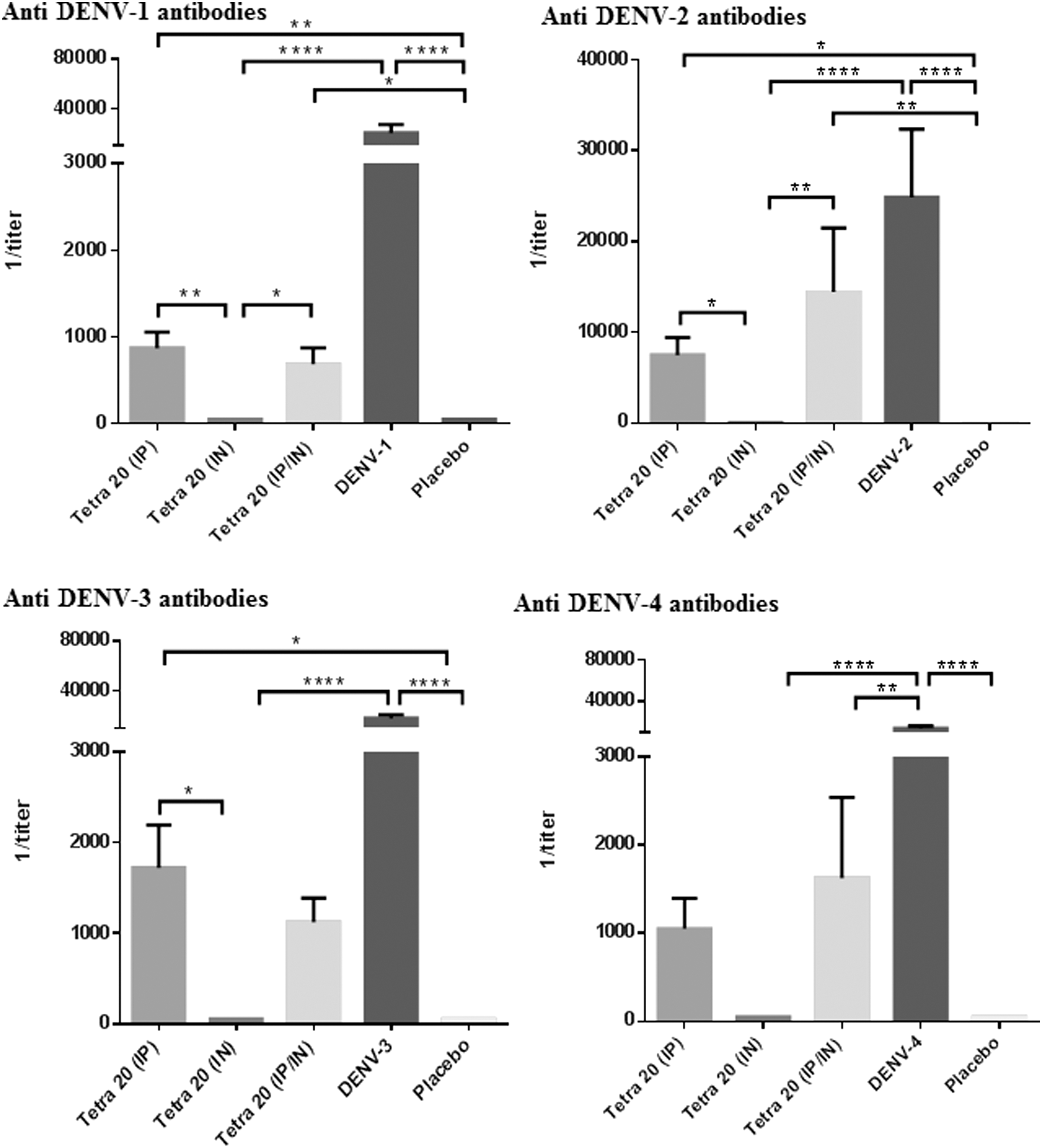
Anti-DENV antibody responses induced in mice. Three groups of BALB/c mice were immunized with a tetravalent formulation of recombinant DIIIC proteins by different immunization routes. A negative control group and four positive control groups were immunized with a placebo formulation and infective DENV-1–4, respectively. One month later, the animals were bled and anti-DENV IgG antibody titers were measured by ELISA. The charts plot mean with standard error media (n = 9). The statistical analysis was performed using Kruskal–Wallis and Dunn's multiple comparison tests (*p < 0.05; **p < 0.01; ****p < 0.0001). The results are representative of two independent experiments.
The functionality of antibodies was evaluated by the in vitro neutralization test. Table 2 illustrates the PRNT50 titers developed by each group. All animals inoculated with Tetra 20 (IP) developed neutralizing antibodies against viral serotypes 1, 2, and 3 with geometric mean titers (GMT) higher than 100. The group immunized with Tetra 20 (IP/IN) developed neutralizing antibodies against DENV-1, 2, and 3, with GMT of 66, 69, and 131, respectively. The lowest response was detected against DENV-4 with less than 50% of positive animals in all immunized groups. The immunization with viral preparations in the control groups did generate an antibody response that bound and neutralized the four viral serotypes (Table 2).
Neutralizing antibody titers are the highest serum dilution that resulted in a 50% reduction in the number of plaques produced by the viruses.
GMT, geometric mean titer; % Pos., Percentage of animals with titers ≥10; DENV, dengue virus.
Cellular immune response
To evaluate the cellular immune response, we measured the IFN-γ levels secreted by spleen cells after in vitro stimulation with DENV. Splenocytes from animals receiving viral preparations were stimulated with the homologous virus, whereas splenocytes from the groups receiving the tetravalent formulation or the placebo were stimulated with each DENV serotype. Immunization with the Tetra 20 (IP) formulation generated detectable IFN-γ secretion against DENV-1 and -2 in more than 60% of animals (Fig. 3). Moreover, animals from group Tetra 20 (IN) showed positive IFN-γ-secreting cell response in 29%, 62.5%, and 100% of animals against DENV-3, -1 and -2, respectively. More than 40% of mice immunized by both immunization routes, simultaneously, showed positive IFN-γ-secreting cell response against DENV-1, 2, and 3. We did not detect any response against DENV-4. As expected, a very low response was detected in the animals receiving the placebo, which contains the highest evaluated dose of ODN 39 M.
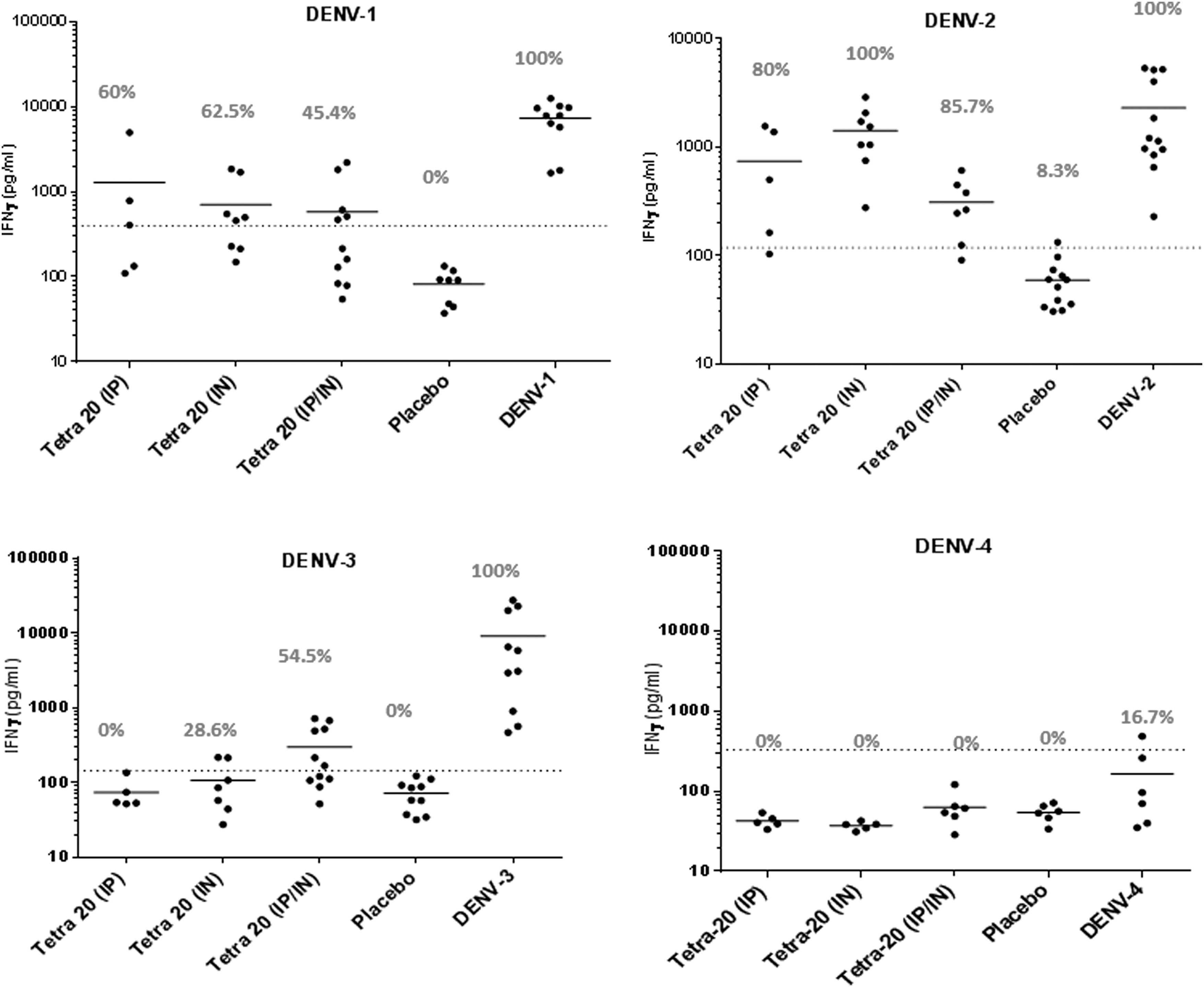
Cell-mediated immune responses induced by Tetra-DIIIC in mice. Three groups of BALB/c mice were immunized with tetravalent formulations of recombinant DIIIC proteins by different immunization routes. A negative control group and four positive control groups were immunized with a placebo formulation and infective DENV-1–4, respectively. One month after the last dose, splenocytes from the animals were stimulated in vitro with each DENV, and we determined the IFN-γ concentrations in the resulting culture supernatants by ELISA. The charts plot mean (n = 5–12). The dashed line indicates the cutoff value (two times the average of the placebo group). Percentages of animals with positive IFN-γ-secreting cells response are shown.
Discussion
The tetravalent formulation Tetra DIIIC inoculated by intraperitoneal route, induces a protective immune response in mice based on neutralizing antibodies and IFN-γ secretion by in vitro-stimulated splenocytes (30). The results of this study indicate that intranasal immunization of Tetra DIIIC, favors the generation of cell-mediated immunity to the detriment of humoral immune response. However, the coimmunization using intraperitoneal and intranasal routes, simultaneously, generate anti-DIIIC, functional anti-DENV antibodies, and an in vitro response of IFN-γ secretion.
To our knowledge, this is the first study in vivo on the evaluation of recombinant proteins against DENV using the intranasal route. Sim et al. (27) evaluated the ability of a recombinant Lactococcus lactis strain producing the envelope domain III from DENV-2 to trigger a systemic antibody response upon nasal or oral administration to BALB/c and C57BL/6 mice. Those results showed that the antibody response depended on the route of administration and on the mouse strain; however, low titers of neutralizing antibodies were detected (27). In another study published recently, Nantachit et al. (23) demonstrated that the domain III of dengue serotype-3 E protein loaded into trimethyl chitosan nanoparticles stimulates a strong innate antiviral response on primary human nasal epithelial cells, used as an in vitro model for nasal responses (23).
Unexpectedly, in the present study animals immunized with Tetra DIIIC only by intranasal route did not develop detectable IgG antibody response in sera. However, several reports have described the capacity of the intranasal route to induce humoral and cell-mediated immunity in systemic compartments (10,36,37). Lobaina et al. (19) evaluated a recombinant capsid protein from Hepatitis B virus employing different mucosal and parenteral immunization routes. Authors detected the highest IgG response in sera from animals intranasally immunized without adjuvant. Moreover, these mice showed the highest ratio IgG2a/IgG1 and the highest lymphoproliferative response, 2 months after the last immunization (19). In a more recent study, Woo et al. (36) evaluated whether intranasal immunization with the recombinant protective antigen (rPA) of Bacillus anthracis induced immunological memory responses in the mucosal and systemic compartments (36). As a result, a significant induction of specific memory B cells was observed in spleen, cervical lymph nodes, and lung after booster immunization. Furthermore, intranasal immunization with rPA plus cholera toxin remarkably generated effector memory CD4+ T cells in the lung. Different antigen dose and immunization schemes could influence on these results.
One important issue of intranasal vaccination is antigen retention period in the nasal cavity to ensure an efficient uptake of antigens before being cleared. Due to the aggregated nature of Tetra DIIIC, we expected a good efficiency of uptake by microfold cells as it has been demonstrated for similar antigens (23). Another concern is that a mucosal adjuvant could be needed for an IN delivery. In this study, the administrations by IN route did not include alum. We hypothesized that the presence of the ODN 39 M (12) could be sufficient to enhance the immunogenicity of Tetra DIIIC. However, the present is a preliminary study to evaluate the immunogenicity induced by our vaccine candidate using the intranasal route and in future studies we will have to deal with these issues.
Limitations of our study are that neither the IgA antibody response, nor the humoral response in mucosal fluids, was determined. However, both aspects do not have relevance for dengue infection, since it is well known that DENV transmission occurs through the bite of an infected mosquito, and innate and adaptive immune responses on skin and blood are the main antiviral barriers.
Accordingly, with a previous study evaluating in mice the Tetra DIIIC formulation (30), the lowest immune response was achieved against DENV-4. The low immunogenicity against DENV-4 has been reported by several authors after the evaluation of vaccine candidates based on recombinant proteins (7,18,29) and even for vaccine candidates based on live attenuated viruses (25,28), suggesting this serotype is poorly immunogenic. Nevertheless, the protective capacity against DENV-4 induced by Tetra DIIIC has been demonstrated in mice using the intraperitoneal route (30).
As we mentioned above, the immunization using intraperitoneal and intranasal routes, simultaneously, generated functional antibodies and IFN-γ secretion by spleen cells. The costimulation of mucosal and systemic immunity has been previously demonstrated in mice using an HBV therapeutic vaccine candidate (20) and a multiantigenic formulation against HIV (17). In both studies, high levels of serum IgG and IFN-γ-secreting cells were achieved. Unlike our study, these authors evaluated subcutaneous, intradermal, and intramuscular as systemic immunization routes instead of the intraperitoneal one. Although the last one is not a vaccine immunization route feasible in humans, it has been successfully employed in the assessment of vaccine candidates against DENV in mice (11,18,30). Further studies will focus on the evaluation of Tetra DIIIC in mice and nonhuman primates using different combinations of immunization routes suitable for human use and its protective capacity.
As we said above, in this study, the intranasal immunization favored the cellular immune response instead of humoral immune response. Neutralizing antibodies have been long considered the main component of a protective response against dengue; however, they can exacerbate disease severity when present at subprotective levels (13,38,39). Therefore, this study points that intranasal route will be more attractive using antigens, inducer of cellular immune response, like that developed by our group that is based only on the capsid protein of dengue virus (Tetra NLP) (11).
In conclusion, data presented in this article suggest that the combination of systemic and mucosal administration routes could be a promising strategy to increase the immunogenicity induced by vaccine candidates against DENV based on recombinant proteins.
Footnotes
Acknowledgment
The authors thank Dr. Eduardo Martínez-Montes for his advice and help in the preparation and revision of the article.
Author Disclosure Statement
No competing financial interests exist.