
Abstract
Preexisting neutralizing antibody (NAb) against human adenovirus serotype 5 (AdHu5) can reduce the immunogenicity of AdHu5 vector-based vaccine, thus inhibiting the host's immune response and utility of other homologous vectors. Common marmoset (Callithrix jacchus), a small new world primate, has attracted considerable attention for its potential as a preclinical research model of vaccine development. However, the prevalence of anti-AdHu5 NAb activity in common marmosets bred in China remains unknown. A recombinant adenovirus expressing luciferase and Zs Green reporter genes were constructed to detect NAb against rAdHu5 by flow cytometry (FCM) and chemiluminescence (CL) assay. Five of 25 marmosets (20%) presented AdHu5 NAb detectable by FCM. Four animals had low titer (1/16), while the fifth one reached 1/64. While by CL assay, 7 of 25 (28%) marmosets were anti-AdHu5 NAb positive. Four animals, two of whom were negative by FCM, also had low titer NAb (1/16), suggesting assay discrepancy at low levels. Two marmosets, 1/32 titer by CL, were at 1/16 by FCM. A single animal showed a high titer with both assays (1/128 and 1/64 by CL and FCM, respectively). The CL method was simpler, more sensitive, accurate, and stable. The low prevalence of preexisting anti-AdHu5 NAb in marmosets provides important background information on the feasibility and applicability of using marmosets as a preclinical research model for vaccine development.
Introduction
Adenoviruses (Ads), nonenveloped icosahedral viruses, have gained considerable attention as vaccine vectors since they are safe, stable, and easy to produce and induce both innate and adaptive immune responses in mammalian hosts (15). Recombinant human adenovirus serotype 5 (rAdHu5) vector-based vaccines have been proven to elicit vigorous cellular immune responses in animal models (14). Candidate rAdHu5 vector has been utilized into clinical trial of HIV, rheumatoid arthritis, and other diseases (13). However, a major limitation of this procedure is the high titer of AdHu5 neutralizing antibody (NAb), which has been found in 40–60% of adults residing in the United States and more than 90% in humans residing in Sub-Saharan Africa (8). Preexisting antibody can reduce the immunogenicity of rAdHu5 vector-based vaccines in mice, rhesus monkeys, and humans (2,11,12), thus inhibiting the host's immune response and the utility of other homologous vectors. Therefore, it is critically important to examine the anti-AdHu5 NAb activity before the administration of rAdHu5 vector-based product.
In recent years, common marmoset (Callithrix jacchus), a small new world primate, has attracted considerable attention as being widely used for studying aging, reproduction, neuroscience, toxicology, and infectious disease (9). Common marmoset belongs to the Callitrichidae family, native to the Atlantic coastal forests of northeastern Brazil (10). Adult common marmoset weighs 350–400 g and has gentle disposition and a compressed life span compared to other nonhuman primates (NHPs), making it suitable for rapid breeding and experimental requirements (3).
To construct and evaluate HCV vaccine candidates with promising adenoviral vectors, such as simian-derived adenoviral vectors (1), rare serotype adenoviral vectors or modified rAdHu5 vectors to mask AdHu5 neutralizing epitopes, evaluation of the immunogenicity, and tolerance of target genes of rAdHu5 vector in the marmoset model should first be done. Before this, the prevalence of preexisting anti-AdHu5 NAb activity in common marmosets bred in China should be done. It will provide important background information for the feasibility and utility of the common marmoset as a preclinical research model of vaccine development.
Materials and Methods
Animal serum samples
Animal serum samples were obtained from 25 common marmosets (Callithrix jacchus) with an average weight of 350 g imported from Tianjin Medical University and individually fed in Laboratory Animal Research Center of Nanfang Hospital, Guangzhou, China. A part of the animals was used for infection with HCV/GBV-B chimeras in our previous studies (7,22); serum samples were collected specifically for this study. Animal experimentation and blood sample collection were approved by Southern Medical University (SMU) Animal Care and Use Committee (permit numbers: SYXK(Yue)2010–0056). All animal care and procedures were in accordance with national and institutional policies for animal health and well-being. All efforts were made to minimize suffering of animals.
Recombinant adenovirus production
Luciferase and Zs green report genes were amplified from pHAGE-CMV-Luc-IZs Green plasmid stored in our laboratory, using primers Luc-Zs-F 5′-CCGGAATTCGCCACCATGGAAGACGCCAAAAACATAAAG-3′ and Luc-Zs-R 5′- CTAGCTAGCTCAGGGCAAGGCGGAGCCGGA-3′ with EcoRI and Kozak in the forward and NheI in the reverse primer. The amplification started with incubation for 2 min at 98°C, then 30 cycles with 20 s at 98°C, 20 s at 55°C, and 3 min at 68°C, extended for 10 min at 68°C, and finished at 4°C. The amplified fragment was inserted into pDC315 plasmid (provided by Dr. JH Zhou, State Key Laboratory of Veterinary Biotechnology, Harbin Veterinary Research Institute of Chinese Academy of Agricultural Sciences) forming shuttle plasmid pDC315-Luc-IZs Green that was afterward cotransfected with pBHGlox(delta)E1,3Cre (provided by Dr. JH Zhou) into 293A cell to package recombinant adenovirus rAdHu5/Luc/Zs Green. Virus was plaque-purified, analyzed for Zs Green expression, amplified in T75 flasks, purified by CsCl gradient ultracentrifugation, and dialyzed into phosphate-buffered saline (PBS) containing 5% sucrose. Purified viruses were stored at −80°C. Virus infectious titer was determined by TCID50. Specific infectivity was assessed by PFU assays. To identify rAd5/Luc/Zs Green after purification, polymerase chain reaction (PCR) or reverse transcript PCR (RT-PCR) was used to amplify Luc-IZs Green from genomic DNA and total RNA. The expression of the protein was verified by cell immunohistochemical experiments using anti-AdHu5 positive human serum (screened in the laboratory from Maoming Blood Center).
Virus neutralization assays
The optimal adenovirus virus infectious titer was determined by detecting the expression efficiency of luciferase and Zs Green reporter gene when doing the neutralization experiments. AdHu5 NAb titers in marmosets' serum samples were assessed by neutralizing assays based on Luc and Zs Green reporter genes. Background parameters were equalized by wells containing cells only; maximum parameter was determined by wells with cells and rAdHu5/Luc/Zs Green, without serum. Neutralization titers were defined as the maximum serum dilution that neutralized 50% of Luc or Zs Green activity (6). Titers equal to or higher than 16 were scored as positive for the presence of AdHu5 NAb.
Luc reporter system
Briefly, rAdHu5/Luc/Zs Green at the infection titer of 1.1 × 10⁸ PFU/mL was mixed with twofold serial dilutions of serum (ranging from 1:16 to 1:32,768) in 100 μL reaction mixture volume, incubated for 1 h at 37°C and 5% CO2, then added into 2 × 10⁴ 293A cells per well in triplicate in 96-well plates. Following a 24-h incubation, Luc activity in the cells was measured using the Bright-Glo™ Assay Reagent (Promega) with a Victor 1420 multilabel counter (Perkin Elmer).
Zs Green reporter system
For experiments with Ads carrying the Zs Green transgene, the supernatant medium was aspirated and 200 μL of PBS was added, and then fluorescence was measured with a fluorescent plate reader.
Statistical analysis
The data were analyzed using the statistical package SPSS version 19.0. The consistency of the results of two different NAb detection methods was analyzed using Fisher's exact probability method. p Values <0.05 were considered statistically significant.
Results
Construction, package, and identification of recombinant adenovirus
The adenovirus shuttle vector pDC315-Luc-IZs Green containing luciferase (1,653 bp) and Zs Green reporter (699 bp) genes linked with internal ribosome entry site to equally translate the two open reading frames (ORFs) were conventionally constructed and preliminarily identified to express labeled proteins in eukaryotic 293A cells using the method of transient transfection. By cotransfection of adenovirus shuttle vectors and packing bone plasmid to 293A, infectious recombinant adenovirus rAd5/Luc/Zs Green was successfully achieved (Fig. 1A). The infection titer finally reached 2.2 × 10¹² PFU/mL after three times plaque formation, large-scale amplification, and purification. To identify the recombinant adenovirus, Luc-IZs Green gene from genomic DNA and total RNA was successfully amplified by PCR or RT-PCR. Virus structural protein could express in 293A cell detecting by cell immunohistochemical experiments using anti-AdHu5 positive human serum (Fig. 1B). The 293A cells were infected with a serial dilution of rAd5/Luc/Zs Green to get the optimal expression efficiency of Luc and Zs Green report gene. Respect to Zs Green expression, when the infection titer was 2.2 × 10¹⁰, 2.2 × 10⁹, and 2.2 × 10⁸ PFU/mL, the fluorescence of cells increased too fast within relatively short period of time, while at the titer of 2.2 × 10⁷ PFU/mL, the protein expressed was relatively stable and convenient to be observed (Fig. 1C). At the same time, the Luc activity was detected at different time points. As shown in Table 1, the expression efficiency of Luc increased slowly with time. When the titer reached 2.2 × 10⁸ PFU/mL, the Luc expression was more stable. Therefore, the overall optimal adenovirus virus infection titer was between 2.2 × 10⁷ and 2.2 × 10⁸ PFU/mL. A titer of 1.1 × 10⁸ PFU/mL was chosen in the following neutralization experiments.

Package and identification of recombinant adenovirus.
Determination of Relevant Luciferase Activity Unit of Luciferase Gene Expression Efficiency
293A cells were infected with a serial dilution of rAd5/Luc/Zs Green to get the optimal expression efficiency of Luc report gene. The Luc activity was detected at different time points.
AdHu5 NAb titers in marmosets' sera assessed by Flow cytometry based on Zs Green reporter gene
A total of 25 common marmosets' serum samples were serially diluted from 1:16 to 1:32,768 and neutralized by recombinant adenoviruses in 293A cells. Flow cytometry (FCM) was used to detect Zs Green protein expressed by the recombinant adenovirus rAd5/Luc/Zs Green. After 1 h neutralization and 24 h incubation, cell fluorescence expression rate was over 90% in negative controls without serum samples visualized by fluorescence microscope. NAb in the sera could prevent entry of rAd5/Luc/Zs greens into the cells, thereby inhibiting Zs green expression and inducing cell fluorescence rate decrease. Figure 2 showed the percentage of 293A cells with green fluorescence compared to virus control detected by FCM in all marmosets. Neutralization titers were defined as the maximum serum dilution that neutralized 50% of Zs Green activity. Figure 2 showed that marmoset CJ16, CJ17, CJ22, CJ28, and CJ31 were NAb positive for adenovirus type 5. Four marmosets CJ16, CJ17, CJ28, and CJ31 scored a NAb titer of 1/16, while one marmoset CJ22 reached a titer of 1/64.
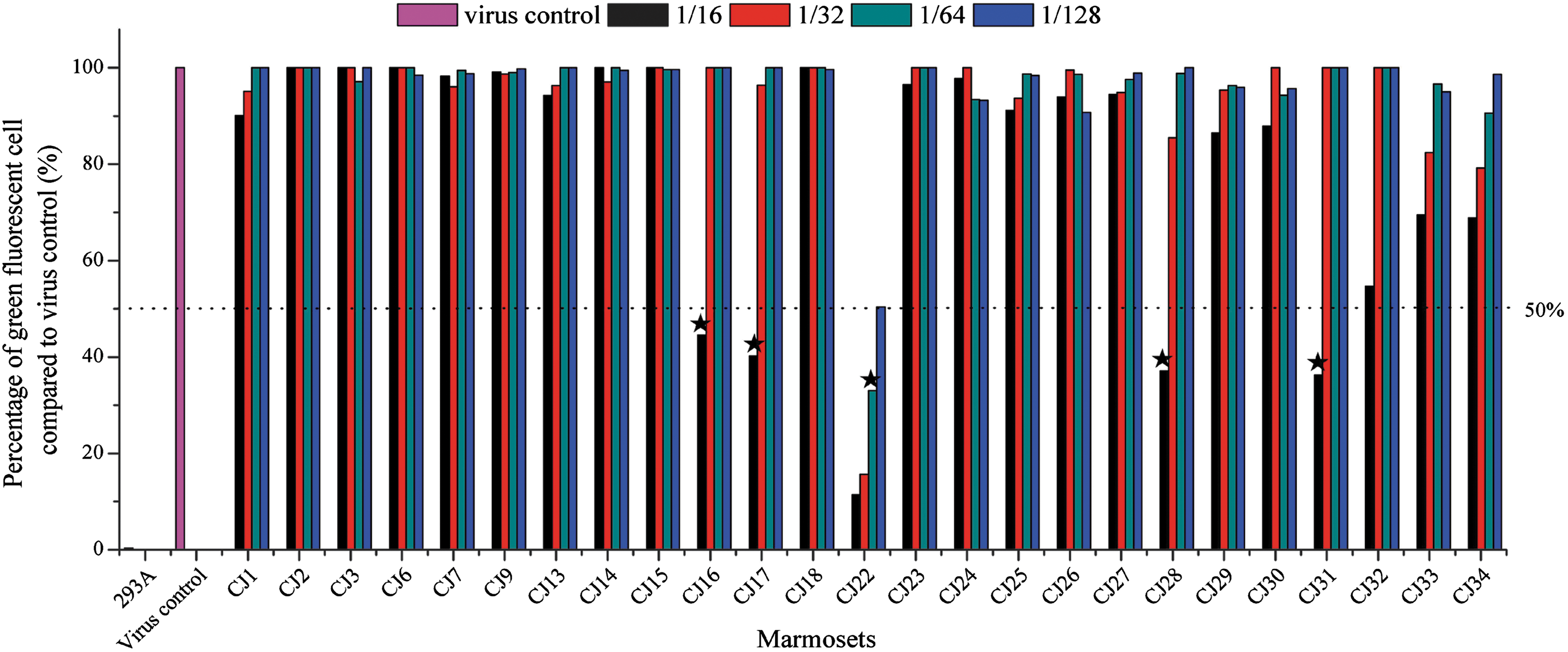
Percentage of green fluorescent cell compared to virus control by FCM and titer judgment. Neutralization titers were defined as the maximum serum dilution that neutralized 50% of Zs Green activity. The dot line indicated 50% of green fluorescent cells of virus control. The bars below dot line with maximum dilution were defined as neutralization titer (indicated with star). FCM, flow cytometry. Color images are available online.
Overlapped histograms of five NAb positive marmosets' sera were drawn (Fig. 3). The red histogram showed that Zs Green was not expressed, while the blue histogram displayed virus infection controls of the Zs Green expression. More than 80% of the cells are located in the Zs Green expression peak. With gradual increase of the sera dilution ratio, the number of cells expressing Zs green increased, and the peak titer migrated from left to right.
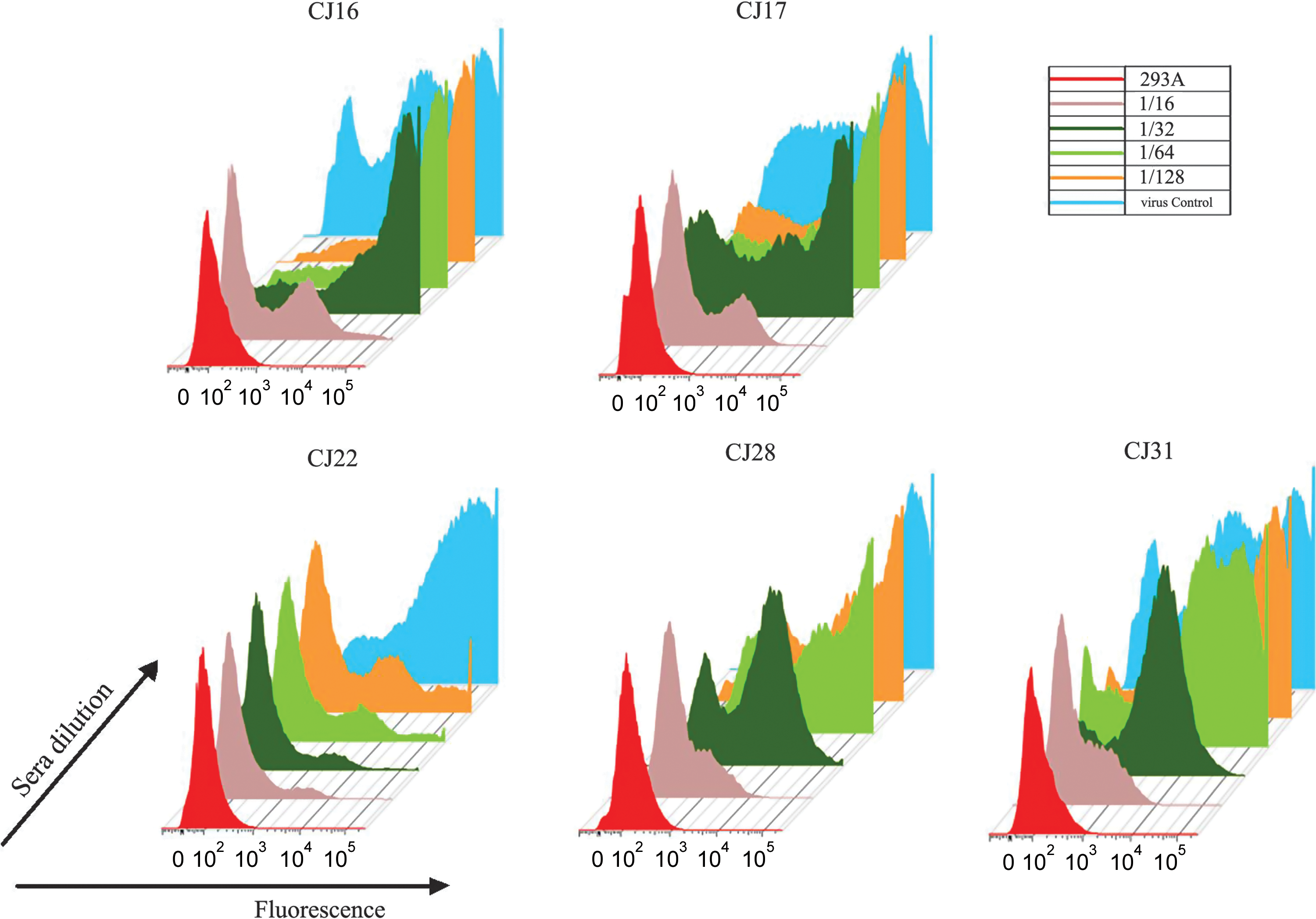
Overlapped histograms of neutralizing antibody positive marmosets' sera. The red histogram showed that Zs Green was not expressed in 293A cells. The blue histogram displayed virus infection controls of the Zs Green expression. With gradual increase of the sera dilution ratio, the number of cells expressing ZS green increased, and the peak titer migrated from left to right. Color images are available online.
AdHu5 NAb titers in marmosets' sera assessed by CL assay based on Luc reporter gene
For the other method, after 24 h neutralization, CL assay was carried out to detect relevant luciferase activity unit (RLAU) determined by Luc expression inhibition. RLAU was compared to virus control in Figure 4. Neutralization titers were defined as the maximum serum dilution that neutralized 50% of Luc activity. Figure 4 showed that seven marmosets of 25 (28%) were anti-Ad5 neutralizing antibody positive (NAb+). Four of the anti-Ad5 NAb+ marmosets CJ16, CJ17, CJ24, and CJ29 showed low titer (1/16). Two marmosets CJ28 and CJ31 were 1/32, and another one CJ22 was 1/128.
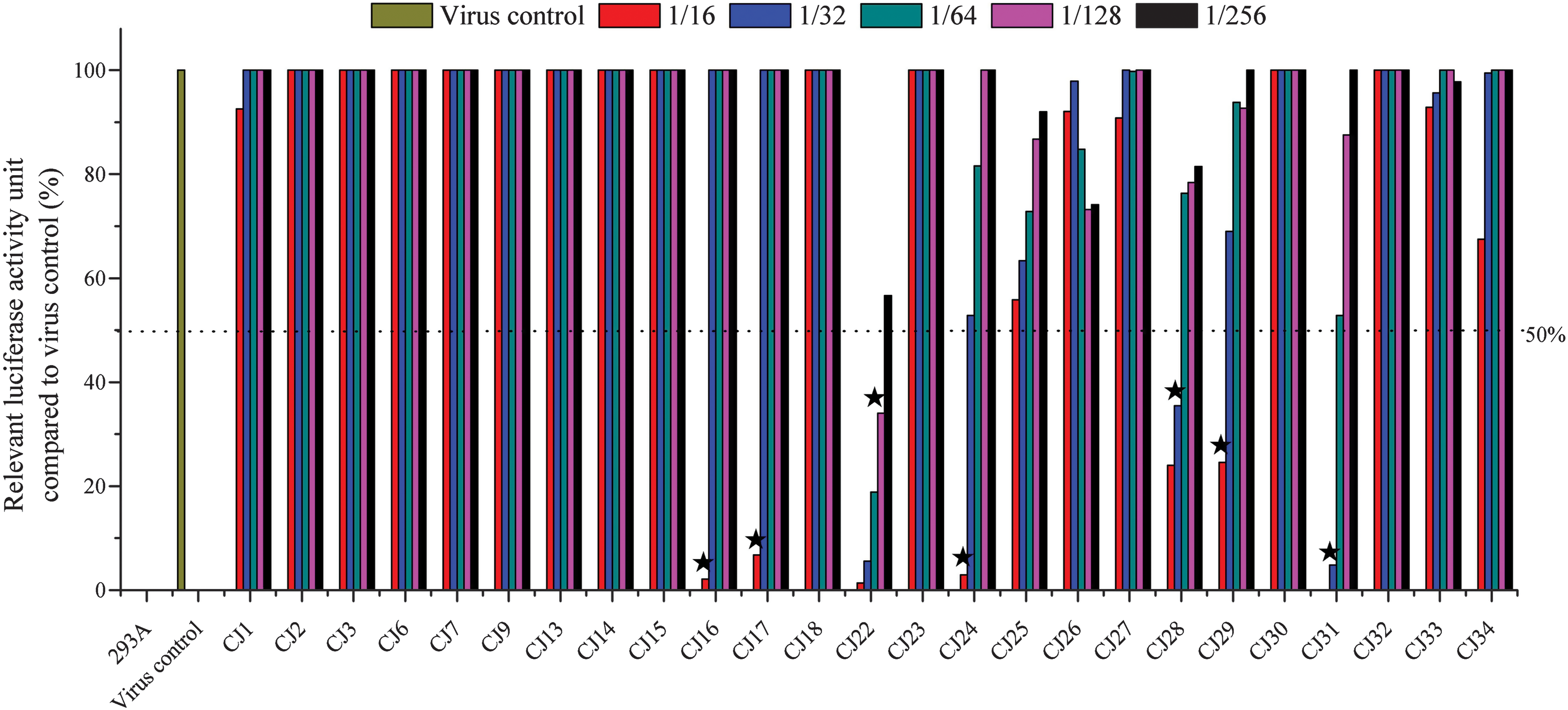
Relevant luciferase activity unit compared to virus control in neutralizing assay and titer judgment. Neutralization titers were defined as the maximum serum dilution that neutralized 50% of Luc activity of virus control (indicated with star). Color images are available online.
Comparison of rAdHu5 based Luc and IZs green neutralizing assays
The detection of NAb in 25 marmosets by two assays is presented in Figure 5. Statistical analysis showed that the results of CL and FCM are generally consistent (p < 0.01, Kappa coefficient = 0.783) with slight differences. Marmoset CJ16 and CJ17 had a completely consistent NAb titer of 1/16 with both methods. Compared with FCM, CL detected higher titers of NAb in CJ22, CJ28, and CJ31. Results for CJ24 and CJ29 are positive with titer of 1/16 by CL but negative by FCM.
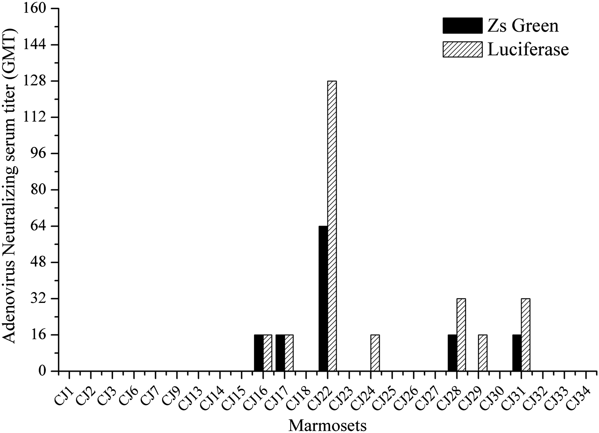
Comparison of different assays in determining AdHu5 neutralizing serum titers.
Discussion
AdHu5 achieved success as vaccine vector in preclinical trials despite having the disadvantage of preexisting immunity (13,21). It is estimated that 30–100% of the adult human population carry specific NAbs to AdHu5 in Europeans, North Americans, Asians, or Africans (18). In epidemiological studies performed in China, seroprevalence of AdHu5 in adults was 72–74.2% (16 –18). AdHu5 vector vaccine can be evaluated for immunogenicity and tolerance of the immunogen that induces the protective immunity in NHP models, which has genetic homology of up to 99.9% with human beings. It might then require changing for other promising adenoviral vectors such as simian derived adenoviral vectors (1), rare serotype adenoviral vectors, or modified rAdHu5 vectors masking AdHu5 neutralizing epitopes. Thus, it is necessary to detect the level of AdHu5 NAb in the NHP that was proposed to be the model in China before embarking into a clinical trial.
We previously reported that HCV/GBV-B chimera infected marmosets might constitute a small primate model suitable for evaluation of vaccines against HCV infection (7,22). In Brazil, 5 of 16 tested marmosets had detectable antibodies to AdHu5 with a conventional assay, but none of the five was tested from the United States (5). The most recent ancestors common to all marmosets used in the present study were 15 paired animals introduced from Australia bred locally for three to four generations before the generation we utilized. No data were generated for those marmosets.
As the use of adenoviral vectors in gene therapies is increasing, a simple, sensitive, accurate, and reproducible titer method needs to be developed. Among multiple methods to titrate antibodies to recombinant adenoviral vector, we chose FCM and CL assay to semiquantify the inhibition of reporter gene expression after deficient adenovirus reacted with antibodies in the serum. By FCM, 5 marmosets out of 25 animals (20%) were positive for NAb to AdHu5. Four marmosets scored a titer of 1/16, while a single marmoset reached a titer of 1/64. With the CL assay, 7/25 marmosets (28%) carried anti-AdHu5 NAb. The majority carried low titer (1/16), two of which being negative by FCM, suggesting higher sensitivity of the CL assay. Two animals presented a 1/32 titer by CL assay, but of 1/16 by FCM. Another animal had a titer of 1/128, but 1/64 by FCM. As seen in Figures 2 and 4, the Luc activity units increased steadily along with the serum dilution, while the percentage of green fluorescent cell increased sharply, suggesting higher accuracy and stability of the CL assay. From assay comparison, the CL method appears simpler, more sensitive, accurate, and stable. From the results of our two tests for measuring NAb, seroprevalence of preexisting NAbs to AdHu5 in common marmosets bred in China was relatively low (28%) and at low level (1/16). Few marmosets presented a titer ranging between 1/32 and 1/128.
AdHu5 NAb+ marmosets were of relatively low prevalence and level, which were conducive to construct vaccine candidate based on AdHu5 vector. A preclinical evaluation of the induced immunogenicity of such vector in marmosets should be examined. Low or no NAb against AdHu5 in marmosets also gave a hint of low prevalence of other human adenovirus serotypes, which might also have importance in preparation and application of other human adenoviral vaccines. At the same time, the preexistence of human adenoviral antibody in primates suggests that it may be related to human adenovirus infection transmitted through animal contact. It has been found that simian adenoviruses had cross-species transmission to human beings, for example, human adenovirus type 4 was a typical case, which was a recombinant virus between simian and human adenoviruses (4,19). The NAbs against simian adenoviruses in African residents might be more common than other people (20). The simians in Africa may have higher human Adenovirus NAb prevalence. The potential risk of cross-species transmission suggested the importance of background information for the development of recombinant human adenovirus vaccine.
Footnotes
Compliance
All institutional and national guidelines for the care and use of laboratory animals were followed.
Authors' Contributions
C.L. and T.L. designed research; Q.W., Y.S., Y.X., Y.W., and T.L. performed research; Q.W., Y.S., T.L., H.W., and Y.F. analyzed data; and T.L., C.L., and J.P.A. wrote the article.
Author Disclosure Statement
The authors declare that they have no competing interests.
Funding Information
This work was supported by grants from the National Natural Science Foundation of China (No. 31500134 and 31770185), Project Supported by Guangdong Province Universities and Colleges Pearl River Scholar Funded Scheme (2017), the Guangzhou major project of industry-university-research cooperation and collaborative innovation (No. 201508020061 and 201704020083), and the Innovative R&D Team Introduction Program of Guangdong (No. 2014ZT05S123).