
Abstract
The clinical outcome of dengue is due to a complex interplay between dengue virus (DENV) and host immune factors, including complement and cytokine systems. Proinflammatory cytokines are mainly produced by monocytes in response to infectious pathogens. This study investigated the levels of proinflammatory cytokines, including tumor necrosis factor-α (TNF-α), interleukin-1 beta (IL-1β), and IL-12 in Vietnamese patients with dengue, and their correlations with the clinical outcome of dengue infection in 156 patients clinically classified as dengue without warning signs (DWS−, n = 87), dengue with warning signs (DWS+, n = 62), and severe dengue (SD, n = 7) patients as well as in 60 healthy controls (HCs). Serum TNF-α, IL-1β, and IL-12 levels were quantified by enzyme-linked immunosorbent assay (ELISA). The results showed that TNF-α, IL-1β, and IL-12 levels were significantly increased in dengue patients compared with HCs (p < 0.0001). TNF-α levels were significantly correlated with white blood cells and platelet counts (r s = 0.52, 0.2; p < 0.0001, p = 0.018, respectively). IL-1β levels were correlated with red blood cells counts and the levels of aspartate aminotransferase and alanine aminotransferase (r s = 0.23, 0.21, 0.23; p = 0.004, 0.012, 0.005, respectively). The results suggest that these three proinflammatory cytokines are associated with the clinical outcome of dengue and could play roles in the pathogenesis of the disease.
Introduction
Dengue is a mosquito-borne disease caused by the dengue virus (DENV) and is a health problem in tropical areas. DENV infection may be asymptomatic or lead to a wide spectrum of clinical symptoms, and in some cases, dengue can be a life-threatening disease (2). Approximately 390 million DENV infections are reported worldwide annually and 96 million cases manifest clinically with severe forms of the disease causing 21,000 deaths (28). More than 3.9 billion people are at risk of infection with DENV. Despite the risk of infection existing in 128 countries (4), 70% of the actual burden is experienced in Asia and South America (28). Dengue has resulted in a significant health burden in Vietnam, with more than 90,000 cases reported annually. The number of dengue cases varies significantly year to year, but dengue, which is an endemic, in Vietnam has no tendency to decline (10).
The DENV belongs to the Flaviviridae family and has four antigenically distinct serotypes (DENV-1, DENV-2, DENV-3, and DENV-4) (18). Although the host immune system can produce specific antibodies against a particular DENV serotype during primary infection, subsequent infection with other serotypes may result in greater severity of clinical symptoms (2). Currently, there is no specific treatment available for severe dengue (SD) due to the gaps in understanding of the pathogenesis of DENV infection (25). The mechanisms that led to severe clinical manifestations of dengue are not fully elucidated. Host immune responses and virus serotypes contribute to disease progression and severity (9,21). The occurrence of dengue without warning signs (DWS−), dengue with warning signs (DWS+), and SD are thought to be the result of a complex interplay between DENV and host immune factors (15). Many studies have been established to identify potential biomarkers of the development of severe forms (dengue shock syndrome [DSS]). The serum levels of several proinflammatory cytokines have been found to be significantly increased in patients with SD compared with other dengue groups, including interleukin-10 (IL-10) (13), C-X-C motif chemokine 10 (CXCL10), tumor necrosis factor-alpha (TNF-α) (16), IL-6, macrophage migration inhibitory factor (6), and interferon-gamma (IFN-γ) (23). These cytokines have been proposed as potential predictors of disease severity.
Dengue pathogenesis involves components from both the virus and the host, and it is widely accepted that dengue presents a critical immunopathological feature. The association between the cytokine profile and the clinical outcome of DENV infection has been established (21,25). Monocytes, but not T or B cells, have been shown to be the main target cells for DENV infection and replication (12). Proinflammatory cytokines such as TNF-α, interleukin-1 beta (IL-1β), and IL-12 are mainly produced by monocytes in response to infectious pathogens and thus may modulate the pathogenesis of dengue (25). In this study, we aim at investigating the serum levels of TNF-α, IL-1β, and IL-12 in dengue patients and their association with the clinical outcome of DENV infection.
Materials and Methods
Ethics statement
Written informed consent from patients and healthy controls (HCs) was obtained. The study was approved by the institutional review board of the Vietnam Military Medical University (VMMU), Hanoi, Vietnam. All biological materials collected were anonymized after completion of clinical data collection.
Patients and controls
One hundred fifty-six (n = 156) Vietnamese patients infected with DENV were recruited for this study. They were admitted to the 103 Military Hospital, VMMU during the dengue outbreak from August to December 2017. These patients were further classified into three different subgroups based on clinical symptoms. Dengue patients were diagnosed and classified according to the clinical practice guidelines of WHO 2009 (27). The clinically classified groups included DWS− (n = 87), DWS+ (n = 67), and SD (n = 7). Accordingly, DWS− is characterized by the rapid onset of fever in combination with severe headache, myalgia, arthralgia, gastrointestinal discomfort, positive tourniquet test, minor hemorrhagic manifestations, epistaxis, and gingival bleeding. DWS+ is characterized by all the symptoms of dengue and in addition liver pain, mucosal bleeding, urine <500 mL/24 h, increased vomiting, increased hematocrit (HCT), and rapid reduction of platelet (PLT) count. SD is characterized by all the symptoms of DWS+ and in addition to hyposphyxia, heavy bleeding and multiple organ failure (27). All dengue patients were confirmed positive for nonstructural protein 1 antigen or/and positive for anti-dengue Immunoglobulin M (IgM) antibodies. We also excluded the dengue patients who had superinfection or other coinfections from this study.
As a control group, 60 Vietnamese blood donors were recruited and these control individuals were confirmed negative for HBsAg, anti-HCV, and anti-HIV by routine serological procedures. These control individuals had no history of any febrile or other illnesses in the previous 3 months. In addition, control individuals also had no history of chronic diseases such as diabetes, hepatitis, heart disease, or any infection, nor any alcohol and/or drug usage. Approximately 5 mL of venous blood was collected from all dengue patients and HCs, and the serum and/or plasma was separated from whole blood. Aliquots were transferred to a fresh polypropylene tube and were stored at −80°C until use.
Measurement of IL-1β, IL-12, and TNF-α levels by enzyme-linked immunosorbent assay
The levels of IL-1β, IL-12, and TNF-α were measured in serum samples of the dengue patients (DWS−, DWS+, and SD) and in HCs by using SimpleStep ELISA® Kit for human TNF-α, IL-1β, and IL-12 (Abcam, Cambridge, United Kingdom) following the manufacturer's instructions.
Statistical analysis
Data were presented as number and percentage, mean with standard deviation, or medians with ranges and Chi-square or Fisher's exact tests. Student's t-test, Analysis of variance (ANOVA), Kruskal–Wallis, or Mann–Whitney U test were used to analyze the differences between/among groups, where appropriate. Spearman's rank correlation coefficient was applied to analyze the correlation between cytokine levels and clinical parameters. The SPSS software v.19 (SPSS Statistics, IBM, Armonk, NY, USA) was used for all statistical analyses, and the significance level was set at p < 0.05.
Results
Baseline characteristics of dengue patients
The demographic characteristics (such as age, gender) and clinical data (such as blood counts and laboratory tests) for 156 dengue patients and 60 control individuals are summarized in Table 1. No significant gender differences were observed between the dengue patients and HCs. PLTs, red blood cells (RBC), white blood cells (WBC), and HCT values of DWS− and DWS+ patients were significantly decreased compared with HCs (p < 0.01). The RBC and WBC counts, and HCT values showed no significant difference between the SD group and HCs (p > 0.05). PLT counts were significantly decreased in the DWS+ group compared with DWS− patients and HCs (p < 0.001). WBC counts were significantly decreased in the dengue group compared with SD and HC groups (p < 0.05) (Table 1).
Characteristics of Vietnamese Dengue Patients and Control Individuals
p-Values were calculated by using Chi-square/Fisher's exact text, Kruskal–Wallis test where appropriate to compare among groups.
AST, aspartate aminotransferase; ALT, alanine aminotransferase; DENV, dengue virus; DWS−, dengue without warning signs; DWS+, dengue patients with warning signs; HC, healthy controls; HCT, hematocrits; IgG, Immunoglobulin G; IU, international unit; NA, not applicable; NS, not significant; NS1, nonstructural antigen 1; PLT, platelet; RBC, red blood cells; SD, severe dengue; WBC, white blood cells.
Levels of TNF-α, IL-1β, and IL-12 in patients and HCs
The levels of TNF-α, IL-1β, and IL-12 in patients (median: 473.8, 26.1, and 54.96 ng/mL, respectively) were significantly higher compared with HCs (median: 11.03, 13.5, and 7.7 ng/mL, respectively) (p < 0.0001) (Fig. 1). We also compared TNF-α levels among the subgroups of patients, and the results showed that TNF-α levels were highest in DWS− patients (median: 498.3 ng/mL) followed by SD (median: 405.6 ng/mL), and DWS+ (median: 394.7 ng/mL) (Fig. 1A). The IL-1β levels were highest in SD patients (median: 121.8 ng/mL) followed by DWS+ (median: 26.7 ng/mL) and dengue (median: 25.2 ng/mL) patients (Fig. 1B). The IL-12 levels were highest in severe dengue (SD) patients (median: 62.1 ng/mL) followed by DWS+ (median: 58.6 ng/mL) and dengue (median: 53.7 ng/mL) patients (Fig. 1C). However, no significant difference in TNF-α, IL-1β, and IL-12 levels among patient groups was observed. These results indicate that TNF-α, IL-1β, and IL-12 are produced to respond to DENV infection and contribute to the pathogenesis of dengue.

Levels of TNF-α, IL-1β, and IL-12 in dengue fever patients and controls. The boxes present median and 25–75% quantiles. p-Values were calculated by Mann–Whitney U test; # p < 0.0001 when compared with other groups, except between HCs and SD for IL-12 levels (p = 0.007). The asterisk and small circles are outline data points. DWS−, dengue without warning signs; DWS+, dengue with warning signs; IL-1β, interleukin-1 beta; IL-12, interleukin-12; HC, healthy control; SD, severe dengue; TNF-α, tumor necrosis factor alpha.
Correlation of TNF-α, IL-1β, and IL-12 levels with clinical parameters
We further investigated the association of TNF-α, IL-1β, and IL-12 levels with available clinical parameters such as aspartate aminotransferase (AST) and alanine aminotransferase (ALT) levels, RBC, WBC, and PLT counts, and HCT levels. In all dengue patients, TNF-α levels were significantly correlated with WBC and PLT counts (r s = 0.52, 0.2; p < 0.0001, p = 0.018, respectively). The IL-1β levels were correlated with RBC counts and the AST, ALT, and HCT levels (r s = 0.23, 0.21, 0.23, and 0.17; p = 0.004, 0.012, 0.005, and 0.037) (Fig. 2). In dengue patients, we observed a correlation between TNF-α levels and WBC counts (r s = 0.42; p < 0.0001), between IL-1β levels and RBC counts (r s = 0.23; p = 0.035) (Fig. 3, upper panel). In DWS+ patients, TNF-α levels were correlated with AST levels (r s = 0.4; p = 0.001), WBC and PLT counts (r s = 0.71, 0.37; p < 0.0001, p = 0.003, respectively) (Fig. 3, lower panel).
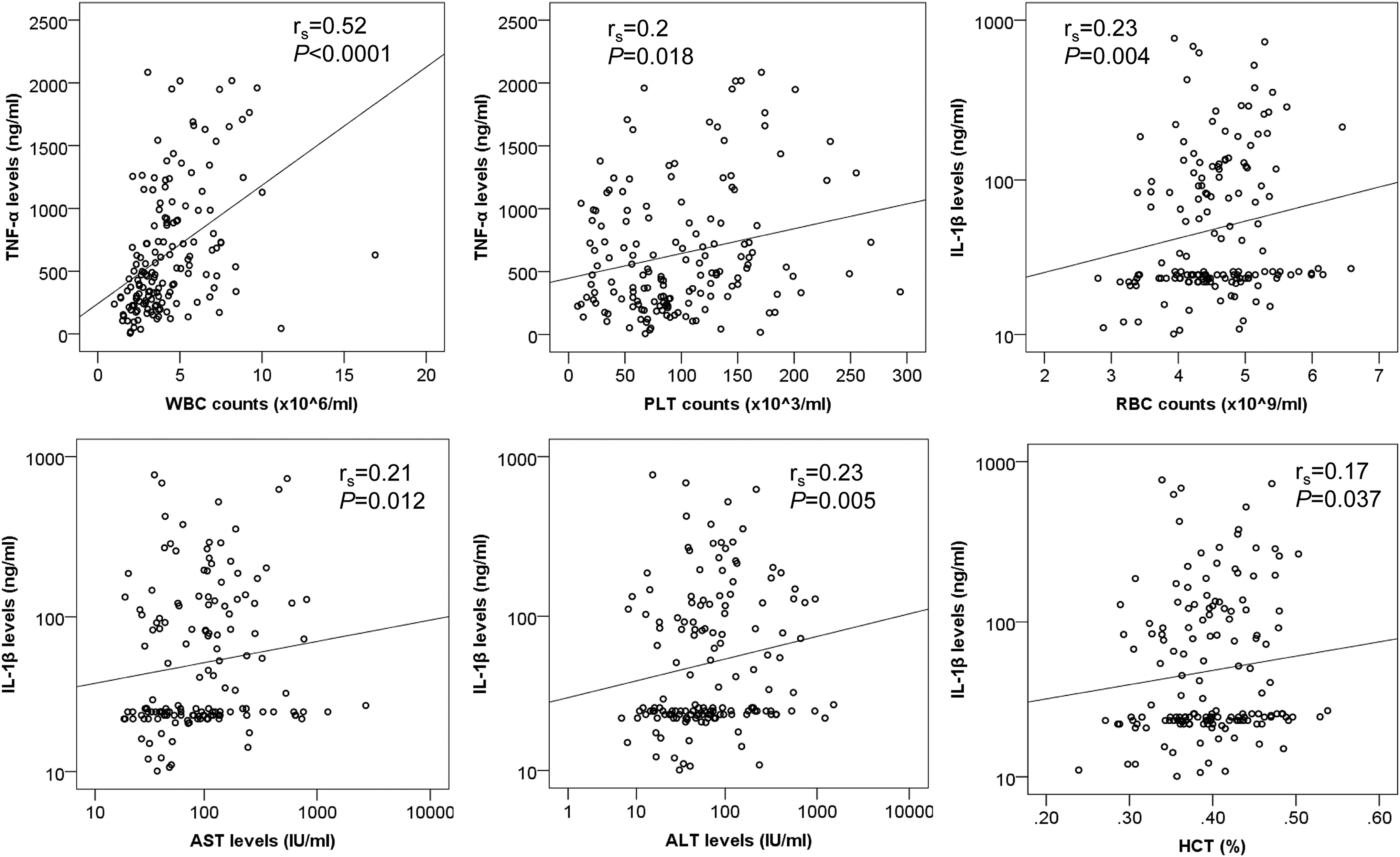
Correlation of cytokine levels with clinical parameters in dengue fever patients r s and p values were calculated by Spearman's rank correlation coefficient.

Correlation of cytokine levels with clinical parameters in DWS− and DWS+ patients. (Upper panel) correlation of TNF-α and IL-1β levels with clinical parameters in DWS− patients; (Lower panel) correlation of TNF-α levels with clinical parameters in DWS+ patients. r s and p values were calculated by Spearman's rank correlation coefficient.
Discussion
The development of a severe form of dengue is known to be the involvement of both viral and host immune factors, including cytokines (21,25). This study has shown that TNF-α, IL-1β, and IL-12 levels are significantly elevated in dengue patients compared with HCs and that these proinflammatory cytokine levels are associated with dengue severity in Vietnamese patients with dengue infection. These findings are consistent with previous studies indicating a crucial role of TNF-α, IL-1β, and IL-12 in dengue (6,16,19,20). Elevated cytokine levels may contribute to the increase of vascular permeability, initiation of coagulation, and fibrinolysis during DENV infection (26). Thus, our data suggest that increased levels of TNF-α, IL-1β, and IL-12 play a key role in the pathogenesis of DENV infection.
It is documented that increased TNF-α levels are associated with facilitating vascular leakage and are a risk factor for the occurrence of SD (8,22). However, the role of TNF-α in the pathogenesis of DSS remains unclear. The finding that TNF-α levels were not significantly different among patient groups (DWS−, DWS+, and SD) is in line with previous studies (6,16). TNF-α levels have been shown to be particularly high in DSS patients (24), and differential TNF-α levels could also be associated with genetic background and infection with different DENV serotypes (14). The elevated TNF-α levels in dengue patients compared with controls suggest that this cytokine could be a predictive factor for the development of DWS+ and SD. The PLTs play a crucial role in the pathogenesis of infectious diseases, especially in vascular injury, thrombosis, and autoimmunity (7). Significant correlation of TNF-α levels with WBC and PLT counts in dengue patients (DWS+ and DWS+) reveals the role of TNF-α in enhancing the severity of dengue infection. Increased plasma TNF-α levels are associated with decreased levels of PLTs in dengue and dengue hemorrhagic fever patients (17), and thus the presence of these cytokines indicates a higher risk of developing severe forms of dengue infection.
IL-1β has been shown to increase vascular permeability, especially in conjunction with TNF-α and IFN-γ, which are increased in patients with SD (3,11). The IL-1β levels were significantly higher in dengue patients compared with HCs in our present study. However, previous studies reported that IL-1β levels did not differ between DENV-infected patients and healthy individuals (1,5). A previous in vitro study has reported that DENV induces the expression of IL-1β in macrophages by activating NLRP3 inflammasome and this finding can be an explanation for the increased IL-1β levels in dengue patients compared with HCs (29). In addition, correlation of IL-1β levels with RBC counts and the levels of AST, ALT, and HCT in dengue patients and in DHF patients suggests that IL-1β is associated with clinical outcome of dengue. Our data reveal higher IL-12 levels in the dengue patients compared with HCs; however, we did not observe a significant difference between dengue patient groups, which might be due to the small number of SD patients included. Nevertheless, high IL-12 levels have been shown to be related to SD (19). IL-12 has a profound effect on Th1 cells and Th1-type cytokines and its levels are elevated in patients with mild dengue but not detectable in the serum of patients with more SD (19). Therefore, IL-12 may play an important role in the progression of severe disease via maintenance of Th1 response.
There were certain limitations in our study, including a small number of SD patients that may lead to a difference in reported cytokine levels compared with other previous studies. Since cytokine levels are dynamically changing during the clinical course of DENV infection, additional follow-up studies are needed to clarify the role of these cytokines in the pathogenesis of SD. In addition, infections with different DENV serotypes may produce different cytokine profiles; DENV serotype data, however, were not available in this study. In conclusion, our study showed the association of TNF-α, IL-1β, and IL-12 levels in DWS+, DWS+, and SD patients and their correlation with clinical parameters. Results suggest that these three key cytokines could play roles in the pathogenesis of the disease.
Footnotes
Acknowledgments
The authors thank all the patients and healthy individuals for their participation.
Authors' Contributions
T.T.T. and N.T.H. performed the experiments and carried out the statistical analyses. N.T.V. and H.V.T. carried out the statistical analyses, interpreted results, and wrote the article. T.T.T., N.T.G., D.T.A., and H.V.H. recruited patients; collected samples and clinical data. D.Q., N.L.T., and H.V.T. designed and supervised the study; contributed materials and reagents. All authors agreed with the results and conclusions.
Author Disclosure Statement
No competing financial interests exist.
Funding Information
This study was funded by the Vietnam National Foundation for Science and Technology Development (NAFOSTED) under grant number 01/2018/ĐX.