
Abstract
A Picornavirus (Ljungan virus [LV]) originally found in bank voles has been associated with type 1 diabetes (T1D) in its wild rodent reservoir, but also associated with T1D in a laboratory rat model for the disease, the diabetes prone (DP) Bio Breeding (BB) rat. Successful treatment of diabetes in this rat model, using experimental antiviral compounds directed against picornavirus, has been reported. In the present study we show significant clinical response in DP-BB rats using antiviral compounds available for human use (Pleconaril, Efavirenz, and Ribavirin). Presence of LV picornavirus antigen has been detected in islets of Langerhans from both human and the T1D rat model with clear morphological similarity. Based on these data it would be of interest to test antiviral treatment in patients with newly diagnosed T1D. Successful outcome will offer both proof of concept regarding the role of virus involvement in the disease and possibly a first generation treatment interrupting a persistent infection and stopping β-cell destruction.
Introduction
Despite extensive research effort, the body of evidence supporting a relationship between viral infections and type 1 diabetes (T1D) remains largely circumstantial. Several viruses have been proposed as an etiological agent for T1D such as cytomegalovirus, parvovirus, retrovirus, and encephalomyocarditis virus (picornavirus), all of which have been subsequently challenged. A large number of reports focus on picornavirus in general and enteroviruses in particular as playing a major role in development of T1D in humans (2,23).
Strains of both enteroviruses and encephalomyocarditis, both belonging to the picornavirus family, have the ability to induce or accelerate diabetes in animal models (9,32).
We have previously reported the association between Swedish small rodent abundance and T1D in humans (16). A novel picornavirus, Ljungan virus (LV), was isolated from wild Swedish rodents, and molecular characterization revealed its relation to encephalomyocarditis (19). Subsequently, we found that both rodents in the wild and colonized wild rodents developed diabetes with signs and symptoms mimicking human T1D, including autoantibodies. In addition LV was visualized in islet cells of diabetic animals using immunohistochemistry (IHC) suggesting that the Bio Breeding (BB) animal colony is persistently infected with a picornavirus (15,17,20). It was also shown that LV could induce glucose intolerance in laboratory mice (21). The spontaneous development of diabetes in the diabetes prone (DP) BB rat bears a striking similarity to human T1D (25). In 2007 we reported the finding of LV antigen in beta cells of BB rats (18). Two years later we reported that antiviral therapy significantly delayed onset of diabetes in BB rats (8) and in 2016 it was found that a combination of two antiviral compounds could protect DP BB rats for the entire period of treatment and that in addition some animals remained nondiabetic after treatment (14).
In this work we add support for the viral T1D hypothesis by analyzing pancreas tissue from a human patient and a BB rat, for each in the phase of T1D development, and analyzed samples for presence of LV by IHC. We have also expanded our previous study on antiviral treatment of DP BB rats now using combinations of antiviral compounds approved for human use (14).
Materials and Methods
Antiviral compounds
Three antiviral compounds were used in the experiments. First, Pleconaril, 3-{3, 5-dimethyl-4-[3-(3-methyl-isoxazole-5-yl)-propoxy]-phenyl}(15)(11)-5-trifluoromethyl-[1, 2, 4] oxazole, is a direct acting broad spectrum antipicornaviral agent originally developed for the treatment of the common cold (24). Ribavirin (1-ß-D-ribofuranosyl-1,2,4-Triazole-3-carboxamide) is a broad-spectrum antiviral nucleoside analog, with activity against a broad range of RNA and DNA viruses. In clinical practice, ribavirin is used, alone or in combination with other drugs, in the treatment of a variety of infections such as hepatitis C virus, respiratory syncytial virus, and Lassa fever virus (28). Efavirenz [(4S)-6-chloro-4-(2-cyclopropylethynyl)-4-(trifluoromethyl)-2,4-dihydro-1H-3,1-benzoxazin-2-one] is a potent non-nucleoside reverse transcriptase inhibitor and widely used in antiretroviral therapy (4). Pleconaril, Ribavirin, and Efavirenz were administered to the animals in a dose of 100 mg/kg BW, of 100 mg/kg BW, and of 60 mg/kg BW, respectively, once daily using a 10, 10, and 6 mg/mL, respectively. Formulation contained the vehicle 0.4% (v/v) Tween 80 + 2% (v/v) glycerol +15% (w/v) Hydroxypropyl-beta-cyclodextrin.
Experimental design
DP BB rats were obtained from the breeding colony maintained at the Karolinska Institutet (13). The animals were housed under Specific Pathogen Free conditions in a temperature- and humidity-controlled room with 12-h light/12-h dark cycles. They were fed R36 diet and water ad libitum. Animals of both sexes were used. The overall incidence of diabetes among the DP rats used in the colony study is 100% with 60 days as the mean age of onset.
Group A (n = 9) is a control group given vehicle only, group B (n = 6) Pleconaril alone, group C (n = 7) a combination of Pleconaril and Ribavirin, group D (n = 5) a combination of Pleconaril and Efavirenz, and groups E (n = 16) and F (n = 12) were given a combination of Pleconaril, Ribavirin, and Efavirenz. Group E was given half the dose of group F (Table 1). The results from Group A and B have previously been reported as part of a different study (14).
Animals Receiving Different Therapeutic Regimes During a Period of 30 Days
Mean day of onset of diabetes and SD of the mean time of onset are given, as well as statistical analysis (unpaired two tailed t-test) of the difference between animals in one particular treatment group compared with control animals.
NS, not significant; SD, standard deviation.
Antiviral compounds and the control vehicle were administrated using an oral gavage once daily. Therapy was initiated between 42 and 48 days of age and maintained for a total of 30 days. All animals were followed until onset of diabetes (blood glucose 270 mg/dL or above measured twice and/or presence of ketones) or until 110 days of age, which ever occurred first.
The studies performed in BB rats were approved by the Animal Experiment Ethics Committees at Karolinska Institutet (N239/15) and analysis of the human specimen approved by Human Committees at Karolinska Institutet 2004/3:10.
Immunohistochemistry
Pancreas tissue was collected for tissue transplantation from a 40-year-old male involved in a fatal accident. As part of the screening process before transplantation, it was discovered that the patient suffered from T1D not yet clinically diagnosed. Formalin-fixed paraffin-embedded pancreatic tissue from this patient (H911) was kindly provided by doctor Gun Frisk, University of Uppsala, Uppsala, Sweden and analyzed for presence of LV antigen by IHC (3). Formalin fixed paraffin embedded pancreas tissue from a 64-day-old male BB rat with recent onset of T1D was stained in parallel.
The tissues were fixed in 4% formalin and embedded in paraffin as described previously with minor modifications (27). Presence of LV specific antigen was visualized using a polyclonal recombinant LV VP1 raised in rabbits (31). As control we used serum from a rabbit immunized using the same protocol but with the carrier GST protein only. Tissues from LV-infected and noninfected animals were included as controls. The specificity of the rabbit antibodies was verified by analyzing control specimens generated by mixing infected tissue culture cells with noninfected cells followed by formalin fixation and paraffin imbedding. The specificity of the reaction was also confirmed by blocking the signal with LV antigen in parallel with control antigen.
Results
Effects of treatments with antiviral compounds in BB rats
The mean age of diabetes onset in the control group was 59 (standard deviation [SD] 7) days of age and not found statistically different from the group B receiving Pleconaril alone with mean onset day 62 (SD 8) or from group C receiving Pleconaril–Ribavirin with mean 61 days (SD 7) with p = 0.5 and p = 0.7, respectively, using unpaired two tailed t-test (Table 1). Group D receiving Pleconaril–Efavirenz had mean day of onset 71 (SD 3), being significantly different versus the control group (p = 0.007). Group F receiving Pleconaril–Efavirenz–Ribavirin had mean day of onset 86 (SD 25), being significantly different from the control group (p = 0.005). Group E receiving Pleconaril–Efavirenz–Ribavirin at half the dose of group F also had mean day of onset 73 (SD 9), being significantly different versus the control group (p = 0.001).
Time to diabetes onset in DP BB rats using the six experimental treatment regime is seen in Figure 1. In group E a total of six rats were nondiabetic at day 110. Three of the six animals were not sacrificed at day 110 but at day 200 when all three rats were still nondiabetic. The rats sacrificed at day 200 were all found LV negative in their pancreas by IHC.
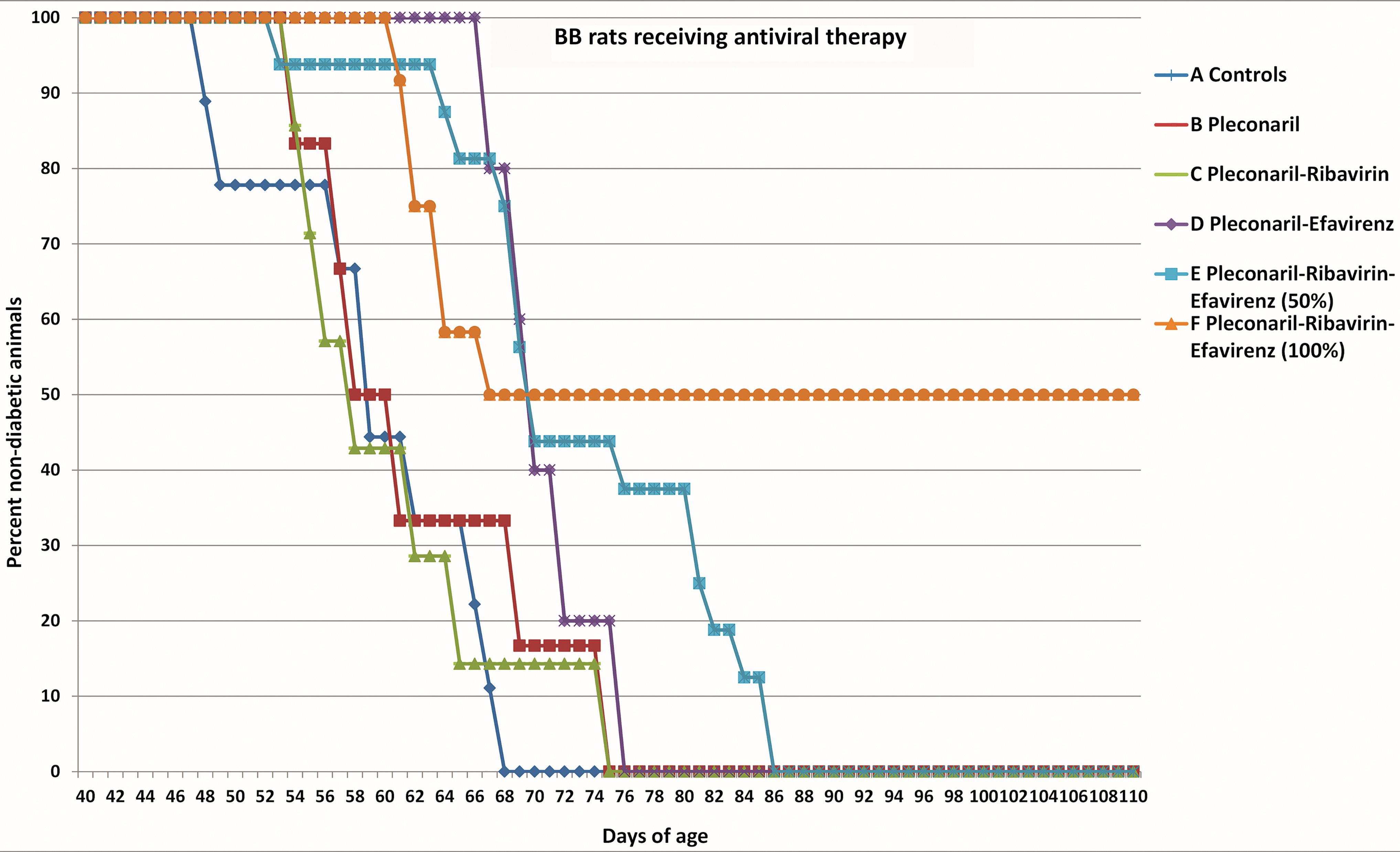
Illustrates the time to onset of diabetes in the six different treatment groups. Control (A) (n = 9), Pleconaril (B) (n = 6), Pleconaril–Ribavirin (C) (n = 7), Pleconaril–Efavirenz (D) (n = 5), Pleconaril–Efavirenz–Ribavirin (E) half dose (n = 16), Pleconaril–Efavirenz–Ribavirin (F) full dose (n = 12).
LV antigen in human and DP BB rat pancreas tissue
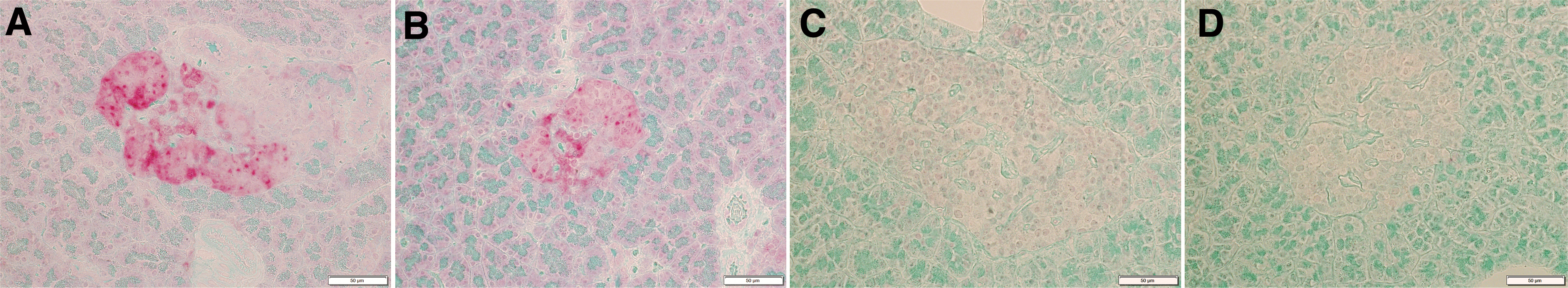
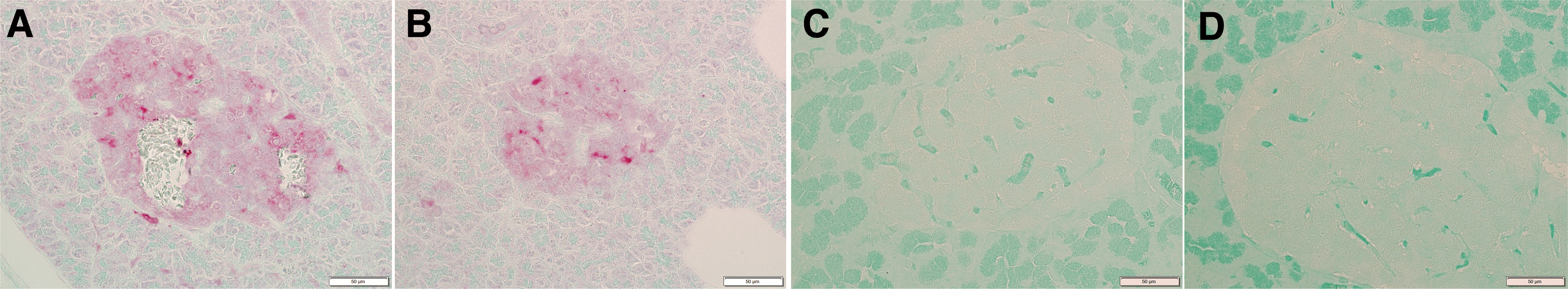
As an illustration of the similarities between T1D in humans and the BB rat animal model, formalin-fixed paraffin-embedded pancreas tissue from both species collected at the time of diabetes onset was analyzed for presence of LV antigen using IHC (human sample H911 provided by Gun Frisk, University of Uppsala, Uppsala, Sweden) (3,27) (Figs. 2 and 3). Presence of LV specific virus protein1 antigen in red was visualized in the islets of Langerhans using a polyclonal recombinant LV VP1 antiserum raised in rabbits (31). It cannot be determined if it is insulin producing β cell or glucagon producing α cell showing LV positive staining. However, the distribution of positive cells suggests that insulin producing cells are infected in both the human and the rat specimen.
Discussion
The BB rat animal model of T1D originates from a Canadian colony of outbred Wistar rats spontaneously developing hyperglycemia that quickly progresses to fatal diabetic ketoacidosis unless treated with exogenous insulin. The DP-BB rat is one of the most engaged animal models for the study on β cell destruction and onset of T1D (13). The fact that all DP-BB rats develop diabetes within a narrow time frame provides an excellent opportunity to study the impact of treatment before, during, or after disease onset.
The observations of LV antigen in the islets of Langerhans in BB rats were followed by the finding that diabetes onset could be postponed using the antiviral compounds Pleconaril and Ribavirin (8,18). We later extended our antiviral experiments using treatment with Pleconaril and an experimental antiviral kinase inhibitor APO-N039. The two compounds showed additive effect when evaluated in tissue culture (14). This finding was repeated in the BB rats with Pleconaril, and APO-N039 administrated alone only showed marginal delay on diabetes onset, while the combination of both drugs protected all animals for the entire 6-week duration of treatment. Some animals remained nondiabetic several months post treatment. Some animals became diabetic when antiviral therapy was terminated but became nondiabetic again when antiviral therapy was reinstated. The BB rat has been perceived as an animal model in which autoimmunity is the main driving force in the pathogenesis (13). However, the observation that Pleconaril–APO-N039 combination could reverse already established clinical diabetes symptoms points to the possibility that diabetes can be treated after onset. The short duration between initiating antiviral treatment and clinical response and the clinical “on-off” effect observed when treatment was terminated and again reinstated suggest a direct effect of the antiviral compounds on a viral infection in the β cells rather than a virus triggering an autoimmune effect. Assuming that the scenario is true, the immune system action of removing the insulin producing β cells would be an appropriate response to an infected cell, which thereby induces diabetes and is not part of an autoimmune process.
In the present study we used Pleconaril, Ribavirin, and Efavirenz, three antiviral compounds selected based on potential effects on a LV related picornavirus and compounds that can be used in humans. Pleconaril binds to a hydrophobic pocket in viral protein 1, the major protein which makes up the capsid of picornaviruses. This renders the viral capsid rigid and compressed and prevents the uncoating of its RNA. As a result, the virus is stopped from attaching to the host cell and causing infection (24). Ljungan belongs to the Parechovirus genus in the picornavirus family, and several members of the Parechovirus, including Ljungan virus, lack a hydrophobic pocket binding structure on the surface of the VP1 protein (11,34). Despite this finding Pleconaril does show antiviral activity to LV in tissue culture (14). Ribavirin, a nucleotide analog of guanosine, has broad-spectrum direct antiviral effect on members of the picornavirus family. Ribavirin was also selected based on antiviral activity to LV in tissue culture and apparently acting through distinct aspects of the viral life cycle (1,8). A dose dependent toxicity resulting in hemolytic anemia is a limiting side effect of this drug. Efavirenz, a non-nucleoside reverse transcriptase inhibitor, is widely used against HIV (33). The antiviral effect on picornavirus is unexpected since picornavirus does not utilize reverse transcriptase for its replication. This suggests an additional antiviral mechanism not yet identified. In the present study the combination of Pleconaril and Efavirenz was superior compared to Pleconaril alone. Pleconaril– Efavirenz and Ribavirin added additional delay of diabetes onset. The general advantage of combination therapy over monotherapy is supported by both theoretical models of virus dynamics and treatment experience (7). Combination therapy applied to human immunodeficiency virus type 1 has been a great success for the control of AIDS and has drastically reduced AIDS-related mortality (12).
The ability to identify and associate a virus with human disease is influenced by its biological characteristics. Viruses that are difficult to cultivate decrease the likelihood of linking the agent to disease. A zoonotic agent has little selective pressure for efficient spread beyond the newly infected host individual, yet the human community is understandably quite concerned with any pathogenic effects regardless of viral transmission. This may be why many zoonotic viruses are identified through more challenging procedures of first being isolated in a natural reservoir. Previously reported observations of LV viral antigen in human tissue have never been definitely confirmed by virus isolation or detection of viral RNA using polymerase chain reaction (PCR). Stool samples from children participating in the Norwegian Environmental Trigger of Type 1 Diabetes Cohort study found all samples to be LV negative (30). A Finnish Diabetes Prediction and Prevention study found no association between LV seropositivity and β cell damaging process (10), while LV antibodies did correlate with insulin autoantibodies and HLA-DQ8 suggesting a possible role in T1D in a Swedish study (22).
Presence of LV antigen using a polyclonal antibody and absence of PCR confirmation may depend on low specificity of the PCR used. This in turn may be a result of the fact that the PCR used was designed based on sequence information from a limited number of LV isolates. The specificity of any PCR usually improves gradually over time, when an increasing number of isolates are being characterized and sequenced and used for primer design.
The human pancreas tissue included in this study was recruited from a fatal accident selected for organ donation of pancreas tissue. Investigations before transplantation revealed that the patient had an undiagnosed recent onset of T1D. This in turn offered a unique opportunity to analyze islets of Langerhans from pancreas tissue in good condition at this critical stage of the disease. We included formalin-fixed paraffin-embedded LV IHC microphotographs from this patient together with a pancreas specimen from a BB rat at the same stage of diabetes development as an illustration of the similarities between the two species (3,27).
The six experimental treatment groups on the BB rats are each of small size. Even if the BB rat T1D model has many similarities to the human condition, it is still an animal model. Information from these experiments on tests of drug dosing is quite limited, and the precise viral agent, although clearly picornaviral, is still uncertain. Against this are the facts that the experimental groups did differ according to the drug and concentration of drugs used, with the one clearly successful treatment (group F) being more successful in delaying and stopping T1D than a lower dose of the same drugs (group G). Furthermore, surviving animals in group F remained diabetes-free long beyond any typical BB rat (i.e., >200 days) and all without symptoms of diabetes onset. The IHC on BB rats and a human sample with evidence of T1D revealed that LV surface coat protein antibodies were positive in each of the pancreas samples, clearly pointing to a picornaviral role in diabetes.
The information suggesting “a picorna viral etiology in T1D” as a whole encourages intensified effort using a variety of virological techniques to generate data that can adequately test this hypothesis. In parallel with attempting to fulfill as much as possible for Koch's postulate the availability of antiviral compounds directed against picornavirus infection may offer a possibility to generate both proof of concept of viral etiology and a first-generation therapy directed against the cause of T1D.
We welcome the initiative and the debate regarding the ongoing clinical trial using Pleconaril and Ribavirin T1D launched in 2018 and await the results with great anticipation (5,6,29).
The present data from DP BB rats suggest that Pleconaril–Efavirenz is another unexpected drug combination that may be a superior antiviral combination that could be tried in patients with recent onset of T1D. Nearly a century has passed since T1D was first identified as a disease of the pancreas and successfully treated with insulin. Despite many decades of intensive biomedical research no underlying cause/causes have been brought forward to explain the disease beyond a characteristic adaptive immune response involving B and T cells that was later attributed to autoimmunity (26). Antivirals are still a relatively new therapeutic possibility and their application to T1D is awaited.
Footnotes
Author Disclosure Statement
No competing financial interests exist.
Funding Information
Funding for this project was received from the Karolinska University Hospital and Karolinska Institutet (No. 91201100915), The Family Erling Persson Foundation, Strategic Research Program in Diabetes at Karolinska Institutet and The Stichting af Jochnick Foundation.