




Abstract
Genotype 2 strains of Porcine Reproductive and Respiratory Syndrome Virus (PRRSV-2) have been reported sporadically in Europe. Even if, PRRSV-2 reported to be genetically homogenous in Europe due to the introduction of an MLV vaccine strain, independent introductions of PRRSV-2 field strains have been reported. The aim of the present study was to report the complete genome sequence and evaluate the histopathological lesions of a PRRSV-2 strain, isolated for the first time in Greece. During a routine blood sampling in a commercial pig farm, the results revealed positive samples in weaners of 40–60 days for the PRRSV-2, using real-time polymerase chain reaction. The clinical picture was characterized from respiratory symptoms in weaners, as well as coughing and poor performance at finishing stage and less than 3% mortality rate from weaning stage to finishing stage. The use of ORF5 for PRRSV phylogenetic analysis of the isolated PRRSV strain, named “x1544-1 strain”, was successfully determined, belonging to the genotype PRRSV-2. Comparison of the obtained sequence revealed nucleotide sequence identity >98% with PRRSV-2 strain VR2332 and other related strains from Denmark and China. The histopathological evaluation revealed diffuse interstitial pneumonia, multifocal interstitial nephritis, while in the lymphoid organs, follicular and paracortical hyperplasia, coexisting with necrosis and depletion of germ cells were detected. The results of current study undersign the importance for veterinary practitioners to have up-to-date access to phylogenetic data linked to phenotypic information to follow-up the control and prevention strategies against PRRSV.
Introduction
Porcine reproductive and respiratory syndrome virus (PRRSV) causes an important endemic disease, imposing important economic impact to the swine industry due to increased secondary infections and high mortality (30). Along with equine arteritis virus, simian hemorrhagic fever virus, and lactate dehydrogenase-elevating virus, PRRSV belongs to the family Arteriviridae in the genus Arterivirus, order Nidovirales (7,21).
Currently, the PRRSV is now subdivided in two species: genotype 1 (European genotype, PRRSV type 1 and PRRSV-1), and genotype 2 (North American genotype, PRRSV type 2 and PRRSV-2) (17). Historically, the prototype strain of PRRSV-1 is the Lelystad virus (LV), representing strains predominately originating from Europe, which is further divided into four genetic subtypes (36). The prototype strain of PRRSV-2 is the VR-2332 representing viruses predominately originating from North America. Both genotypes display significant degree of molecular and antigenic variation and some morphological and structural similarities due to antigenic heterogeneity (20,23,36,37,39). The first PRRSV-2 case in Europe reported in 1995, due to use of a PRRSV-2 MLV vaccine in Denmark (4). Since then, PRRSV-2 strains were isolated in various European countries, which display similar sequences (>98% identical in ORF5 or ORF7 nucleotide sequence) to that PRRSV-2 MLV vaccine (35,36). However, virulent PRRSV-2 field strains, which are not related to PRRSV-2 MLV vaccine, have been reported in Hungary and Germany (2,13,35). The available data in Europe for PRRSV-2 diversification and evolution are limited, in contrast to North America (USA, Canada) (5,24,36). In Greece, there are no reports for PRRSV-2 detections, and the vaccination protocols include only PRRSV-1 vaccines, killed or MLV (28 –30). In a recent study on PRRSV molecular characterization in Greece from more 50 commercial pig farms, only PRRSV-1 strains were isolated from 70% of the tested farms (8). The aim of this study was, for first time, to describe the pathogenesis of a PRRSV-2 strain, isolated in Greece.
Materials and Methods
Ethical statement
All applied procedures in animals during this trial were in accordance with National and European Animal Welfare requirements (11,12). All animal care and handling procedures were approved by the Ethics Committee of the Faculty of Veterinary Medicine, School of Health Sciences, University of Thessaly (Approval number 84/2019).
History/Farm
The current study was carried out on a farrow-to-finish commercial swine farm, located in Central Greece [Koskinás (Thessaly, Nomós Kardhítsas): coordinates: N 39° 29’ 31” and E 22° 0’ 43”]. This farm had a capacity of 550 sows with the same genetic background of (commercial hybrids of Large White × Landrace, Topigs Norsvin), maintaining its own artificial insemination (AI) laboratory for the application of “Do it yourself AI.” Moreover, the farm had its own grandparent nucleus of 40 sows for the production of gilts, which were separately housed, but in the same premises as the commercial herd. Sporadically, semen was introduced from boar stud for AI in sows of its own grandparent nucleus. The vaccination scheme of breeding stock included vaccinations against PRRSV-1 with killed vaccine (Progressis®, Ceva Santé Animale), Aujeszky's disease virus, parvovirus, Erysipelothrix rhusiopathiae, atrophic rhinitis, Escherichia coli, and Clostridium perfringens.
During a routine blood sampling on February 2018 in breeding stock, weaning, growing, and finishing stage, the results revealed positive samples in weaners of 40–60 days for the PRRSV-2 (Ct value: 26.61), using real-time polymerase chain reaction (PCR). All samples were negative for PRRSV-1. The clinical picture of animals was characterized from mainly respiratory symptoms, including cough, pyrexia, sneezing, periocular edema, and oculonasal discharge in weaners (40–60 days of age), as well as coughing and poor performance at finishing stage. The mortality rate from weaning stage to finishing stage was less than 3%.
Sampling
Based on the positive results for PRRSV-2 of routine sampling, a new blood sampling was designed to investigate further the previous detection of PRRSV-2. Blood samples were collected for laboratory examinations (RT PCR and phylogenetic analysis) from: (a) breeding stock (pool 1 = 5 samples-parity 1, pool 2 = 5 samples-parity 2, pool 3 = 5 samples-parity 3, and pool 4 = 5 samples-parity 4), (b) weaning stage (pool 5 = 5 samples from piglets 30–35 days of age, pool 6 = 5 samples from piglets 45–50 days of age, and pool 7 = 5 samples from piglets 55–65 days of age), (c) growing stage (pool 8 = 5 samples from pigs 80–90 days of age and pool 9 = 5 samples from pigs 110–120 days of age), and (d) finishing stage (pool 10 = 5 samples from pigs 140–150 days of age and pool 11 = 5 samples from pigs 160–165 days of age).
Laboratory examinations
All blood samples were examined by RT PCR for PRRSV-1 and PRRSV-2 infection. RNA was extracted by using the QIAamp cador Pathogen Mini Kit (INDICAL BIOSCIENCE GmbH, Leipzig, Germany) according to the manufacturer's instructions. In total, 200 μL of serum from each pool (five samples per pool) was added to 20 μL ready-to-use proteinase K and 100 μL of lysis buffer. Carrier RNA was added to each sample as it enhances the adsorption of viral RNA and DNA and bacterial DNA to the silica membranes, which is especially important when the target molecules are not abundant. Internal positive control RNA (RNA IPC Target; Ingenetix) was added to all samples during RNA extraction and purification to ensure recovery of RNA and prove the functionality of the reaction mix for correct amplification of the pathogen target. After purification, RNA was tested promptly using a commercial real-time RT-PCR kit (ViroReal® Kit PRRS Virus EU & NA 1.1; Ingenetix) for detection of RNA of both viral lineages PRRSV European EU (including Lena strain) and PRRSV North America (NA) using one-step reverse transcription real-time PCR. The aforementioned kit detects the open-reading frame 7, including the 3′-untranslated region of PRRSV EU and NA (PRRSV type 1 and PRRSV type 2). The specific gene targets of the commercial assay are proprietary and not publicly available. The PRRSV EU lineage is detected in FAM channel, while the PRRSV NA is detected in VIC/HEX channel. We used an internal RNA positive control system (detection in Cy5 channel) that allows control of RNA extraction and excludes false-negative interpretation of results due to inhibition of reverse transcription real-time PCR.
One step RT-qPCR was performed and each reaction were run in duplicates. A volume of 20 μL PCR reaction contains 3 μL of nuclease-free water, 10 μL of Luna Universal Probe One-Step RT-qPCR kit (New England Biolabs GmbH, Frankfurt, Germany), 1 μL of each fluorescent probes, and 5 μL of extracted serum RNA. The reaction conditions involved a reverse transcription step at 55°C for 10 min and RT inactivation and initial denaturation at 95°C for 1 min, followed by 45 cycles of denaturation at 95°C for 10 s and annealing/extension at 60°C for 1 min. The final results were analyzed using LightCycler® Software 4.1. The assay was considered valid if the positive control had a cycle threshold (Ct) 23–26 and the negative control had a Ct value of zero. In this examination, the reverse-transcription method of the RT PCR was carried out in the Roche Light Cycler 2.0 instrument, aiming at a highly conserved virus sequence that ensures its detection. The sensitivity of the method was maximized by the use of specifically designed probes in relationship to other RT-PCR techniques.
One real-time RT-PCR-positive sample for PRRSV-2 was selected for subsequent complete ORF5 (Gp5) gene sequencing, using Sanger dideoxy chemistry. The obtained sequence was phylogenetically analyzed along with other PRRSV-2 ORF5 sequences available in GenBank (38). In this examination, the reverse-transcription method of the RT PCR in the Roche Light Cycler 2.0 application was applied, aiming at a highly conserved virus sequence that ensures its detection. The sensitivity of the method was maximized by the use of probes in relationship to other RT PCR techniques.
In addition, three of the aforementioned piglets at weaning stage (at 35, 50, and 65 days of age), displaying respiratory signs and enlarged lymph nodes, were euthanized. Necropsy for the evaluation of gross lesions and collection of samples (lung, heart, kidney, liver, spleen, and lymph nodes) was applied at the laboratory. The tissue samples were fixed in 10% buffered formalin and embedded in paraffin routinely. Finally, dewaxed 3–5 μm-thick sections were cut and stained with hematoxylin and eosin for histopathological evaluation (34).
Results
PCR results
The RT-PCR results show that piglets aged between 45 and 65 days were positive for PRRSV-2.
Phylogenetic analysis
Phylogenetic trees were constructed using Neighbor-Joining and Kimura-2 parameter method (K2P) with 1000 Bootstrap replication on CLC software. A representative strain of each PRRSV genotype and common vaccines strains are included in the following phylogenetic tree (Fig. 1). The isolated PRRSV strain (named “x1544-1 strain”) was successfully determined based on a full length ORF5 sequence and belonged to the genotype PRRSV-2. Comparison of the “x1544-1 strain” with respective sequences of other PRRSV-2 strains, revealed nucleotide sequence identity >98% with PRRSV-2 strain VR2332 and other related strains from Denmark and China.
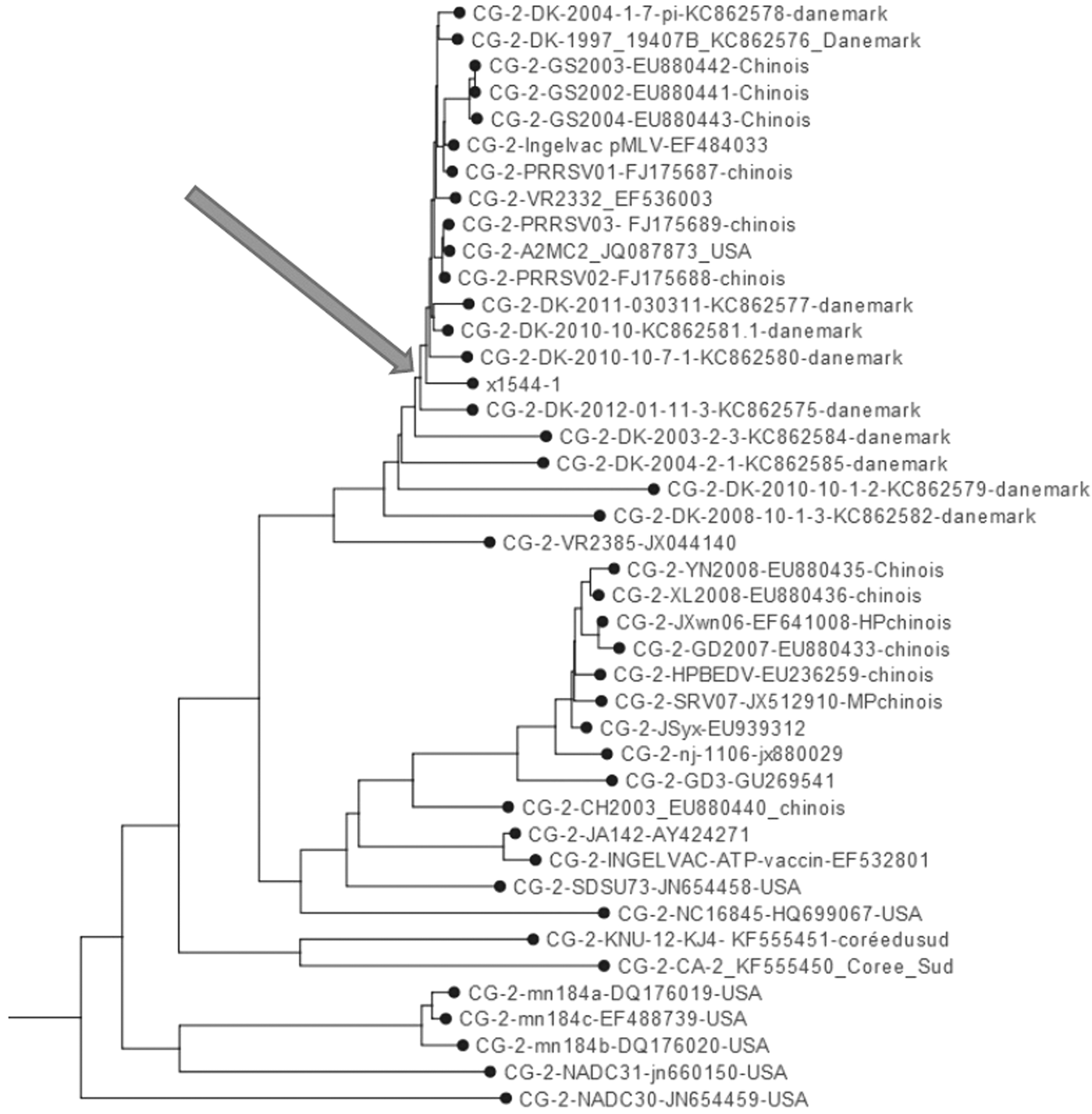
Phylogenetic tree inferred with ML analysis, based on Gp5 sequences of selected genotype PRRSV strains. The sequence obtained from the x1544-1 strain is indicated with an arrow. PRRSV, porcine reproductive and respiratory syndrome virus; ML, maximum likelihood.
Histopathological results
During the necropsy, enlargement and hyperplasia of lymph nodes (mediastinal and inguinal), which appeared firm and whitish, were revealed. The lungs were diffusely mottled tan and red and failed to collapse. The parenchyma was moderately resilient, firm, and moist.
Histopathologically, the lung sections showed diffuse interstitial pneumonia characterized by thickening of the alveolar wall and alveolar septa, mainly due to the presence of histicytes, lymphocytes, and plasma cells (Fig. 2). Multifocal areas with vasculitis and hyperplasia of the bronchi were also described. In the kidney, diffuse parenchymatic hemorrhages and subcortical edema were seen. The renal corpuscles were characterized by hyperemia, hemorrhages, sclerosis of the glomerulus, as well as thickening of Bowman's capsule (Fig. 3). Morphologically, there was multifocal interstitial nephritis, accompanied by peritubular lymphohistiocytic aggregates and hydrophobic degeneration of epithelial cells of juncta glomerulus apparatus and tubules. In the heart, diffuse necrosis and vascular hyperemia were detected. In the liver, activation of Kupffer cells, hyperemia of portal areas vessels and sinusoids and focal thickening of periportal connective tissue were described. In lymphoid organs (mediastinal lymphnodes and spleen), follicular and paracortical hyperplasia, coexisting with necrosis and depletion of germ cells, were the highlights of the lesions (Fig. 4).
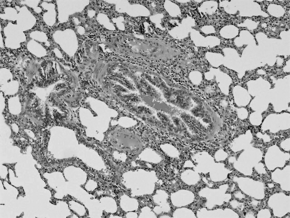
Lung: Thickening of the alveolar wall and septa. HE × 10.

Kidney: Glomerulus sclerosis with mild interstitial nephritis. HE × 10.
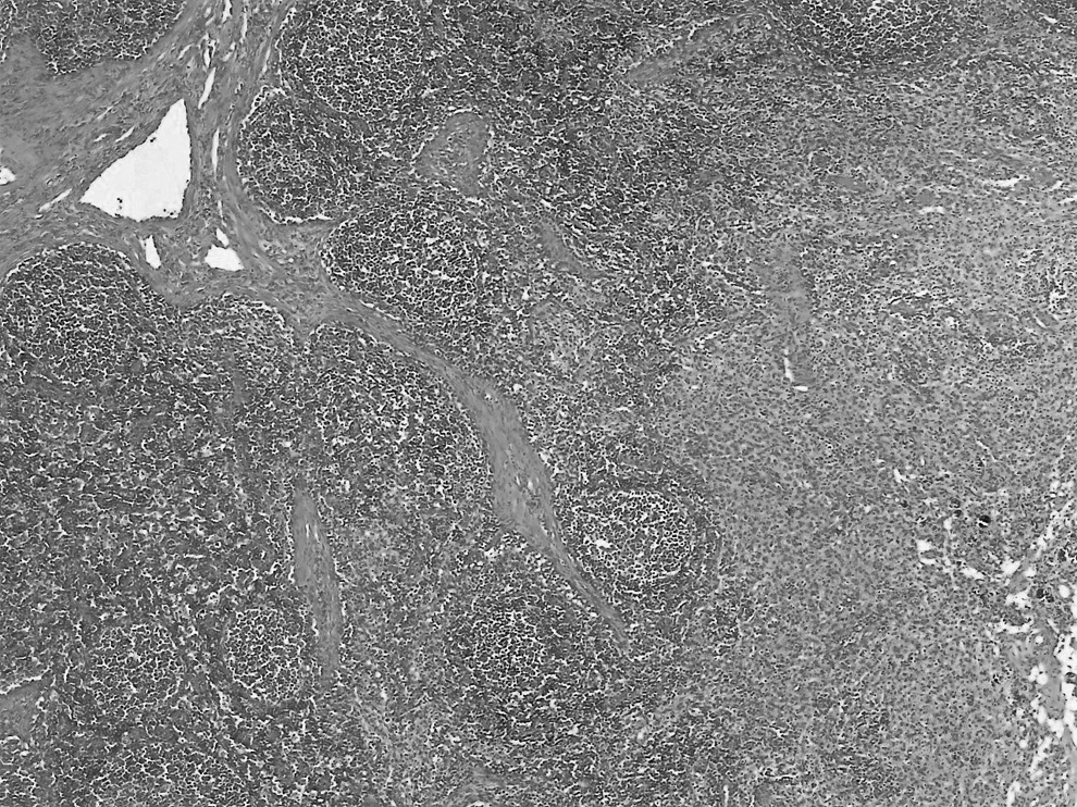
Lymph nodes: Necrosis and depletion of germ cells of mediastinal lymph node. HE × 10.
Discussion
The characterization of PRRSV-2 genetic diversity is a description of ORF5 genetic variation. Global analysis of ORF5 sequences revealed important insights into the evolving geographical diversity of PRRSV (22,35,36). In Europe, it appears that almost all PRRSV-2 diversity is related to reisolation of PRRSV-2 MLV vaccine progeny since its first use in Denmark in 1996 (4). Phylogenetic studies classified all Danish isolates to a single cluster, which comprised strains closely related to the PRRSV-2 prototype isolate VR2332 (18,24). Sparse sequence data from Europe and Russia, primarily in Germany, Lithuania, Poland, and Slovakia, show that independent introduction of virulent field viruses 8 has also occurred (6). Global PRRSV-2 genetic diversity is probably due to the use of MLV vaccines, as there is a possibility for replication and transmission of MLV virus among nonimmune hosts, resulting in new vaccine-related genotypes and reversion to virulence (25 –27). In our study, the detected PRRSV-2 strain, named “x1544-1 strain” perform an identity >98% with PRRSV-2 strain VR2332 and other related strains from Denmark and China. There are no available data about the introduction of this strain in the farm, but we hypothesize that the frequent introduction of replacement animals for its own nucleus herd from other European countries and especially from Denmark in the last decade could be a possible source. Generally, it is difficult to determine whether the isolated strains are evolved from a MLV vaccine introduction or field virus related to the parental strain of the MLV vaccine due to the high similarity between MLV vaccine and parental strains (6).
Our results for the microscopic lesions in lung (diffuse interstitial pneumonia and alveolar septa were infiltrated with histiocytes, lymphocytes, and plasma cells) are characteristic of PRRSV infection and are in accordance with previous studies (10,29). Previous studies reported mild to moderate multifocal lymphohistiocytic vasculitis and perivascular myocarditis in the heart (15,31 –33), while our findings revealed mainly diffuse necrosis and vascular hyperemia. In our study, lesions in kidneys are similar to other studies (9,31), but the main lesions included diffuse parenchymatic hemorrhages and subcortical edema, multifocal interstitial nephritis, and thickening of Bowman's capsule. Moreover, our findings in lymphoid organs (follicular and paracortical hyperplasia, coexisting with necrosis and depletion of germ cells) are characteristic of PRRSV infection and in agreement with other studies (14,33). However, in previous studies, microscopic lesions were predominantly in germinal centers, including mainly necrotic and depleted germinal centers, as well as cortices containing small cystic spaces lined by endothelium and containing proteinaceous fluid, lymphocytes, and multinucleate prokaryocytes (32,33). In a recent PRRSV-2 field case study in Denmark, no histopathological findings were observed in the liver (18). In contrast, in our study, we noticed activation of Kupffer cells, hyperhemia of portal area vessels, and sinusoids and focal thickening of periportal connective tissue were described.
Many studies reported that PRRSV-2 strains induce more severe respiratory disease than PRRSV-1 strains (14,19,37). It is interesting that a recent study reported in highly pathogenic PRRSV-infected pigs not only characteristic PRRSV-2 lesions in lymph nodes and lungs, but also primarily hemorrhagic microscopical lesions in other organs (including spleen, liver, heart, and kidney) (16). The lesions in our study are quite similar to the aforementioned study, contributing that the isolated strain “x1544-1 strain” seems to display highly pathogenesis. However, “x1544-1 strain” induced severe lesions mainly in lung and lymphoid organs, but no significant increase of mortality.
The coexistence of the two genotypes has been reported in many countries around Europe, North America, and Asia (1,2). The current first report of PRRSV-2 in Greece indicates how “colorful” disease is the PRRSV disease (3). The evolutionary and epidemiological dynamics of PRRSV is highly, as the virus is rapidly evolving, contributing to the production of “x1544-1 strain” and exhibiting high diversity. At the same time, the globalization of the modern swine industry acts as a super-spreader, contributing to the distribution of PRRSV strains worldwide (35). For this reason, it is important for veterinary practitioners and other field personnel to have up-to-date access to publicly available phylogenetic knowledge linked to phenotypic information to follow-up the control and prevention strategies against PRRSV based on the virus dynamics (6). The surveillance and monitoring of PRRSV evolution are fundamental to all stakeholders in the herd health management approach. Further epidemiological and molecular studies are needed to investigate the presence of PRRSV-2 strains in Greece.
Footnotes
Author Disclosure Statement
No competing financial interests exist.
Funding Information
The authors received no specific funding for this work.