
Abstract
Hepatitis B viral infection is one of the most important infectious diseases of the liver worldwide. Chronic infection with HBV often leads to cirrhosis and hepatocellular carcinoma. The currently licensed hepatitis B vaccine consists of recombinant hepatitis B surface antigen adsorbed into aluminum adjuvant and administered in three doses over the course of 6 months. However, this vaccine requires invasive administration and requires multiple booster doses. To avoid these limitations, nanoparticle (NP)-based vaccines lent itself as efficient adjuvants and delivery systems for the development of new generation vaccines. The biodegradable synthetic polymeric NPs poly(lactide-co-glycolide) (PLGA) was used in this study to formulate PLGA NPs encapsulated with hepatitis B surface protein to evaluate immune response in human peripheral blood lymphocytes in vitro. Formulation of HBP (HBV surface protein)-encapsulated PLGA (HB-nanovaccine [HB-NV]) was conducted by using double emulsion solvent evaporation technique (water–oil–water), which resulted in 94% encapsulation efficiency and 24% protein loading capacity. The resulted HB-NV had typical characteristics of spherical shape at an average size of 71.08 nm with higher densities and high stability dispersion of negatively charged NPs as assessed by atomic force microscopy, scanning electron microscopy, ultraviolet absorption spectrophotometry, zeta potential, and Fourier-transform infrared. The immune response to HB-NV was measured in vitro in lymphocytes, showed significant increase in levels of IL-2 and IFN-γ, as well as in CD4+ and CD8+ T cell counts, with a dose-dependent effect, examined by enzyme-linked immunosorbent assay and flow cytometry, respectively.
Introduction
Hepatitis viral infection is one of the most important infectious diseases of the liver occurring worldwide where ∼350 million people are chronically infected with hepatitis B virus (HBV). Chronic infection with HBV often leads to cirrhosis and hepatocellular carcinoma. Immune response is required for viral clearance, but it failed to protect ∼10% of HBV cases. The current treatment options for HBV infection are limited to 25% response rate with pegylated IFN-α and nucleos(t)ide analogs such as Lamivudine 36 and Entecavir. Therefore, a final elimination of HBV infection appears not achievable due to the virus ability to integrate into host genome of hepatocytes.
Alternatively, current health care strategy is aimed to reduce viral load and induction of seroconversion through developing effective HBV vaccine targeting the immunogenic surface antigen protein of the viral envelope (HBsAg) (25). The hepatitis B surface antigen (HBsAg) had three proteins, including a major polypeptide of 226 amino acids designated small HBs or major surface protein, in nonglycosylated (p24) and glycosylated (gp27) forms. The middle-sized protein (MHB) is called preS2 (gp33 and gp36). The large HBs protein (LHB) contains the pre-S1 domain (p39 and gp42) as well as the S and pre-S2 domains. HBV is classified into four major serotypes (adr, adw, ayr, ayw) according to antigenic epitopes presented on the envelope proteins, and into eight major genotypes (A–H) (17). Vaccination is mainly done to control HBV infection. Available HBV vaccines contain the hepatitis B envelope, which comprised of adw and A2 genotype (6). The most prevalent HBV genotype in Iraq was genotype D (1). On the contrary, adw serotype has been associated with all genotypes except “D” and “E,” whereas the serotype ayw contains all genotypes except “C.” Therefore, it is imperative to consider a serotype containing genotype D to be used as effective vaccine against the most common D genotype in Iraqi patients. The currently licensed hepatitis B vaccine consists of recombinant HBsAg adsorbed into aluminum adjuvant and administered in three doses over the course of 6 months. This vaccine proved to be both safe and effective with >90% protection upon completion of a full three-dose-vaccination course. However, this vaccine requires invasive administration and requires multiple booster doses to elicit an effective immune response. These efficacy limiting factors can be overcome by using an efficient vaccine delivery system to evoke enduring immune responses into the target sites with minimal side effects and less doses (if possible). Also, there is an increasing demand to develop a new generation molecular vaccine that will simultaneously act as immunogen and adjuvant. For this purpose, nanotechnology-based formulations lent itself for the development of new generation vaccines. Nanocarrier-based delivery system can protect the vaccines from premature degradation, improve stability, provide good adjuvant properties, and enhance immunogenicity to the antigen presenting cells (APCs). Nanovaccine immunogen can be encapsulated within the nanocarrier molecules, which can protect the antigen from premature protease degradation and elicit sustainable release. Nanocarrier-based delivery systems provide a suitable route of administration of vaccine molecules and enhance cellular uptake to evoke robust innate, humoral, cellular, and mucosal immune responses when compared with unconjugated antigens (21). A biodegradable polymer such as poly(lactide-co-glycolide) (PLGA) is considered safe, nontoxic, and biocompatible with great potential applications in tissue engineering, drug and gene delivery, and new vaccination strategies. Accordingly, PLGA is one of the primary candidates for use in nanovaccine preparation using peptides, proteins, viruses, and other macromolecules providing sustained immunological or immunotherapeutic responses in animals, thus avoiding the need for multiple injections (11). Therefore, the study plan of this work was designed to formulate PLGA nanoparticles (NPs) employing HBsAg protein derived from ayw serotype as a source for nanovaccine preparation.
Materials and Methods
Formulation of PLGA-loaded HBV surface antigen protein
PLGA 50:50 mol/wt was obtained from Sigma Aldrich (Germany; molecular weight of 38,000–54,000), which was encapsulated with HBsAg (ayw subtype recombinant protein [commercially available from MyBioSource, Germany]). The formulation of NPs was prepared by a water–oil–water (w/o/w) double emulsion method, according to the method described earlier (9) with some modifications. In brief, 250 μg of HBV surface protein (HBP) was emulsified in 2.5 mL of dichloromethane representing organic phase at different concentrations of PLGA (250, 0.25, 0.5, and 1 mg), by means of sonication using a water bath sonicator for 15 min using span-80 as emulsifier, to form a primary water/oil emulsion. The primary emulsion was further emulsified in 12.5 mL of PVA (polyvinyl alcohol) solution (1%, w/v) and then sonicated for 15 min, to obtain a double w/o/w emulsion. The resultant double emulsion was then stirred at 600 rpm to evaporate the organic solvent. HB-NPs were recovered by ultracentrifugation (at 21,000 rcf, × g) for 20 min at 4°C. The supernatant was removed, and NP sediments were washed twice with Milli-Q sterile water to remove free protein and excess surfactant, followed by lyophilization. It is worth noting that lyophilization did not affect the encapsulation efficiency (EE) of the NPs when it was tested before or after the lyophilization process. However, all characterization processes of NPs were performed after lyophilization.
EE and protein loading capacity
Protein EE was measured by indirect quantification methods using ultraviolet (UV) spectroscopy at 595 nm. The EE was calculated by free protein concentration in the supernatant according to Bradford's method (1976) (8), and the loading capacity (LC) was measured in the precipitate, which was obtained from the ultracentrifugation of NPs. The EE and LC of HBV (HBP) protein were calculated using equations: (A − B/A∗100) and (A − B/C∗100), respectively, where A is the amount of total protein, B is the amount of free protein, and C is the quantified NP weight (9). The EE and LC were tested at various concentrations of PLGA (0.25, 0.5, 1, 100, and 250 mg/mL) against HBP of 250 μg/mL. Pilot studies revealed no considerable differences between 100 and 250 mg/mL of PLGA (data not shown), therefore the highest concentration was used in results.
Characterization of PLGA-loaded HBs-protein NPs
Atomic force microscope
Atomic force microscopic (AFM) analysis was used to determine the surface morphology and particle size of the NPs. Samples for AFM were prepared by casting drops of hepatitis B surface protein (HBsP)-NPs solutions onto glass slides, and left to air dry at room temperature. These slides were analyzed using a NTEGRA (NT-MDT, Russia) device.
Field emission scanning electron microscope
Scanning electron microscope (SEM) was used to examine the morphology and the size of the encapsulated NPs. Samples for SEM were prepared by casting drops of HB-NV solutions onto glass slides, and left to air dry at room temperature. SEM images were acquired using a VEGA 3 (Tescan, Czech Republic) instrument. SEM images were processed to calculate the mean diameter of length and area of nanoparticles.
UV absorption spectrophotometry
The absorption of the HBsP-NPs was monitored by using Shimadzu UV–Vis absorption spectrophotometry (Shimadzu, Japan). The specific absorption spectrum was acquired within the range from 200 to 700 nm for HBsP-NPs. The absorption spectrum for deionized water was used as a reference.
Fourier-transform infrared spectroscopy
The functional group of the HBsP-NP and PLGA alone was determined by using a Fourier-transform infrared (FTIR) spectroscopy (Shimadzu-8400S) at a spectral range of 4,000–400 cm−1 with a resolution of 4 cm and in attenuated total reflection mode. The HBV-NPs suspension was dropped onto the glass slides using a sterile pipette and allowed to dry at 30°C in an incubator.
Zeta potential
The surface charge of HBsP-NPs in colloidal solution was determined by zeta potential analytical technique. NP charges were measured by using Zeta PLUS Analyzer. NPs were first diluted in ddH2O, filtered using a 0.22 μm Millipore filter unit, and then placed in ultrasonic chilled bath and subjected to ultrasonic force for 5 min and finally analyzed at room temperature.
Isolation of peripheral blood lymphocytes and assessment of immunological response to HBV-nanovaccine
Peripheral blood lymphocyte (PBL) cultures were collected from healthy volunteers, prepared and maintained to be used for in vitro evaluation of cytokine release (IL-2 and IFN-γ) and proliferation capacity of CD4+ and CD8+ cells that are induced by the PLGA-encapsulated HBV-NPs. The secretion of IL-2 and IFN-γ was measured by enzyme-linked immunosorbent assay (ELISA) (human IL-2 and IFN-γ ELISA kit; KomaBiotech, Korea), and the cell proliferation of CD4+ and CD8+ was sorted by flow cytometry (BD). PBL cultures were treated at various concentrations (20, 50, and 100 μg/mL) of HBsP, PLGA polymer, PLGA-HBP (which was named HBV-nanovaccine [HBV-NV]), in addition to the locally used HBV commercial vaccine (Euvax-B).
Statistical analysis
A one-way analysis of variance (ANOVA) and two-way ANOVA were performed to test whether group variance was significant. Statistical significance was defined as *p ≤ 0.05 or **p ≤ 0.01. All samples analyzed statistically were run in triplicates. Data were expressed as mean ± standard deviation, and statistical significance was determined using Graph Pad Prism version 6 (Graph Pad Software, Inc., La Jolla, CA).
Results
Results of this study presented here provide data on investigation on the choice of PLGA as a nanocarrier to encapsulate the HBP for possibility of nanovaccine preparation and in vitro evaluation of its cellular immune response in PBL cultures.
Formulation of PLGA-loaded HBV surface protein NPs
PLGA-loaded HBVsAg protein NPs were prepared by using a w/o/w emulsion solvent evaporation method for encapsulation. As a result, the final mixture of PLGA-loaded HBV surface protein nanoparticles (HBP-NPs) appeared as a homogeneous emulsion with milky color. The encapsulated NPs were named HBV-nanovaccine (HB-NV), which has been characterized further below.
EE and protein LC
The protein containing supernatants resulted from the double emulsion (w/o/w) solvent evaporation method was indirectly measured to find out the EE of HB-NP, which ranged between 69% and 94% and was significant (p = 0.01); whereas the estimation of protein LC (%wt/wt) of these NPs (after ultracentrifugation and lyophilization) ranged between 5.79% and 24.6% and was significant (p = 0.0001) (Fig. 1). These ranges of EE (69%, 77%, 83%, and 94%) and LC (5.76%, 7.56%, 9.27%, and 24.6%) were correlated with the PLGA concentrations used (0.2, 0.5, 1, and 250 mg), where the highest concentration of PLGA (250 mg) resulted in highest efficiency for both measured parameters (Fig. 1). It is worth noting that the loss of protein after encapsulation was ∼6%. Therefore, this concentration (250 mg) was used for further characterization and biological activities.
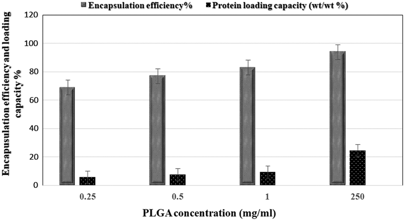
Measurement of the ratio of EE and protein LC using different concentrations of PLGA polymer (0.25, 0.5, 1.0, and 250 mg). EE ratio reached its highest of 94% and calculated by (A − B/A∗100); while LC was 24.6% and calculated by (A − B/C∗100) at 250 mg of PLGA. The p values of EE and LC were shown to be significant (p = 0.01 and p = 0.0001). EE, encapsulation efficiency; LC, loading capacity; PLGA, poly(lactide-co-glycolide).
Characterization of polymeric HBP-NP
The physical and chemical characteristics of the prepared HBP-NPs were evaluated to assess the size, shape, zeta potential, and surface characteristics of HBP-NP following standard techniques (outlined below).
HBP-NP characteristics by using AFM
The size, shape, and surface characteristics of free PLGA and the encapsulated HBP-NP were examined by AFM. The results showed a typical spherical shape with a size distribution of PLGA-encapsulated HBV protein (HBP-NP) (Fig. 2). The average size of free PLGA alone was 41.30 (Fig. 2A), while that of HBP-NP product was 72.08 (Fig. 2B) nm with smooth surface morphology, which were presented here in a two-dimensional and three-dimensional shape NP.
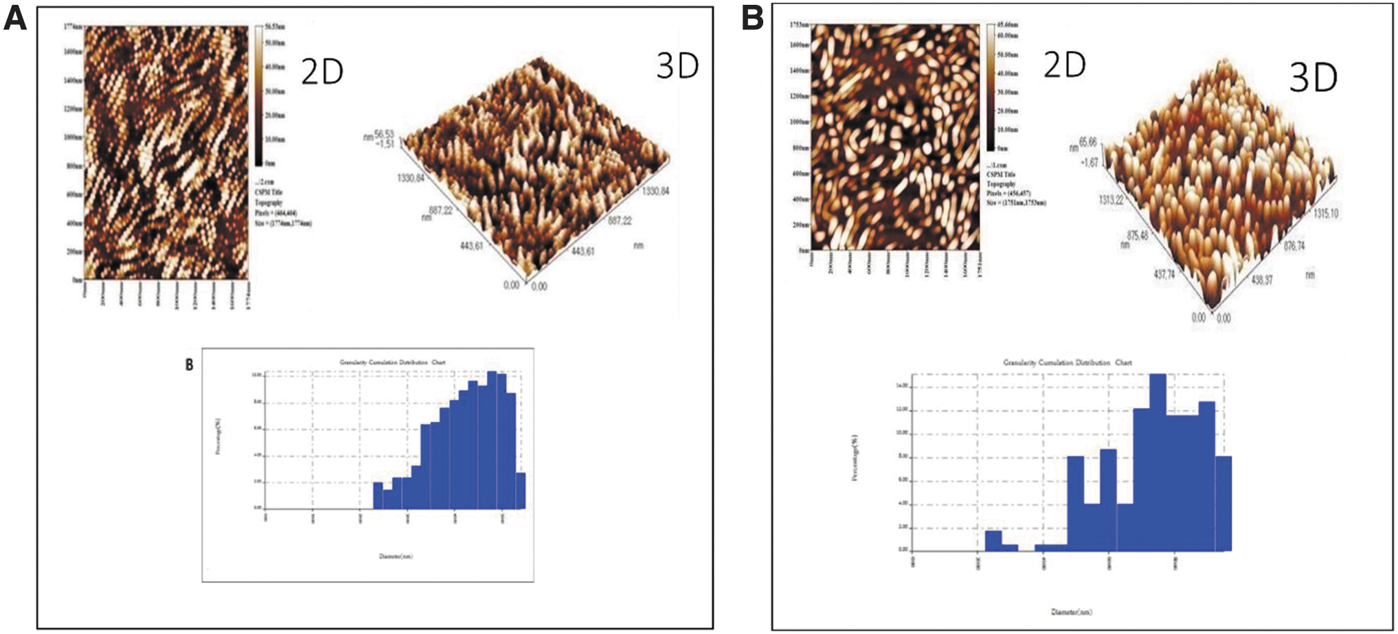
Morphology of free PLGA and HB-NV tested by AFM.
Examination of HBP-NP morphology by field emission scanning electron microscopy
The surface morphology of the formulated HB-NV was determined by using scanning electron microscopy (Mira-3; Tescan) at a beam intensity of 700 and working distance of 4.95 mm. The NPs had variable sizes ranging from 31.27, 53.12 to 67.93 nm, which resulted from different readings of the SEM (Fig. 3A), but when the mean and standard deviation were calculated it appeared to range between 16 and 20 nm (Fig. 3B).
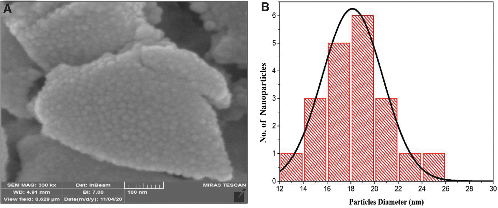
Morphology of HBP-loaded PLGA nanoparticles emulsified by the double emulsion solvent evaporation technique.
Measurement of the optical properties of HB-NV by ultraviolet absorption spectrophotometry
The UV absorption spectroscopy profile of HB-NV, HBP, and PLGA appeared as single peaks at 265, 230, and 355 nm, respectively. The absorbance spectrum for the HB-NV produced a unique new band at 265 nm among other spectra (Fig. 4).
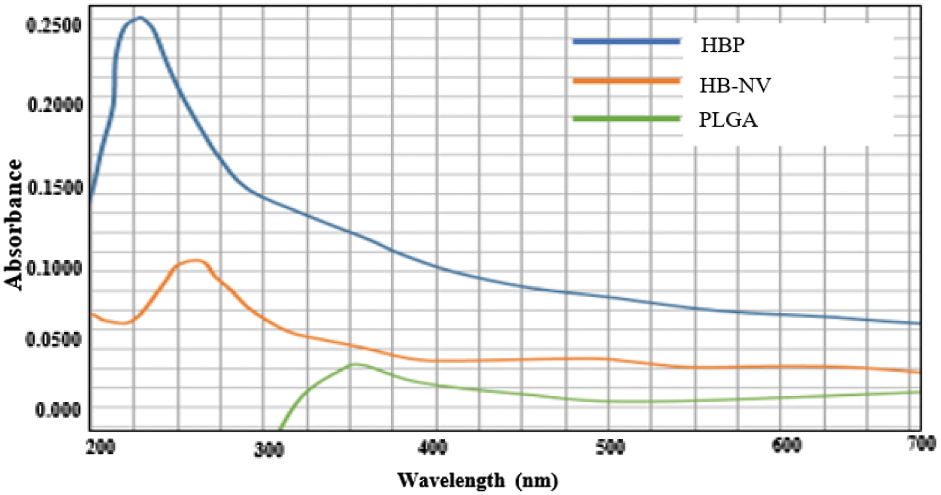
Measurement of the optical properties of HB-NV compared with free PLGA (PLGA) and HBP, by using UV-visible absorption spectrophotometry. The optical HB-NV (265 nm), HBP (230 nm), and PLGA at 355 nm. UV, ultraviolet. Color images are available online.
Identification of functional group of free PLGA and HB-NV by FTIR spectroscopy
The stable formulation of the current HB-NV was evaluated for FTIR spectra and shown to have three absorption peaks within the range 3504.42–3448.49, 1323.08–1245.93, and 850.55–628.75 cm−1 compared with the PLGA alone, which had four absorption peaks (2997.17–2950, 1766.6, 1272.93, and 738.69–702.04 cm−1) (Fig. 5).

Measurement of the absorption spectra of free PLGA and HB-NV by using FTIR spectroscopy.
Range of stability dispersion of HB-NV by zeta potential
The zeta potential of the free PLGA and HBP-NP showed negative charges of both the free PLGA and HBP-NP, which were positioned within the high stability dispersion range of −64.00 and −69.00 mV with mobility −1.23 and −1.39 (μ/s)/(V/cm), respectively (Fig. 6). These data indicate that the stability of the encapsulated HB-NV ended up to be more stable than the PLGA alone.

Distribution graphs of ZP (in mV) and ELS (in V/cm) mobility showing negative ZP value measured by using zeta sizer of free PLGA
Cellular immune response of HB-NV in vitro
Addition of HB-NV to PBL in vitro cultures resulted in induction of IL-2 (Fig. 7) and IFN-γ (Fig. 8) secretion after 72 h post-treatment, as measured by ELISA. The T cell immune responses of the optimized HB-NV also resulted in induction of CD4+ and CD8+ (data not shown) cell proliferation by flow cytometry. HB-NV had significantly higher induction levels of the studied immunological and cellular markers, which superseded those of free PLGA or HBP alone. It should be mentioned that the induction levels of IL-2 and IFN-γ were significantly higher by 3.3% than their levels in untreated cultures (Figs. 7 and 8, respectively). The levels of IL-2 and IFN-γ were also accompanied by stimulation of CD4+ (7.8%) and CD8+ (23.9%) counts in comparison with the untreated PBL cultures, particularly at the high concentration (100 μg/mL) of HB-NV (data not shown). Regardless of the concentration of HB-NV used, the induction responses of studied markers (IL-2, IFN-γ) and CD4+ and CD8+ (data not shown) were always greater in magnitude than these individually added PLGA, HBP, or Euvax (Figs. 7 and 8).

IL-2 levels in PBL suspension treated at various concentrations (20, 50, and 100 μg/mL) of HB-NV, PLGA polymer (PLGA), HBV-protein (HBP), and HBV-Euvax-B (Euvax) compared with control-untreated cultures (control). **p ≤ 0.01, *p ≤ 0.05, NS, not significant; PBL, peripheral blood lymphocytes.
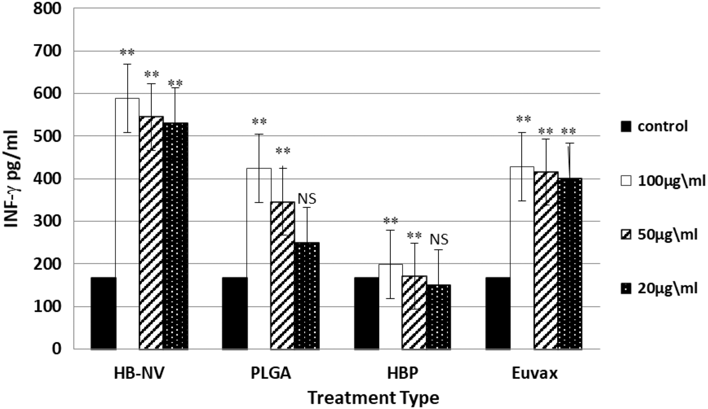
INF-γ level in PBL suspension treated at various concentrations (20, 50, and 100 μg/mL) of HB-NV, PLGA polymer (PLGA), HBV-protein (HBP), and HBV-EuvaxB (Euvax) in comparison with the control-untreated cultures (control). **p ≤ 0.01, *p ≤ 0.05.
Discussion
The physical and chemical characteristics of the prepared HBV-NPs were evaluated to assess the size, shape, zeta potential, and surface characteristics of HBV surface protein-PLGA nanovaccine (HB-NV). Polymeric particles encapsulating antigen can be synthesized by a variety of methods, including emulsion-based methods, salting out, nanoprecipitation, and spray drying (18,28). The most common method used for antigen encapsulation was double emulsion (4). In this study, the average particle size of HB-NV was found to be much higher than that of free PLGA alone (Fig. 2). The average particle size was shown to be within the range between 20 and 100 nm, which has the ability to directly enter the lymphatic system within a few hours of administration, while 200–500 nm particles must be internalized by APCs to reach the lymphatic system (19). In clinical applications, it has been shown that the microparticles have a tendency to promote humoral responses, whereas the NPs are able to penetrate into cells and eventually induce specific cellular immune responses (13).
Regarding the shape configuration, AFM and SEM images revealed that the encapsulated NPs were typically smooth and spherical in shape with higher densities (Figs. 2 and 3). Results about the NP shapes were controversial where a spherical shape was shown in this study and others (5,31). This shape was considered more efficient as antigen depot and eventually controlled antigen release. Conversely, the rod-shaped NPs had greater rates of internalization by dendritic cells compared with spherical NPs (30).
The UV absorption spectroscopy profile of HB-NV, protein, and PLGA appeared as a single peak at 265, 230, and 355 nm, respectively. The absorbance spectrum for the HBV-NPs produced a unique new band at 265 nm among other spectra (Fig. 4). This indicates a successful encapsulation of the HBP with the PLGA. These results were similar to those reported earlier (7,8).
The stable formulation of the current HB-NV was evaluated for FTIR spectra and shown to have three absorption peaks within the range 3504.42–3448.49, 1323.08–1245.93, and 850.55–628.75 cm−1 compared with the PLGA alone, which had four absorption peaks (2997.17–2950, 1766.6, 1272.93, and 738.69–702.04 cm−1) (Fig. 5). The new peak pattern of the HB-NV was obviously either shifted or diminished compared with the free PLGA, indicating a successful uniform encapsulation of PLGA with HBP. Such activity makes PLGA-based vaccine delivery carriers more active to reach the target sites. Results of this study are similar to those described earlier (24).
As zeta potential is considered as an essential characteristic of NP stability, especially when the PLGA NPs were emulsified with PVA, they are mostly ended up with negative surface charges (4,9,23). These reports are consistent with the findings of this study where HBV-NPs had typical negative zeta potential (−69.00 mV) due to the presence of terminal carboxylic groups (–COOH) on the surface of NPs (Fig. 6). This value is positioned within the range of excellent anionic zeta potentials, which effectively contributes to the colloidal stability, but its influence on the stability needs further investigation. Since most cellular membranes are negatively charged, zeta potential can affect the permeability of cell membranes to NPs, with anionic NPs being generally nontoxic. According to the classification described by Müller and Hildebrand (20), the stability of NPs was classified according to the values of the zeta potential scale of that over −60 mV, −30–60 mV, and below −20 mV, as excellent, neutral, and limited physical colloidal stability, respectively. A zeta potential value within the range between −30 and +30 mV is generally considered to have sufficient repulsive force to attain better physical colloidal stability of the emulsion (2). On the contrary, a small zeta potential value can result in particle aggregation and flocculation due to the van der Waals attractive forces limiting its physical stability (12,16,22).
T cell immune responses of the optimized HB-NV were measured by the induction of CD4+ and CD8+ cell proliferation by 40% than PLGA alone and by 36% over that observed with Euvax as well as enhancement of IL-2 and IFN-γ secretion by using flow cytometry and ELISA in in vitro PBL cultures. The induction levels of these immune cellular markers due to HB-NV addition have superseded those resulted from the individual addition of free PLGA or HBP alone. It should be mentioned that the induction levels of IFN-γ were accompanied by stimulation of CD8+ counts by sixfold, particularly at the high concentration (100 μg/mL) of HB-NV. Regardless of the concentration of HB-NV used, the induction responses of studied markers (IL-2, IFN-γ, CD4+, and CD8+) were always greater in magnitude than other treatment types (data not shown). This HB-NV was able to induce T cell responses, which is the characteristic of Th1 type cytokines such as IL-2 and IFN-γ. The enhanced Th1 response for both cytokines may provide indication for a strong cellular immune response that is capable of viral clearance and can adequately control viral replication. Moreover, flow cytometry data of T cell subsets, especially those for CD8+ and its correlation with elevation levels of IFN-γ, may support their effective role in HBV eradication and minimizing the term of chronic HBV (26). It is worth noting that the formulation, production, and stability of nanovaccines would facilitate its use in developing countries such as Iraq. It was also shown that CD4+ Th1 cells enhance defense against the intracellular protozoal pathogen Leishmania major (27), whereas Th1 cell production of IFN-γ activates macrophages to kill intracellular pathogens. On the contrary, Th2 cytokines usually contribute to reduction of chronic hepatitis, due to their immunomodulatory role (15). The correlation between immune responses of T cell subsets and Th1/Th2 cytokine production is complicated and not well understood (29). Furthermore, it has been shown that nanovaccines were able to produce better humoral and cellular responses in mice when administered intramuscularly (3,10). As a matter of fact, the microparticles have a tendency to promote humoral responses, whereas the NPs are able to penetrate into cells and eventually induce specific cellular responses (14).
Conclusions
In summary, successful development and characterization of HBV surface antigen of HBV carrier were performed using PLGA aiming to produce HB-nanovaccine (HB-NV). HB-NVs were formulated by using double emulsion solvent evaporation technique (w/o/w), which showed 94% EE and 24% protein LC. The produced HB-NV had high densities, spherical shape with an average size of 71.08 nm with high stability dispersion and negatively charged nanomolecules as assessed by AFM, SEM, zeta potential, FTIR, and UV. The in vitro cell culture of PBL treated with HB-NV compared with controls induced secretion levels of IL-2 and IFN-γ, and enhanced absolute number of CD4+ and CD8+ T lymphocytes. As the newly designed HB-NV effectively stimulated the in vitro cellular immune response with a therapeutic potential to be pursued further. This latter properties deserve further in vivo investigations.
Footnotes
Authors' Contributions
H.M.N. and M.M.M. have contributed equally to this study.
Acknowledgments
The authors acknowledge the support of the Department of Molecular and Medical Biotechnology, College of Biotechnology, Al-Nahrain University for using their facility and project supervision. They also appreciate the support of Mr. Mahmoud Mohammad for partially funding the experimental cost of this study.
Ethics Approval
The article does not contain clinical studies or patient data.
Author Disclosure Statement
No competing financial interests exist.
Funding Information
No funding support was received for this study.