
Abstract
Pseudorabies (PR), the causative agent of Aujeszky's disease, has rapidly increased in recent years and has caused significant economic losses. To understand the seroprevalence and epidemiological characteristics of PR in Tianjin, China, a total of 23,627 blood and 1,093 tissue samples were collected from 228 pig farms during January 2010 to December 2018. The Pseudorabies virus (PRV) glycoprotein E (gE) antibody was tested by enzyme-linked immunosorbent assay (ELISA), and wild-type PRV (WT PRV) was detected by gE-polymerase chain reaction (PCR). Macroscopic and microscopic lesions were observed in tissue samples. The results showed that 46.70% of the serum samples and 49.76% of pig farms were seropositive for PRV gE antibody based on the ELISA results, and 13.54% of the tissue samples were positive for WT PRV detected by PCR. The positive rate of serum samples increased rapidly after 2011 and reached 62.40% in 2013. Although it gradually decreased from 2014 to 2018, the positive rate of serum samples remained at a high level. The positive rate of pig farms showed the same trend. Moreover, after 2011, the detection rate of WT PRV was increased rapidly and was significantly higher than in 2010 and 2011. Macroscopic and microscopic lesions were observed in various tissues during histopathological examination. Based on univariate analysis, the increased risk of seropositivity was associated with the immune status and infection in sows and fattening pigs. These findings demonstrate that PR was prevalent in the region of Tianjin, China. These epidemiological data can assist in the control of PR.
Introduction
Pseudorabies (PR
In 1902, PR was first discovered by Aujeszky in Hungary, and then the disease spread to most pig-raising countries and regions of the world. Owing to effective eradication programs and the use of glycoprotein E (gE)-deleted PRV vaccines, PR has been eradicated from domesticated pigs in the United States and many European countries (3). In China, an outbreak of PRV was first reported in the 1950s. From the 1990s to 2011, the number of PR cases increased rapidly in pig farms. The Bartha-K61vaccine was adopted in most pig farms in China, and PR was effectively controlled in most areas (2,12,20). Since late 2011, severe PR outbreaks occurred in PRV-vaccinated swine herds in some provinces and regions of China (1,14,16), posing a new threat to the pig industry (1,15,17,21). Studies indicated that this re-emergence was caused by PRV variant strains (1,6,13,14,19,26). Thus, epidemiological data of PR are necessary for the prevention and control of the disease.
Tianjin is China's third largest city, one of the municipalities in China. To the east of Tianjin province lies the Bohai Gulf, Beijing is in the northwest of Tianjin, and the border of Hebei encircles the north, south, and west sides of Tianjin. Tianjin has 16 subordinate districts, 10 of which are agriculture-related districts. Jizhou, Baodi, Wuqing, and Ninghe districts are four main pig-breeding areas. According to the official statistics of the Tianjin Statistical Bureau, there were ∼2.972 million pigs and 0.29 million sows at the end of 2017 in Tianjin.
Epidemiological data for PR in Tianjin are limited. Therefore, we designed the current study to investigate the seroprevalence and the infection rates of the PRV in Tianjin between January 2010 and December 2018 in Tianjin.
Materials and Methods
The animals used in this study were cared for in accordance with established guidelines developed by the China Council on Animal Care. The animal experimental protocols were approved by the Animal Welfare and Ethical Censor Committee at Tianjin Academy of Agricultural Sciences (Tianjin, China).
Investigation region and sample collection
The investigation area encompassed the major districts in Tianjin, excluding urban districts (Fig. 1). From January 2010 to December 2018, a total of 23,627 blood samples and 1,093 tissue samples were collected from 228 pig farms (no. of sows ≥50), where the pigs had been immunized with PRV live vaccine (Bartha-K61 strain). Among the pig farms, 20 pig farms were located in Jizhou (JZ), 36 in Wuqing (WQ), 17 in Baodi (BD), 45 in Ninhe (NH), 18 in Beichen (BC), 12 in Dongli (DL), 10 in Xiqing (XQ), 12 in Jinnan (JN), 43 in Jinhai (JH), and 15 in Binhai (BH). Blood samples for this study were randomly collected from boars, sows, replacement pigs, nursery pigs, and fattening pigs (Table 1). Tissue samples including fetuses, lungs, spleens, and brains were collected from pigs of different ages with signs suspected of PRV infection (Table 3). Data were recorded for each sample, including age, production phase, animal identification, date, and geographical location of origin.
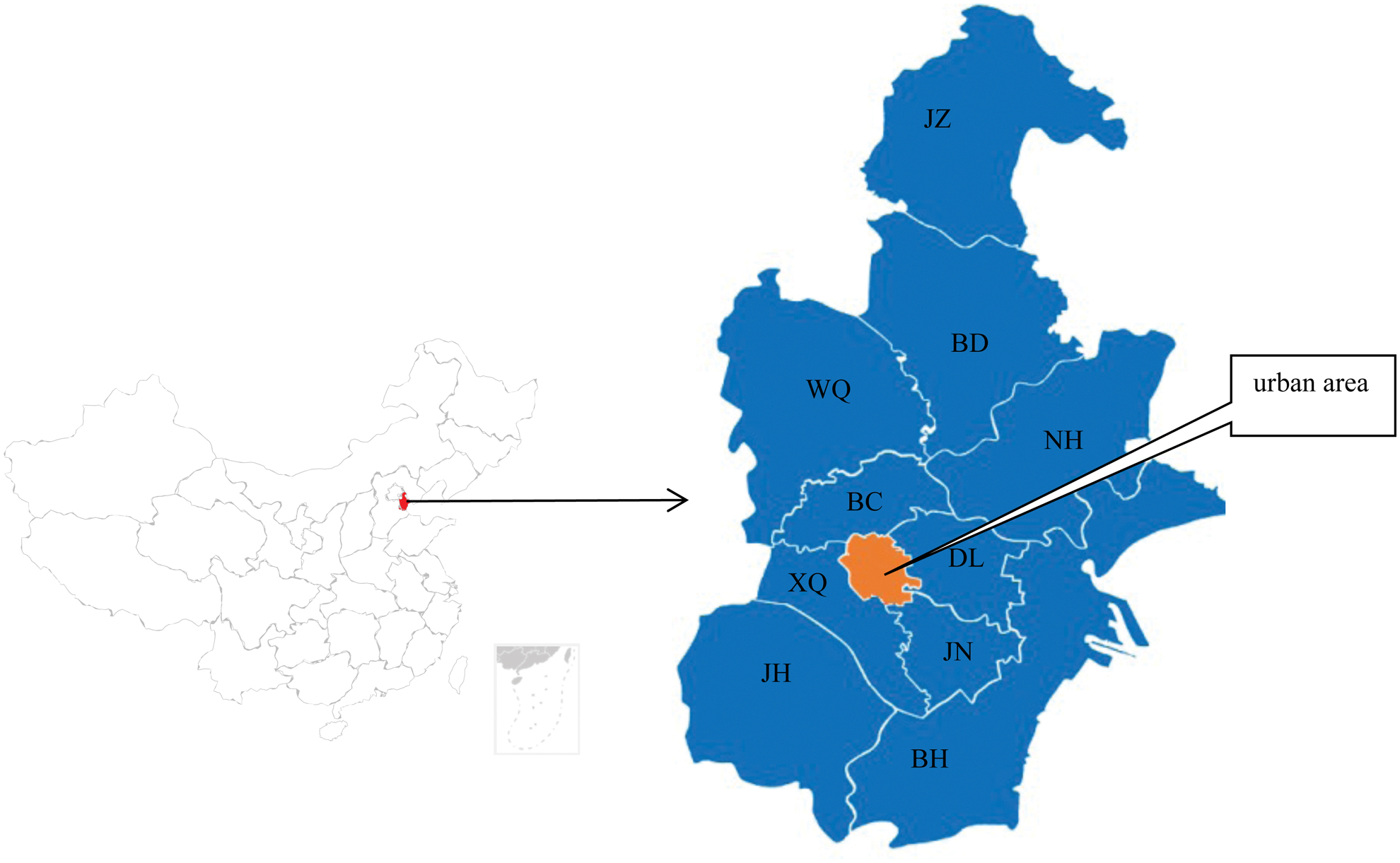
Map of Tianjin: The 10 regions are: JZ, Jizhou; BD, Baodi; WQ, Wuqing; NH, Ninhe; BC, Beichen; DL, Dongli; XQ, Xiqing; JN, Jinnan; JH, Jinhai; BH, Binhai. Color images are available online.
Pseudorabies Virus Glycoprotein E Antibody-Positive Rate Among Different Pig Herd Types
CI, confidence interval; OR, odds ratio; ref., references.
Serological detection of gE antibody by enzyme-linked immunosorbent assay
Blood samples were collected under sterile conditions, and serum was separated and kept at −20°C before testing. The samples were tested by PRV gE antibody enzyme-linked immunosorbent assay (ELISA) kits (IDEXX) according to the manufacturer's instructions, The test differentiates between vaccinated and infected pigs. Serum samples with an S/N ratio ≤0.60 were considered positive for the PRV anti-gE antigen. In this study, a farm was considered to be a positive farm if at least one positive sample was detected. Otherwise, the farm was considered to be a negative farm.
PRV detection of gE gene by polymerase chain reaction
After collection, tissues were minced, immersed in phosphate-buffered solution, and homogenized using the Stomacher 80 Biomaster (Seward, United Kingdom). After centrifugation at 4,000 g for 5 min, the supernatants were harvested for DNA extraction. DNA extraction was performed using the Tissue DNA/RNA Extraction Reagent Kit (TACO BIO, Xiamen, China) following the manufacturer's instructions. Polymerase chain reaction (PCR) assays targeting the gE gene of PRV were conducted using primers P1 (5′-CCGGCCCATCTGGTGAACGT-3′) and P2 (5′-CCCACCGCCACAAAGAACACG-3′) (23). The PCR reaction was conducted in a 50 μL mixture including 25 μL 2 × GC buffer (TaKaRa BIO, China), 4 μL 2.5 mM of each dNTPs, 2.5 U of rTaq DNA polymerase (TaKaRa BIO), 2 μL of each 10 μmol primers, 5.5 μL of extracted DNA, and distilled water up to 50 μL of final volume.
The amplifications were performed in a thermal cycler (Bio-Rad) under the following conditions: after initial denaturation at 95°C for 5 min, 30 cycles were conducted at 94°C for 30 sec, 56°C for 30 sec, and 72°C for 30 sec, followed by a final extension at 72°C for 10 min. The PCR product was detected by electrophoresing through 1.0% agarose gel in 1 × TAE (40 mM Tris-aceate, 1 mM EDTA, pH 8.0). The estimated amplicon size of PRV gE was 337 bp. The PCR method used in this experiment has high specificity (Supplementary Fig. S1).
Clinical signs, gross lesions, and histopathological lesions
Clinical cases were observed and dissected during 2010 to 2018 from 228 pig farms in Tianjin that had vaccinated using PRV live vaccine (Bathar-K61 strain) and in which pigs presented clinical signs of PR, such as high temperature (40°C to 41.5°C), abortion, and severe neurological disorders in piglets. Brain, tonsils, trigeminal ganglion, liver, and spleen tissues were collected for histological examination. These sections were fixed with 10% phosphate-buffered formalin for 24 h and embedded in paraffin wax, after which 3-μm-thick slices were cut and stained using a hematoxylin and eosin (H&E) staining kit (Solarbio, Beijing, China) according to the manufacturer's instructions..
Statistical analysis
Positive rates of PRV gE antibody and wild-type PRV (WT PRV) were compared by analyzing years, different herds, and different regions. Possible risk factors associated with seropositivity and virus infection were analyzed by univariate logistic regression models using SPASS 21.0 (IBM, Inc.). For each date the odds ratio (OR) and 95% confidence interval (CI) were calculated. If the 95% CI of a variable was >1, it was considered an influencing factor. Chi-square test was conducted to compare the differences between regions using the simple cross tables in the descriptive analysis of SPSS software. A one-way analysis of variance was used for the determination of differences in measurements between groups using GraphPad Prism® 7.00 (GraphPad Software, Inc.).
Results
Seroprevalence of PRV gE antibody
The average positive rate of PRV gE antibody of serum samples and pig farms was 46.70% between 2010 and 2018. The lowest positive rate and highest positive rate of serum samples were 1.14% (2010) and 62.40% (2013), respectively (Table 1). The seroprevalence of serum samples varied with different herds and years (Fig. 2 and Table 1). The positive rates of boars, sows, nursery pigs, replacement pigs, and fattening pigs showed similar trends with lower rates in 2010 and 2011 than in 2012 to 2018. Among the five herds, the sample seroprevalence of sows was highest (51.10%) with the peak occurring in 2014 (65.23%). The sample seroprevalence of boars and fattening pigs was also highest in 2014, with rates of 51.72% and 62.61%, respectively. However, the positive rates of replacement pigs and nursery pigs were peaked in 2013.
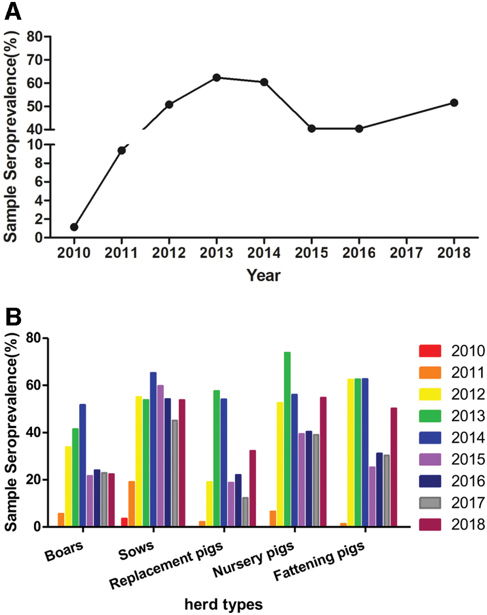
Sample seroprevalence in different years
The survey results of the pig farms indicated that the positive rate increased rapidly from 2012 to 2014; the rate reached the highest level in 2014 (68.22%), gradually decreased to 46.09% in 2017, and then slightly increased to 48.98% in 2018 (Table 2). The pig farms were distributed in 10 regions of Tianjin, and the positive rate of pig farms varied with geographical location. The average positive rate of pig farms in BC district (54.88%) was the highest, whereas that in BD district (40.30%) was the lowest. Chi-square analysis showed that the differences between regions were not significant (χ 2 = 9.108, p = 0.427).
Proportion of Pseudorabies Virus Glycoprotein E Antibody Positive Pig Farms in Different Regions
JZ, Jizhou; BD, Baodi; WQ, Wuqing; NH, Ninhe; BC, Beichen; DL, Dongli; XQ, Xiqing; JN, Jinnan; BH, Binhai.
PCR detection for PRV gE gene
The average positive rate of samples was 13.54% between 2010 and 2018 (Table 3), ranging from 5.11% (2011) to 30.50% (2012). The positive rate of samples in 2012 was significantly higher than those in other years (p < 0.001). In 2010 and 2011, the positive samples were mainly from suckling piglets and nursery pigs. By 2012, the overall positive rate of WT PRV in pigs suddenly increased to 30.50%, and the positive rates of different pigs increased, especially in fattening pigs at 58.83%. The results showed that PR was prevalent in various pig groups and caused clinical cases. From 2013 to 2018, the average positive rate of WT PRV in pigs decreased from 20.70% to 9.88%, indicating that prevention and control measures had a significant effect.
Wild-Type Pseudorabies Virus-Positive rate in Tianjin from 2010 to 2018 by Polymerase Chain Reaction Method
Clinical signs, gross lesions, and histopathological lesions
The main signs in suckling piglets were fever, diarrhea, and nervous system disorders. The incidence rate was <20.0%, and mortality was 50.0–100.0%. The clinical characteristics of nursing pigs were similar to those of suckling piglets, with an incidence rate of 5.0–80.0% and a case fatality rate of 10.0–30.0%. Sows mainly showed abortion, mummified fetuses, stillbirth, and weak piglets, and the incidence rate differed between pig farms (1.0–30.0%). Fattening pigs were characterized by fever and respiratory signs, with an incidence rate of 13.5–50.0% and a low mortality rate. In severe cases, infected pigs developed vomiting, frothing at the mouth, tremors, and eventually died (Fig. 3). After dissecting the aborted fetuses and dead pigs among the PR cases, scattered bleeding spots on the lung surface and grayish-white necrotic foci on the surface of the liver were observed (Fig. 3). In addition, pathological changes could be seen in the brain, tonsils, trigeminal ganglion, liver, and spleen when histopathological examination was performed (Fig. 4).
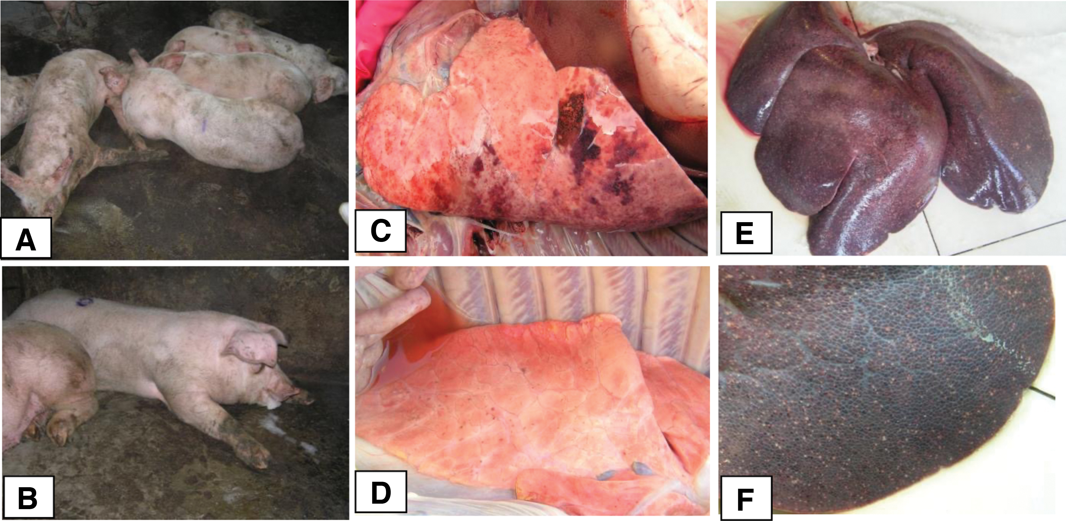
Clinical signs and gross lesions observed in fattening pigs infected with PRV.
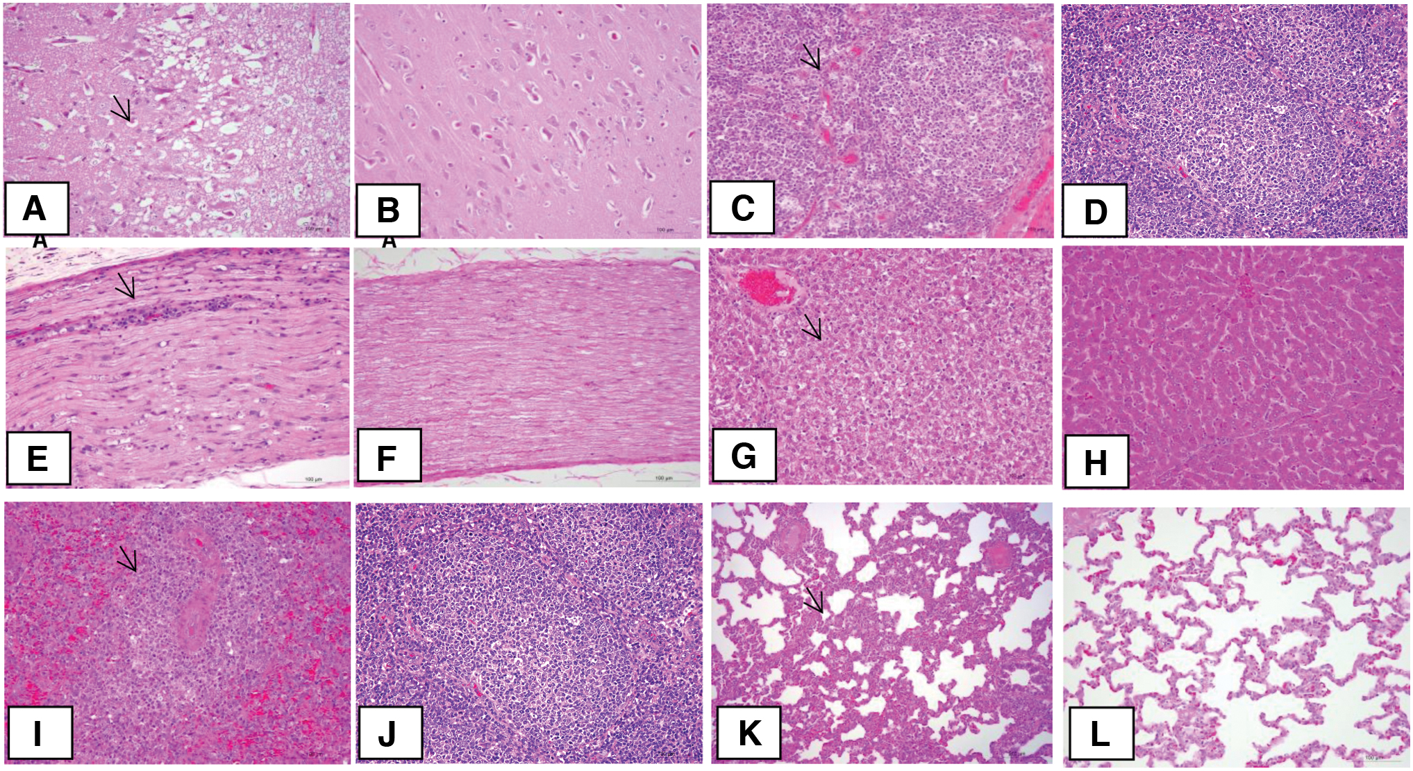
Histopathological lesions in the tissues of fattening pigs infected with PRV.
Discussion
PR is one of the most serious diseases threatening the pig industry. At the end of 2011, severe PR outbreaks occurred in several provinces of northern China, and the epidemic spread rapidly, causing huge economic losses to the Chinese pig industry (4,12). PR outbreak also occurred in many pig farms immunized with live PRV vaccines in Tianjin, resulting in a rapid increase in seropositivity for WT PRV. However, there were few systematic studies of the prevalence or infection of PRV in Tianjin. This study is the first epidemiological investigation focusing on PRV seroprevalence and infection rates in Tianjin, China. The results can be useful for initiating control measures against PR for pig farms in Tianjin.
Based on the fact that the PRV gE gene deletion live vaccine was widely used in Tianjin pig farms, detection of the PRV gE gene and PRV gE antibodies can distinguish immunized pigs from WT PRV-infected pigs. The PRV gE gene detection by PCR and PRV gE antibody detection by ELISA played important roles in the PR diagnosis and immune monitoring of pig farms; these data can provide us with detailed and reliable data for PRV infection (25).
According to the serological investigation, the prevalence of PR in Tianjin could be divided into three stages for the period from 2010 to 2018. In the first stage, from 2010 to 2011, the positive rates of PRV gE antibody were the lowest: 1.14% and 9.38%, respectively. In this period, the proportion of PRV gE antibody-positive pig farms was also low, indicating that the PRV infection rates were low in this area. The second stage was from 2012 to 2015. The positive rate of PRV gE antibody rose sharply to 62.40% in 2013 with an average positive rate of 54.92% during the 4 years. This period was the peak of PRV infection and incidence in Tianjin pig farms. The period 2016 to 2018 comprised the third stage of PRV infection. The PRV gE antibody-positive rate of pig farms and serum samples showed a gradual downward trend. In 2017 and 2018, the proportions of pig farms positive for PRV gE antibody were 46.09% and 48.98%, respectively. This indicated that the number of pigs carrying the WT PRV in the investigated areas was still large, and the risk of PR in the third stage was much higher than in the first stage (22).
There are many reports on epidemiological investigations of PR, but there are few reports on the prevalence of the PRV gE antibody and WT PRV in different herds. In this study, 23,627 blood samples and 1,093 tissue samples from different herds were collected for analysis. As given in Table 1, in 2010, antibody-positive pigs were mostly sows, whereas all other herd groups were negative. In 2011, the positive rates of gE antibody in the five herd groups were all increased. After 2012, the positive rates of serum samples in different herd groups were higher than in 2010 and 2011, and the positive rate order was fattening pigs > sows > nursery pigs > boars > replacement pigs. The univariate analysis indicated that sows (OR, 3.054; 95% CI, 2.505–3.722), nursery pigs (OR, 2.941; 95% CI, 2.412–3.586), and fattening pigs (OR, 2.319; 95% CI, 1.902–2.827) were at higher risk than the boars or replacement pigs. As given in Table 3, the WT PRV-positive rate of tissues increased rapidly and was significantly higher than in 2010 and 2011. After 2011, the infection rates of fattening pigs increased significantly. The univariate analysis indicated that fattening pigs (OR, 2.479; 95% CI, 1.403–4.380) were at higher risk than other herd groups. These studies suggested that the immune status and infection of sows and fattening pigs have an important influence on PRV seropositivity in pig farms.
For different herds, corresponding measures were taken starting from 2012: vaccine immunization for sows and boars was changed from 2–3 to 3–4 times per year; fattening pigs were vaccinated at 9–11 weeks of age. Before 2012, they were generally not vaccinated, and the boars and replacement pigs positive for PRV gE antibody were eliminated. From 2013 to 2017, based on the adjustment of vaccination procedures and the strengthening of biosafety control measures, the overall positive rate of pigs gradually decreased, and the positive rates of different herds also decreased. These findings indicated that the corresponding comprehensive measures adopted since 2012 to control PR have been effective in Tianjin since 2012.
We also investigated the PR prevalence of pig farms in different areas. Ten regions of Tianjin were surveyed. Based on the geographical location and significant differences in economic development level, the development of pig production in the 10 regions was unbalanced. As given in Table 3, seroprevalences of pig farms in different regions varied significantly depending on geographical location. The positive rate of pig farms in BC district was the highest, whereas that in BD district was the lowest. The subsequent univariate analysis indicated that the BC district (OR, 1.590; 95% CI, 0.813–3.110) had higher risk for PRV herd seropositivity than other districts. This was probably because of the pig farms in BC district being mainly small scale, imperfections in vaccination, differences in management procedures, and biosecurity measures that led a higher risk of PR incidence and epidemics.
In addition, our results demonstrated that after 2011, the infection rates of fattening pigs increased significantly. It is noteworthy that the fattening pigs infected with PRV usually had obvious clinical manifestations of PR, and the severe cases usually died. After dissecting the dead pigs, the livers had obvious gray-white necrotic spots, and the lungs had interstitial pneumonia with bleeding spots, which were rarely seen before 2012. Since most pig farms had been inoculated with the PR vaccine, the above results indicated that the infectivity and pathogenicity of the novel epidemic PRV increased significantly. In fact, this conclusion has been confirmed in pathogenicity experiments with PRV isolated strains in our laboratory (11). As the protective effect of the current vaccine is declining, the original vaccination program for pig farms cannot effectively protect pigs against infection with PRV variant strains (18,24). Although the prevalence of PR has been controlled to some extent by strengthening vaccine immunization, traditional PR vaccines such as Bartha-K61 do not provide ideal immunity protection. It is gratifying that significant progress has been made in the research on new PR vaccines (3,14,15,16,22), which creates favorable conditions for the prevention of PR in the future.
In conclusion, this study investigated the seroprevalence, incidence rate, and clinical features of PR in Tianjin, China, between January 2010 and December 2018. These findings indicate that we are facing a serious situation in the prevention and control of PR. Monitoring antibody detection, adjusting immunization programs, and strengthening biosafety measures should be conducive to the prevention and control of PR.
Footnotes
Authors' Contributions
Conceptualization: L.Z., W.R.; Methodology: J.C., X.L., C.L., L.W.; Formal analysis: W.R.; Data curation: J.C.; Investigation: X.T., F.L., Z.D.; Writing-original draft: L.Z., W.R.; Funding acquisition: L.Z., M.Y.; Data curation: X.L., C.L., S.J.; Writing-review& editing: M.Y., L.Z.; Project administration: M.Y., L.Z.. All authors have read and agreed to the published version of the article.
Acknowledgment
The authors thank the pig farmers for their contribution in sample collection.
Author Disclosure Statement
No competing financial interests exist.
Funding Information
This study was supported by Tianjin Academy of Agricultural Sciences young researchers innovative research and experimental project (Grant No. 201905), and the Science and Technology Project of Tianjin (Grant no. 18YFZCNC01110), and Tianjin Pig industry technical system pig disease prevention and control post project (Grant No. ITTPRS2017003). The funders did not play any role in the design, conclusions, or interpretation of the study.
Supplementary Material
Supplementary Figure S1
References
Supplementary Material
Please find the following supplemental material available below.
For Open Access articles published under a Creative Commons License, all supplemental material carries the same license as the article it is associated with.
For non-Open Access articles published, all supplemental material carries a non-exclusive license, and permission requests for re-use of supplemental material or any part of supplemental material shall be sent directly to the copyright owner as specified in the copyright notice associated with the article.