
Abstract
Hepatitis B Virus (HBV) is posing as a serious public health threat mainly due to its asymptomatic nature of infection in pregnancy and vertical transmission. Viral sensing toll-like receptors (TLR) and Interleukins (IL) are important molecules in providing an antiviral state. The study aimed to assess the role of TLR7-mediated immune modulation, which might have an impact in the intrauterine transmission of HBV leading to mother to child transmission of the virus. We investigated the expression pattern of TLR7, IL-3, and IL-6 by RT-PCR in the placentas of HBV-infected pregnant women to see their role in the intrauterine transmission of HBV. We further validated the expression of TLR7 in placentas using Immunohistochemistry. Expression analysis by RT-PCR of TLR7 revealed significant downregulation among the Cord blood (CB) HBV DNA positive and negative cases with mean ± standard deviation (SD) of 0.43 ± 0.22 (28) and 1.14 ± 0.57 (44) with p = 0.001. IL-3 and IL-6 expression revealed significant upregulation in the CB HBV DNA-positive cases with p = 0.001. Multinomial logistic regression analysis revealed that TLR7 and IL-3 fold change and mother HBeAg status are important predictors for HBV mother to child transmission. Immunohistochemistry revealed the decreased expression of TLR7 in CB HBV DNA-positive cases. This study reveals that the downregulation of TLR7 in the placenta along with CB HBV DNA-positive status may lead to intrauterine transmission of HBV, which may lead to vertical transmission of HBV.
Introduction
Hepatitis B virus (HBV) infection affects over 296 million people and has become a significant public health problem worldwide (WHO, 2022). The primary cause of chronic HBV infection in endemic places such as Asia and sub-Saharan Africa is vertical transmission of HBV from mother to infant (Marjenberg et al., 2022; Shan et al., 2018; Shimakawa et al., 2016). Maternal Immunity contributes greatly to mother to child transmission (MTCT) of HBV (Vyas et al., 2019), therefore identifying predictive maternal indicators for vertical HBV transmission is of utmost importance in preventing transmission from mother to child.
In the innate immune response to viral infections, toll-like receptors (TLRs), a class of transmembrane receptors that recognize pathogen-associated molecular patterns, are essential. TLR7 has a significant role in immune responses during HBV infection (Sepehri et al., 2016). It has been demonstrated that activating TLR7 inhibits HBV replication in vitro and improves the immune response against viruses in animal models (Isogawa et al., 2005). A study by Tian et al. (2015) examined TLR7 expression in the placentas of pregnant women with and without HBV, where the author hypothesized that the downregulation of TLR7 links with the pathophysiology of intrauterine transmission of HBV. The findings demonstrated that the placenta of HBV-positive mothers had considerably lower TLR7 expression than those of HBV-negative controls (Tian et al., 2015). TLR7 has an important and crucial role in HBV infection which has been evident in previous reports (Sepehri et al., 2016).
Vyas et al. (2019) found that downregulation of MYD-88 and TLR7 expression is evident in the cases where the HBV transmission is more common. Further, the study also found that the downstream signaling molecules of TLR signaling pathway were also downregulated in the HBsAg-positive mothers group which gave birth to HBsAg-positive babies (Vyas et al., 2019). However, the direct role of TLR7 in pregnancy in HBV cases has not yet been fully understood. A robust study is firmly necessary to gain a comprehensive understanding of the mechanisms underlying TLR7 downregulation during HBV infection and to determine the potential viability of TLR7 as a therapeutic target in preventing the vertical transmission of HBV from mother to child.
Moreover, the downstream molecules of the TLR signaling pathway, that is, IL-6 an important pleiotropic cytokine for providing an antiviral state in HBV infection and reduction of HBV replication (Alison et al., 2011; Kishimoto, 2006; Kopf et al., 1994; Kuo et al., 2009). IL-6 levels are reported to be upregulated during HBV infection (Hösel et al., 2009; Kuo et al., 2009). Interleukin-3 (IL-3) is evident to increase antiviral immunity, but there is much scope for research into the expression of IL-3 in HBV, which is still not studied (Bénard et al., 2023). Earlier research has evidenced the role of cytokines in the maintenance of pregnancy as well as placentation (Bowen et al., 2002), but their role in intrauterine transmission of HBV is not yet studied.
The present study aimed to analyze the placental expression pattern of TLR7 and interleukins IL-3 and IL-6, which are correlated with vertical transmission pattern of HBV. Further, the analyses of mother HBeAg status, Cord Blood (CB) HBeAg, and HBV DNA status are also evaluated along with the expression study.
Methods
Sample collection and screening
The present study screened a total of 3,587 pregnant women attending the antenatal outpatient department (OPD) of Gauhati Medical College and Hospital, Assam for HBsAg for a period of 2 years (December 2020 to December 2022). Those found positive for HBsAg and negative for other viruses such as HCV, HEV, HIV, and any other autoimmune diseases were recruited in our study and were followed up until delivery. We have excluded any participant receiving antiviral therapy for HBV. After the samples were found to be HBsAg positive, 5 cc peripheral blood, umbilical CB, and a placental tissue measuring 1 × 1 cm sample was collected during delivery using standard procedure.
A total of 72 antenatal subjects with HBsAg-positive status and 23 healthy pregnant women were recruited in the study irrespective of their gravida status. HBsAg and HBeAg were detected by ELISA using the kits – (HbsAgMonolisa™ HBs Ag ULTRA, Catalog no: 72346) and (DIAPRO: Diagnostic Bioprobes srl, Italy, Cat no: HBE.CE), respectively. The biochemical test is done as a routine procedure of the hospital after the cases were found to be HBsAg or HBV DNA positive. The study is conducted according to ethical Principles for Medical Research Involving Human Subjects outlined in the 2013 Declaration of Helsinki, and it is approved by the Institutional Ethics Committee of Gauhati University, Assam and Gauhati Medical College and Hospital. The demographic details of the patients were collected using a standard proforma. Written informed consent was taken for including the patients and healthy controls in this study.
Molecular detection of HBV
Viral DNA was extracted using QIAamp DNA Blood Mini Kit (QIAGEN, Inc., Valencia, CA). The extracted DNA was subjected to PCR amplification for HBV detection. Twenty-five microliters PCR mixture was set up which contained template DNA, PCR master mix, primers 10 mM PreS2 (GGGACACCATATTCTTGG), and S1R (TTAGGGTTTAAATGTATACCC). DNA amplification was performed for 35 cycles. The condition of each cycle was denaturation at 94°C for 3 min and 94°C for 45 sec, primer annealing at 53°C for 1 min, and extension at 72°C for 1 min and 30 sec. The PCR product was further amplified with the inner nested primer set, 10 mM forward primer YS1 (GCGGGGTTTTTCTTGTTGA) and YS2 (GGGACTCAAGATGTTGTACAG). The same PCR conditions were used for the second round of PCR. The final product size obtained was 585 bp.
Expression analysis of TLR7, IL-3, and IL-6 by RT-PCR
Total mRNA was extracted from placental tissue using the TRIzol Reagent (Invitrogen; cat# 15596018). Samples were treated with DNAse I (Invitrogen; cat# AM2224), and 1–5 μg of total RNA was reverse transcribed using iScript cDNA synthesis kit (BIORAD). Gene expression was measured by quantitative real-time PCR (qPCR) using the QuantStudio 3 SYBR Green real-time PCR system (Applied Biosystems) and normalized to glyceraldehyde 3-phosphate dehydrogenase (GAPDH). The fold change in TLR 7, IL-3, and IL-6 gene expression was calculated by 2–ΔΔCt method as defined by (Livak and Schmittgen, 2001). Oligonucleotide used was for TLR7 forward (AATGTCACAGCCGTCCCTAC) having, TLR7 reverse (GCGCATCAAAAGCATTTACA) having Tm = 58.5°C, IL-3 forward (AGGACGGTGACTGGAATGAA) having, IL-3 reverse (TTTTGATGTCCCGAGGCTCT) having Tm = 58.1°C, IL-6 forward (ATTCCAAAGATGTAGCCGCC) and IL-6 reverse (AGTGCCTCTTTGCTGCTTTC) having Tm = 58.1°C.
The product sizes for the oligonucleotides used were 223 bp, 162 bp, and 151 bp, respectively, for TLR7, IL-3, and IL-6. The housekeeping primer (GAPDH) used has sequences for the forward (GGGTCATCATCTCTGCCCC) and for reverse (TGAGTCCTTCCACGATACCA) with a product size of 171 bp.
Immunohistochemistry
Immunohistochemistry was performed to assess the expression of TLR7 in HBV-infected placentas and healthy controls as described by (Pudney and Anderson, 2011). Fresh placental tissue samples were obtained from both HBV-positive patients and healthy control during delivery. Tissues were immediately fixed in 10% neutral buffered formalin for 24 h, followed by dehydration and paraffin embedding. Four-micrometer-thick sections were cut using a microtome and mounted on glass slides. A rabbit monoclonal antibody against protein TLR7 (Abcam; catalog Ab45371, dilution 1:200) was used as the primary antibody. Standard blocking solutions and reagents were used, including 3% hydrogen peroxide to quench endogenous peroxidase activity. Slides were viewed under a light microscope (Leica DM 75; Leica Microsystems, Wetzlar, Germany) with overall magnifications up to 400 × by two independent observers blinded to clinical data.
A cell was determined as immunopositive, when it demonstrated distinctive brown stain on the cell membrane and/or cytoplasm around a nucleus. Images were taken using a CCD camera (Leica Microsystems), mounted on the microscope, and controlled by computer software. Negative controls for the immunohistochemistry were processed in the absence of the primary antibodies. The number of positive cells was evaluated on a scale of (+) to (+++) for TLR7 staining evaluation. (+ for <25% of cells stained; ++ for 25% to 75% of cells stained; +++, > 75% of cells stained). If TLR7 scores of 2 and 3 are achieved, the expression is categorized as high, and if scores 0 and 1 are obtained, the expression is classified as low (Utami et al., 2022).
Statistical analysis
Statistical comparisons were performed using SPSS 21.0 (IBM Corporation) for windows with p-value <0.05 considered as significant. The descriptive data are represented as mean ± standard deviation (SD). Independent t-tests and ANOVA were used for the association study. Multinomial logistic regression was used to perform the regression analysis.
Results
Analysis of different clinicopathological parameters among HBsAg +ve mothers based on CB HBV DNA status
In our study majority of the pregnant women belonged to the age group of 21–30 years (N = 49, 68%). Our study indicated that majority of the patients (73.6%) were from rural areas. The analysis did not reveal any significant difference when comparing among the different area of inhabitation of the patients. Majority of the patients (84.7%) belonged to the low family income group but comparison among the CB HBV DNA-positive and -negative groups did not reveal any significant differences among the income groups. The level of aspartate aminotransferase (AST) was found to be significantly higher in CB HBV DNA +ve group with mean ± SD of 148.3 ± 59.2 and 104.7 ± 45.7 in the negative group with p < 0.005.
Similarly the Alanine transaminase (ALT) level was also found to be significantly raised with mean ± SD of 125.5 ± 46.3 in the positive group and 73.9 ± 38.7 in the CB HBV DNA-negative group (p = 0.001). The Alkaline phosphatase (ALKP) was found to be 270.4 ± 100.5 in the positive CB HBV DNA group and 203.4 ± 73.1 in the negative group, which was also significantly associated (Table 1). Moreover, the platelet count was significantly decreased in CB HBV DNA-positive cases having mean ± SD of 123.92 ± 31.96 compared to 165.48 ± 42.06, p = 0.001.
Comparison of Different Clinicopathological Parameters Among HBsAg +ve Mothers Based on Cord Blood HBV DNA Status
Values are represented as mean ± SD, ANOVA is considered significant when p < 0.05, bolded p-value represents statistical significance, SD stands for standard deviation, N represents number of samples.
AST, aspartate aminotransferase; ALT, alanine transaminase; ALKP, alkaline phosphatase; CB, cord blood.
The analysis revealed that albumin, total protein, creatinine, hemoglobin, and white blood cell counts did not have any significant association p > 0.05. Further, the age groups also did not have any significant association with CB HBV DNA in the studied population. Total bilirubin was found to be 1.21 ± 0.46 in case of CBHBV DNA-positive cases, whereas it was found to be 1.00 ± 0.47 in the negative group, but it is not found to be statistically significant p = 0.061.
Twenty-four (77.4%) of the CB HBV DNA-positive mothers were HBeAg positive, whereas 19 (41.3%) were negative, which is statistically significant p = 0.001. Out of the 41 HBeAg-negative mothers, 37 (90.2%) had CB HBV DNA-negative status. Similarly, 27 (58.6%) out of 46 mothers who had HBV DNA-positive status had CB HBV DNA-positive status and 19 (41.3%) had CB HBV DNA-negative status (p = 0.001). Out of the 12 CB HBeAg-positive mothers, 9 (75%) of them had CB HBV DNA-positive and 3 (25%) have CB HBV DNA-negative state with p-value = 0.008 (Table 1).
The study observed downregulation of TLR7 in the CB HBV DNA positive compared to the negative group having mean ± SD 0.43 ± 0.22 (28) versus 1.14 ± 0.57 (44) with p-value = 0.001. IL-6 expression was increased with mean ± SD of 3.97 ± 1.54 (28) versus 2.03 ± 1.36 (44) in positive and negative groups, respectively, with p-value = 0.001. Also, the IL-3 expression was higher with fold change values 3.77 ± 1.41 (28) and 1.79 ± 0.78 (44) with p-value = 0.001 (Table 1).
Analysis of association between TLR-7, IL-3, and IL-6 expression pattern in placental tissue and HBV replication markers among HBsAg +ve mothers
The mRNA expression of TLR7 reduced significantly upon comparing the HBsAg-positive and -negative groups having mean ± SD of 0.91 ± 0.60 (72) and 2.40 ± 0.48 (22), respectively, with p-value 0.001. Further, within the HBsAg-positive mother groups, HBeAg-positive mother had significantly lower expression of TLR7 with mean ± SD of 0.58 ± 0.47 (31) in the positive group compared to 1.15 ± 0.6 (41) in the negative group (p = 0.001). Similarly, mother HBV DNA-positive group exhibit decreased expression of TLR-7 with mean ± SD of 0.64 ± 0.49 (44) in the positive group and 1.33 ± 0.57 (28) with p-value = 0.001 (Table 2).
Comparison of Mean Expression of TLR-7, IL-3, and IL-6 in Placental Tissue Among HBsAg +ve Mothers Based on Different Replication Marker and Also with Healthy Control
Values are represented as mean ± SD for mRNA expression fold change value by using real-time PCR, ANOVA is considered significant when p < 0.05, bolded p-value <0.05 represents statistical significance, N represents number of samples.
While comparing the expression pattern of IL-3 among the HBsAg-positive and -negative group, the expression was significantly upregulated in the HBsAg positive group which had mean ± SD of 2.23 ± 1.35 (72) compared to healthy controls 0.81 ± 0.54 (22) with p-value = 0.001. Further, among the HBsAg-positive mothers, the HBeAg-positive group had high expression of IL-3 with mean ± SD of 2.94 ± 1.72 (31) compared to 1.70 ± 0.59 (41) in the negative group with p-value = 0.001. Also, the HBV DNA-positive group had higher expression of IL-3 in the positive group with mean ± SD of 2.66 ± 1.55 (44) compared to 1.57 ± 0.44 (28) in the negative group with p = 0.001 (Table 2).
While comparing the expression of IL-6 in HBsAg cases and healthy mothers, IL-6 was found to be elevated having mean ± SD values of 2.40 ± 1.66 (72) in the positive group compared to 0.46 ± 0.17 (22) in the negative group, p = 0.001 (Table 2). Further, in the group within the HBsAg-positive group, the IL-6 expression was upregulated in the positive HBeAg group having mean ± SD of 3.17 ± 1.94 (31) and 1.81 ± 1.12 (41) in the negative group with p = 0.001. The expression of IL-6 was also elevated in the positive mother's HBV DNA group with mean ± SD of 2.97 ± 1.86 (44) and 1.50 ± 0.60 (28) with p-value = 0.001 (Table 2).
Analysis of association between altered TLR-7, IL-3, and IL-6 expression in placental tissue samples with diverse CB HBV DNA status
Upon further stratification based on CB HBV DNA-positive and -negative status among the HBeAg-positive mothers, the placental expression of TLR7 was significantly downregulated with mean ± SD values of 0.40 ± 0.22 (24) compared to the negative group 1.06 ± 0.56 (7) with p = 0.001. In the HBeAg-negative mothers only, 3 had CB HBV DNA-positive status with a mean of 0.63 ± 0.20 compared to 1.20 ± 0.46 (28) with p = 0.048 (Table 3). In the mother HBV DNA-positive status group, the expression of TLR7 was downregulated with mean ± SD values of 0.43 ± 0.23 (27) and 0.88 ± 0.55 (19) with p-value = 0.001.
Analysis of Association Between Altered TLR-7, IL-3, and IL-6 Expression in Placental Tissue Samples with Diverse Cord Blood HBV DNA Status Stratified Based on Viral Replication Marker
Mean ± SD values represents the mRNA fold change values of genes done by using real-time PCR, ANOVA is considered significant when p < 0.05, bolded p-value <0.05 represents statistical significance, N represents number of samples.
However, the negative HBV DNA group also showed a downregulation pattern in the CB HBV DNA group with mean ± SD of 0.45 ± 0.00 (1) in the positive group and 1.34 ± 0.50 (22) in the negative group, but it was found to be insignificant (p = 0.104). Similarly, CB HBsAg-positive group with CB HBV DNA-positive status group showed downregulation having mean ± SD of 0.45 ± 0.22 (26) and 1.18 ± 0.50, p = 0.001 in the negative group. Similarly in the CB HBeAg-negative group, there is a downregulation with mean ± SD of 0.33 ± 0.21(9) in the positive group and 0.83 ± 0.38 (3) in the negative group, p = 0.016. The CB HBeAg negative group also exhibit reduced expression with mean ± SD 0.49 ± 0.22 (19) in the positive group compared to 1.16 ± 0.58 (41) with p-value = 0.001 (Table 3).
Further stratification based on the CB HBV DNA status, IL-3 expression was found to be upregulated in the mother HBeAg-positive cases with mean ± SD value of 3.99 ± 1.37 (24) and 2.34 ± 1.42 (7), p = 0.010. However, in the HBeAg-negative group, although an upregulation pattern is found, it is not significant with mean ± SD of 2.35 ± 1.01 (3) in the positive CB HBV DNA group compared to 1.64 ± 0.57 (28) with p = 0.066. In the mother HBV DNA-positive group, the IL-3 expression was upregulated in the CB HBV DNA-positive group with mean ± SD of 3.81 ± 1.42 (27) and 2.13 ± 1.03 (19) in the negative group with p = 0.001. Further, the CB HBsAg-positive group had higher expression of IL-3 in the CB HBV DNA-positive group 3.78 ± 1.46 (26) and 1.73 ± 0.91 (22) with p = 0.001.
On the contrary, the CB HBsAg-negative group had a similar higher expression pattern of IL-3 in the CB HBV DNA-positive group compared to the negative group with mean ± SD 3.57 ± 0.29 (2) in the positive group and 1.85 ± 0.65 (22) with p = 0.002. Further, the CB HBeAg-positive group although showed higher expression of IL-3 in the +ve CB HBV DNA, it is not statistically significant. While in the negative CB HBeAg group, the IL-3 is found to be upregulated in the positive CB HBV DNA group with mean ± SD of 3.93 ± 1.29 (19) in the positive group compared to 1.83 ± 0.79 (41) with p = 0.001 (Table 3).
Further, while comparing the expression of IL-6 in the CB HBV DNA group, the expression is found to be upregulated in the mother HBeAg-positive group with mean ± SD 4.29 ± 1.39 (24) compared to 2.91 ± 2.00 (7) in the CB HBV DNA-negative group with p = 0.045. While in the HBeAg-negative group, although the IL-6 expression is upregulated in the CB HBV DNA-positive group, the comparison with the CB HBV DNA-negative cases did not reveal significant upregulation. Further, while analyzing the expression among mother HBV DNA-positive group, IL-6 is found to be upregulated in the CB HBV DNA-positive cases with mean ± SD 4.02 ± 1.55 (27) in the positive group and 2.84 ± 1.70 (19) in the negative with p = 0.018.
Similarly in the mother HBV DNA-negative group, upregulation was observed in the CB HBV DNA positive cases 2.70 ± 0.00 (1) compared with 1.42 ± 0.52 (25) in the negative group with p-value = 0.025. Further, in the CB HBsAg-positive group, the IL-6 expression was higher in the CB DNA-positive cases with mean ± SD of 3.91 ± 1.54 (26) and 1.99 ± 1.30 (22) in the negative with p = 0.001. Similar results were obtained while comparing the CB HBV DNA-positive cases among the CB HBsAg-negative cases, the IL-6 expression was elevated, 4.85 ± 1.83 (2) in the positive cases and 2.07 ± 1.45 (22) in the negative group with p = 0.018.
Upon comparing the cases among the CB HBeAg-positive mothers, IL-6 expression did not reveal any significant upregulation between the CB HBV DNA-positive and -negative cases having mean ± SD values of 3.90 ± 1.69 (9) and 2.55 ± 0.82 (3), respectively, with p = 0.205. Whereas, the CB HBeAg-negative group showed an upregulation (p = 0.001) with a mean 4.01 ± 1.59 (19) in the CB HBV DNA-positive group and 1.99 ± 1.39 (41) in the negative group (Table 3).
Validation of altered TLR-7 expression in placental tissue based on CB HBV DNA status using immunohistochemistry
In the current study, the TLR7 stain was located on the cell surface as well as the cytoplasm. Immunohistochemistry revealed reduced TLR7 expression among the HBsAg-positive mothers and healthy controls. Upon comparing the expression pattern of TLR7 in the placenta of HBsAg-positive mothers and healthy control, we found 43 (93.5%) low scores in HBsAg-positive group compared to 3 (6.5%). However, in the HBsAg-positive group, 29 (59.1%) had a high score compared to 20 (40.8%) in controls. The chi-square test revealed an odds ratio of 9.3, 95% CI (2.5–34.6) with a p-value of 0.001. Upon comparing among the HBsAg-positive mothers, CB HBV DNA-positive group had the highest number of 25 (67.6%) low scores of TLR7 expression, while 12 (32.4%) in CB HBV DNA –ve group.
Further, in the high score of expression group, the CB HBV DNA −ve group had 7 (77.8%) high expression compared to 2 (22.2%) in the CB HBV DNA +ve group (Table 4 and Fig. 1).
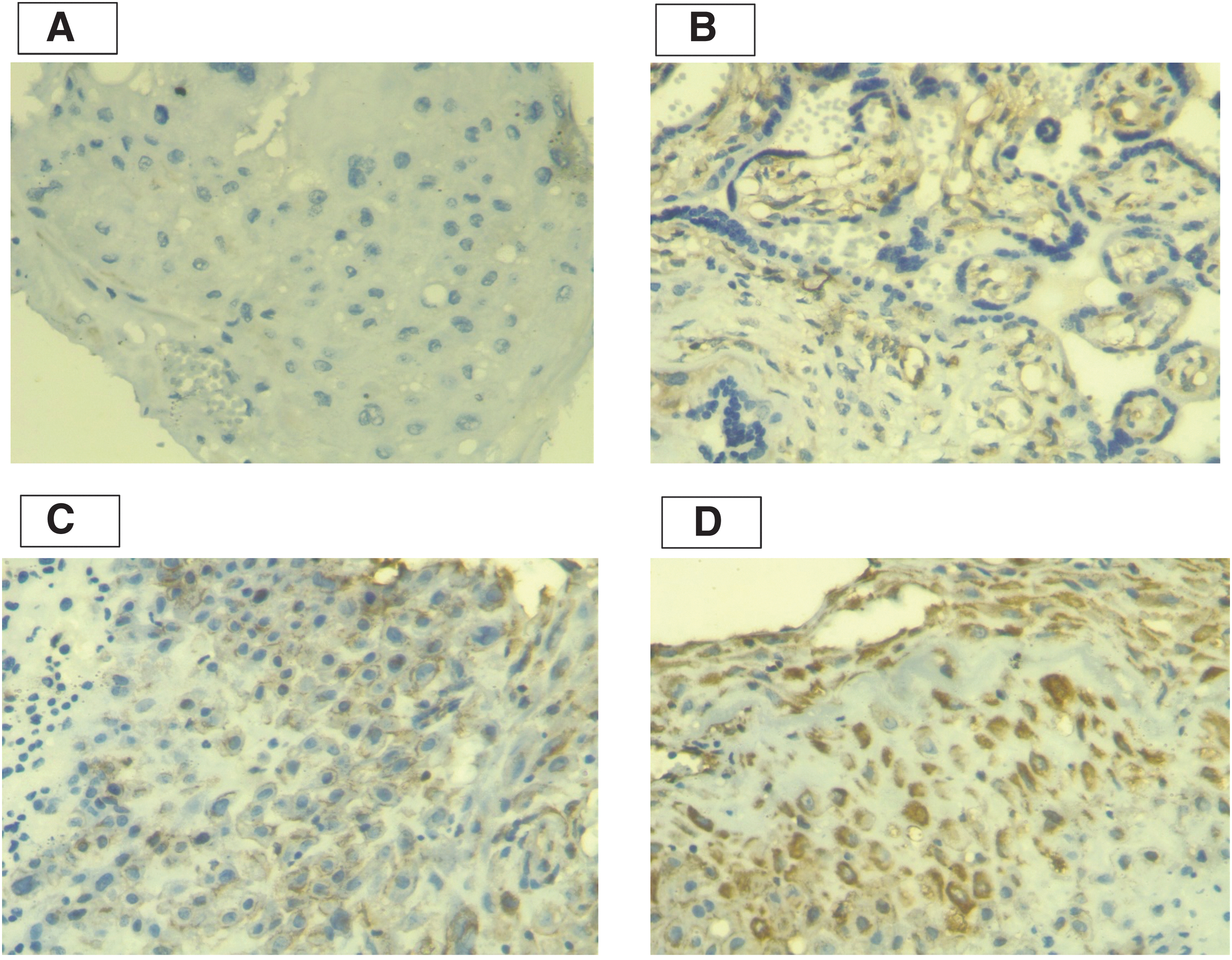
Immunohistochemical analysis to study the expression of TLR7. IHC staining for TLR-7 in HBV-infected placentas (magnifications up to 400 × ),
Validation of Altered TLR-7 Expression in Placental Tissue Based on Cord Blood HBV DNA Status Using Immunohistochemistry
Scoring is done as described in the methods section by using immunohistochemistry analysis, score of 0–1 is considered as low score, score of 2–3 is considered as high score.
Chi-square test is considered significant when p < 0.05 at 95% CI: represented as bold, OR stands for Odds ratio, 95% CI stands for confidence interval, N represents number of samples.
Analysis of probable risk factors in CB mediated intrauterine transmission of HBV
Multinomial logistic regression analysis was performed using SPSS between CB HBV DNA status and other variables such as TLR7, IL-3, and IL-6 fold change, mother HBeAg, and HBV DNA status to find the key factors responsible for the intrauterine transmission of HBV. TLR7-fold change was found to be significantly associated with the CB HBV DNA status with odds ratio (OR) and 95% CI of 0.054 (0.004–0.679) with p = 0.024. Similarly, IL-3 expression was also found to be significantly associated with CB HBV DNA status giving OR and 95% CI of 3.56 (1.035–12.267) with p = 0.044. Mother HBeAg status was also reported to be associated (p = 0.036) having an OR of 0.122 and 95% CI (0.017–0.870). However, the analysis did not reveal any significant association between IL-6-fold change and mother HBV DNA status (p > 0.05) (Table 5).
Multinomial Logistic Regression Analysis for Factors Associated with HBV DNA Positive Cord Blood Among Mothers
p-Value <0.05 was considered significant.
Statistics considering CB HBV DNA negative group as reference.
Multinomial logistic regression is performed and bolded p-value <0.05 considered as statistically significant.
Discussion
HBV-induced liver disease in pregnancy is very poorly understood. Liver disease due to HBV can be fatal to both the mother and newborn and can cause vertical transmission mainly due to its asymptomatic nature of infection. The global prevalence of HBV infection is 3.61%, whereas in India it ranges from 2% to 7% (Chang and Nguyen, 2017; Narayanswamy, 2011). Recent report suggested that Asia is the hub of chronic HBV with more than 75% cases that appears to be a matter of serious concern (Liaw, 2009; Sarin et al., 2016).
Our study screened a total of 3587 pregnant women attending the OPD from which ∼2% (72) were found to be HBsAg positive. The majority of the patients were found in the age group 21–30 years, which falls under the recommended age for giving birth. Since the presence of HBV DNA in the CB is indicative of intrauterine HBV infection (Isogawa et al., 2005), we further investigated the presence of CB HBV DNA along with expression pattern of TLR7, IL-3, and IL-6 in placentas of HBV-positive mothers to assess their role in intrauterine transmission of HBV.
Analysis of key serum biochemical parameters in our study revealed that bilirubin was not significantly higher in the CB HBV DNA-positive cases, which may be possible due to pregnancy-related serum hemodilation (Shekhar and Diddi, 2015; Westbrook et al., 2016). Further, the significant increase of AST, ALT, and ALKP among the CB HBV DNA-positive group compared to the negative group may be due to the presence of replicative and active HBV infection, which may have caused a rise of liver injury markers. Our investigation reveals that 77.4% of the mothers who were positive for HBeAg had CB HBV DNA-positive status, which indicates that the replicative phase of HBV has an impact on the intrauterine transmission of HBV.
Further Kalita et al. (2023) suggested that HBeAg seropositivity may act with altered TLR expression toward MTCT of HBV. Consistent with this finding, Shao et al. (2011) also found an increased risk of intrauterine transmission with maternal HBeAg and HBV DNA positivity. A quantitative analysis of HBeAg in mother's blood and CB may provide a relative picture of HBV DNA level in HBeAg-positive cases (Chen et al., 2017), which can be considered a limitation of the present study.
Our study indicated the overall decreased expression of TLR7 in cases compared to healthy controls, which is consistent with the existing literature states the downregulation pattern of TLR during HBV infection (Das et al., 2017; Sarkar et al., 2015; Vyas et al., 2019; Wu et al., 2009; Xu et al., 2008). IL-3 and IL-6 mRNA expression was found to be significantly upregulated in placentas of HBV-positive mothers compared to healthy controls, which is found to be consistent with other reports (Elefsiniotis et al., 2011; Lan et al., 2015). A decreased level of TLR7 was observed in a previous study on chronic hepatitis B infection compared to healthy control (Kayesh et al., 2021), while Das et al. (2017) observed an inverse relationship in TLR7 expression and HBV DNA highlighting antiviral role of the TLR7.
The proinflammatory cytokine like IL-6 play vital role in HBV replication (Dimitriadis et al., 2023), whereas IL-3 regulates the production and function of various immune cells, particularly hematopoietic stem cells, and myeloid lineage cells. As TLRs and cytokines are involved in inflammatory pathways, the changes in TLR7 expression might lead to altered inflammatory responses, influencing the expression of cytokines such as IL-3 and IL-6 via possible feedback mechanism. The downregulation of TLR7 and overexpression of IL-6 suggested the possible activation of mechanism that suppress the inflammation during HBV infection. T cells are identified as the primary inducers of IL-3 activation, and it is suggested that effector T cells experience significant proliferation during the acute phase of HBV infection (Lantz et al., 1998).
However, after the clearance of antigens, the T cell population reduces as exhausted T cells display impaired effector functions (Wang et al., 2023). Consequently, the current research suggests that an upregulation of IL-3 may indicate that a significant subset of the patient population experiences acute HBV infection. Indeed, the alternate expression of TLR-7 with IL-3 and IL-6 may also suggest an independent expression during HBV infection (Table 6). The exact role of TLR-7 in modulation of IL-3 and IL-6 expression needs further robust study. Understanding the modulating TLR signaling or targeting specific interleukins could be explored as potential strategies for immunomodulation. The upregulation of interleukins may be influenced by other pattern recognition receptors such as Nod-like receptors or RIG-I-like receptors (Das et al., 2017).
Association of Overall TLR Expression Profile with Altered Cytokine Status in Placental Tissue
Fold change of mRNA is done using real-time PCR and values are written as mean ± SD (N) where N represents number of samples.
Bolded p-value <0.05 considered as statistically significant.
Further stratification of the study which compared the HBeAg- and HBV DNA-positive groups also had significant downregulation of TLR7 mRNA in placentas of HBV positive women compared to healthy controls, This findings suggest that impaired TLR response may be a driving factor for intrauterine transmission of HBV. The placental mRNA expression of both IL-3 and IL-6 are found to be significantly higher when there is HBeAg- and HBV DNA-positive status, which is indicative of the fact that interleukin-mediated antiviral response in HBV infections is active but TLR signaling is impaired. TLR7 revealed significant downregulation among the CB HBV DNA-positive groups in both mother and CB HBeAg and HBV DNA, which is consistent with previously reported literature on TLR7 expression in blood (Kalita et al., 2023).
Immunohistochemistry analysis revealed significantly low TLR7 expression among the CB HBV DNA-positive group compared to the negative group suggesting their role in vertical transmission of HBV. These findings are in concordance with the reported reduced expression of TLR7 (Vyas et al., 2019). IL-3 and IL-6 expression showed similar higher expression patterns in CB HBV DNA-positive cases (Table 3). Exploring broader array of cytokines to understand cytokines balance during HBV infection and vertical transmission of HBV could be a potential avenue for future research.
The above mentioned study is limited to 72 numbers of HBsAg-positive samples, which could be more in numbers so as to project a better understanding of our study. The samples were collected by trained phlebotomist in consultation with a clinician which ultimately reduces the potential bias that may be introduced. Further, the collection procedure is strictly done according to the Ethics and Research Committee experts, and we have adhered to the standard guidelines for collection of samples, which have reduced the chances of introducing any potential biases during sample collection. The collection of demographic and patient details is done using a pretested standard questionnaire. Gene expression study which was done using GAPDH as housekeeping gene only provides relative quantification but not absolute. In future, a study using primer-probe may be used to obtain absolute quantification of the data.
Further immunohistochemistry may cause some nonspecific background staining which was minimalized using monoclonal antibody and adhering strictly to the protocol. In future, exploring the therapeutic interventions to modulate the expression of viral sensing TLRs could be undertaken. Furthermore, the study of the role of other TLRs and their downstream molecules in the signaling pathway would have led to a conclusive statement on the study.
Conclusion
Decreased expression of TLR7 in placentas may lead to intrauterine transmission of HBV, which may further lead to vertical transmission of HBV.
Footnotes
Acknowledgment
We thank Department of Science and Technology, Govt. of India, Ministry of Science and Technology, New Delhi- 110016, for providing DST INSPIRE Fellowship to Mr. Simanta Kalita for pursuing his doctoral studies vide sanction number: No. DST/INSPIRE Fellowship/2019/IF190163.
Approval of the Research Protocol
The study is approved by Ethics Committee of Gauhati Medical College & Hospital, Guwahati, Assam and Gauhati University Ethics Committee vide number GMCH MC/190/2007/pt-II/DEC-2020/42 and from Gauhati University -GUIEC-47/2021, respectively.
Informed Consent
Both verbal and written informed consent was obtained.
Disclosure Statement
No competing financial interests exist.
Funding Information
This study received no specific grant from any funding agency.