
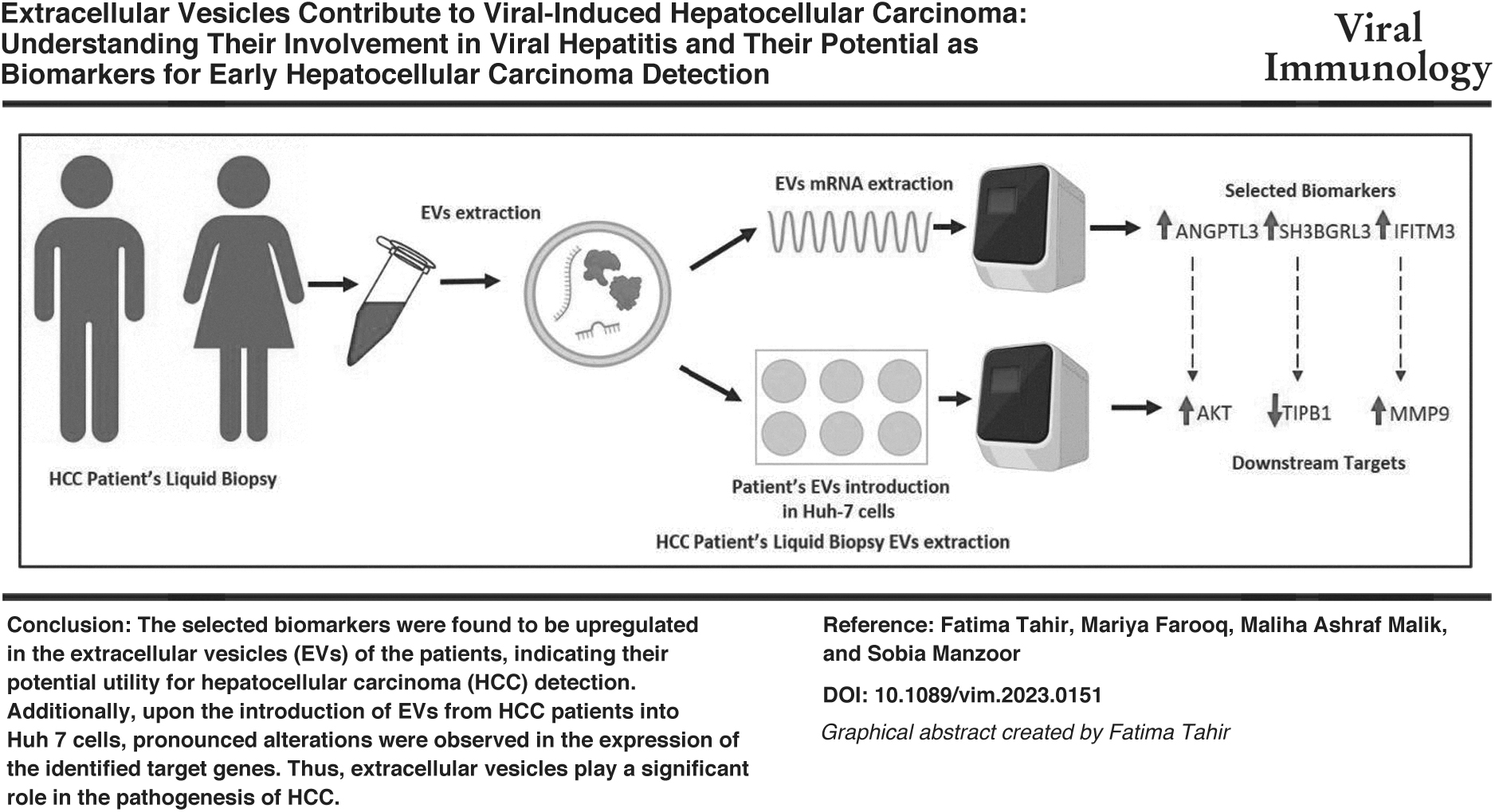
Abstract
The high global prevalence of hepatitis B and hepatitis C and the poor prognosis of hepatitis B and hepatitis C-associated hepatocellular carcinoma (HCC), necessitates the early diagnosis and treatment of the disease. Recent studies show that cell-to-cell communication via extracellular vesicles (EVs) is involved in the HCC progression. The objective of the following study was to explore the role of EVs in the progression of viral-induced HCC and investigate their potential for the early diagnosis of cancer. First, the mRNA derived from EVs of HCC patients was compared to the mRNA derived from EVs from the healthy controls. Expression analysis of ANGPTL3, SH3BGRL3, and IFITM3 genes from the EVs was done. Afterward, to confirm whether hepatocytes can uptake EVs, HuH7 cells were exposed to EVs, and the expression analysis of downstream target genes (AKT, TNF-α, and MMP-9) in Huh7 cells was done. Transcriptional analysis showed that in the EVs from HCC patients, the expression levels of ANGPTL3, SH3BGRL3, and IFITM3 were significantly increased by 2.62-, 4.3-, and 9.03-folds, respectively. The downstream targets, AKT, TNF-α, and MMP-9, also showed a considerable change of 4.1-, 1.46-, and 5.05-folds, respectively, in Huh7 cells exposed to HCC EVs. In conclusion, the following study corroborates the role of EVs in HCC progression. Furthermore, the significant alteration in mRNA levels of the selected genes demonstrates their potential to be used as possible biomarkers for the early diagnosis of HCC.
Introduction
Hepatocellular carcinoma (HCC) being the fifth most common malignancy in men and ninth most common malignancy in women, is globally the second leading cause of cancer-related deaths. Annually, around 800,000 deaths occur due to HCC, and it accounts for almost 75% of all liver cancer-related deaths (Sayiner et al., 2019). Among various factors, cirrhosis plays the primary role in development of HCC resulting in ∼80–90% of cases of HCC. The major cause of liver cirrhosis is viral hepatitis, in particular Hepatitis B virus (HBV) and Hepatitis C virus (HCV) (El-Khazragy et al., 2019; Zamor et al., 2017; Zhang and Friedman, 2012). Therefore, viral-mediated HCC is the most prevalent form. Other causes of HCC include nonalcoholic steatohepatitis, alcohol abuse, aflatoxins, cirrhosis, and hereditary hemochromatosis (Ghouri et al., 2017; Saalim et al., 2016).
Tissue biopsy is regarded as the gold standard for diagnosing HCC. Yet, it is quite complicated. Furthermore, cancer is often heterogeneous. Liquid biopsy, a minimally invasive method, is carried out by drawing patients' blood and utilizing it for HCC diagnosis via transcriptomic analysis. Thus, it seems to be a very promising alternative to tissue biopsy (Ali et al., 2018; El-Khazragy et al., 2020; Quandt et al., 2017). With the advancement of biomarker research, extracellular vesicles (EVs) have gained attention in this context (Feller and Lewitzky, 2016; Yi et al., 2017).
It has been recently discovered that EVs serve as a medium of intercellular communication in normal physiological phenomena as well as in pathological conditions. This communication involves the exchange of proteins and nucleic acids cargo among cells (Yang et al., 2017). Moreover, the alterations in EVs' cargo can lead to carcinogenesis, immune dysfunction, and metastasis. Published literature demonstrates that abnormal cells release a higher number of EVs as compared to normal cells (Kogure et al., 2011; Sugimachi et al., 2015).
The current study aims to investigate the role of EVs in the progression of viral-induced HCC and evaluate the potential of EVs to be used as prognostic biomarkers. For that purpose, the expression of selected genes was analyzed in HCC patients and compared with the normal individuals. To study the effect of EVs on the downstream targets of the selected genes in the cancer cells, the cells were treated with plasma, isolated from the HCC patients' blood, and enriched with EVs. Our research indicates that EVs significantly contribute to the pathogenesis of virally caused HCC. In addition, expression analysis of cells treated with EVs from patients highlights their potential use as prognostic biomarkers.
Materials and Methods
Sample collection and inclusion exclusion criteria
Blood samples collected from the Gastrointestinal Ward of Holy Family Hospital and Military Hospital, Rawalpindi, Pakistan, were transferred to Virology Lab II at Atta-ur-Rahman School of Applied Biosciences (ASAB), National University of Science and Technology (NUST).
For blood sampling, ethical approval was taken by the internal review board (IRB) from the Ethical Committee Board of Rawalpindi Medical University (RMU) and IRB Committee ASAB, NUST (Reference No. 38IRB). Moreover, each patients' consent was received on a consent form where they were asked to fill the form before the blood collection. To compare the patients' samples with healthy controls, blood samples were obtained from healthy individuals as well. Patients with HBV/HCV positive HCC (detected by enzyme-linked immunosorbent assay/polymerase chain reaction [PCR]) and Barcelona Clinic Liver Cancer staging done by gastroenterologist were included in the study. Patients with any other co-infections like Tuberculosis and HIV were excluded. The HCC patients were not receiving any antiviral therapy. However, they were receiving Sorafenib (Brand Name: Nexavar), a kinase inhibitor anticancer drug for liver cancer.
EVs enrichment
Protocol for EVs enrichment was obtained from Virology Lab II. The protocol was optimized based on the requirement of the current study. The modified protocol for the enrichment of EVs is shown in Figure 1.
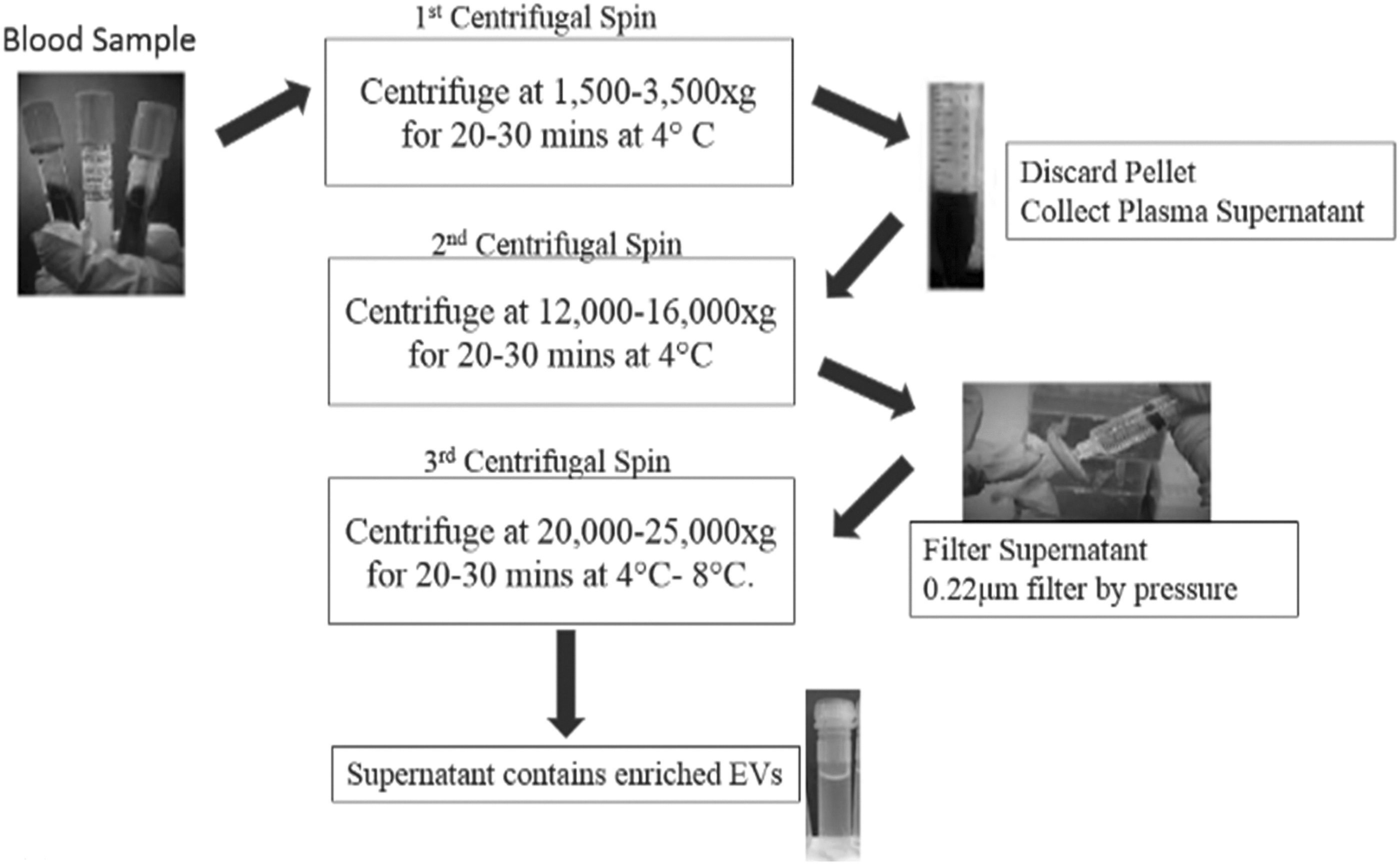
The differential centrifugation-based protocol of enrichment of EVs in plasma. Step 1: PRP is obtained first. Step 2: PPP+apoptotic bodies are obtained through centrifugation. Step 3: Plasma obtained from previous step is syringe filtered through 0.22 μm filter, this removed particles >220 nm. Step 4: EVs including microvesicles and exosomes <220 nm in size are obtained in a resuspended form. EV, extracellular vesicle; PPP, platelets poor plasma; PRP, platelets rich plasma.
Detection of EVs through scanning electron microscopy
For morphological and size-based detection of EVs the plasma sample was fixed on glass cover slips by 25% glutaraldehyde solution. The slides were first washed with plenty of 1 × phosphate-buffered saline and then with different concentrations of ethanol (i.e., 20%, 40%, 60%, 80%, 90% and 100%) for 5 min each. The slides were then left to air dry in a laminar hood cabinet overnight. Scanning electron microscope (SEM) analysis was carried out at the U.S. Pakistan Center for Advanced Studies in Energy (USPCASE), NUST at different parameters to detect EVs from the processed samples. The conductive layer coating done before SEM analysis was of gold.
RNA extraction, quantification, and detection
RNA extraction was carried out via Trizol Reagent (Invitrogen) by following manufacturer's standard method. Proteinase K was added to obtain a better yield of RNA. The extracted RNA was analyzed on a Nanodrop spectrophotometer to determine its quality and quantity. Absorbance at a wavelength of 260 nm shows the concentration of RNA and ratio of 260/280 nm displays the quality of product being analyzed. Quantification of RNA was assessed by using only 1 μL of RNA sample. RNA quality was further confirmed using denaturing urea polyacrylamide gel electrophoresis (PAGE) 8% polyacrylamide gel was used for this experiment (Image shown in Supplementary Data).
Complementary DNA synthesis and expression analysis
Complementary DNA (cDNA) of the extracted RNA samples was synthesized to further assess specific expression levels of our target genes. Standard manufacturer protocol was followed for the cDNA synthesis.
Real-time PCR was carried out by using Maxima® SYBER green/ROX qPCR Master Mix SYBR green master mix. Primer sequences used for the quantitative PCR (qPCR) are mentioned in Table 1. Real-time PCR analysis was done by comparative cycle threshold (Ct) method for the calculation of relative quantification of the target genes.
List of Primer Sequences Used for Quantitative Polymerase Chain Reaction
Uptake of enriched EVs in Huh7 cell line
Maintenance of Huh7 cell line
Hepatocyte-derived carcinoma cells (Huh7 cells) were maintained in Dulbecco's modified Eagle's medium (containing
EVs uptake through human plasma
FBS was replaced by human plasma (HP) and heat-inactivated human plasma (HIHP). Huh7 cells were grown in media containing 10% plasma. This was carried out for the uptake of EVs from HP.
Cellular RNA extraction, cDNA synthesis, and qPCR
After addition of HP containing growth media to Huh7 cells, the total cellular RNA from the cells was extracted using TRIzol reagent (Invitrogen), and the cDNA was synthesized according to the protocol mentioned above. qPCR was performed with the help of primers mentioned in Table 1.
Statistical analysis
For analysis of data obtained from the experiments, Graph pad prism was used. Two-way ANOVA was applied on the data to determine significance by analyzing the p-values. * Showed a p-value of <0.05, ** showed a p-value of <0.005, *** showed a p-value of <0.001, with 95% confidence interval. All the reactions were performed in duplicates.
Results
Detection of EVs through scanning electron microscopy
SEM was performed to detect the presence of EVs. The prepared slides of each EV type (exosomes and microvesicles) were observed under scanning electron microscope simultaneously where the size of these vesicles was observed between 40 and 100 nm. Specifically, mean size of EVs derived from healthy controls and patients' samples were 41.63 and 26.14 nm, respectively. Significantly, higher number of EVs were observed in the plasma of HCC patients in comparison to EVs enriched from healthy controls (Fig. 2).
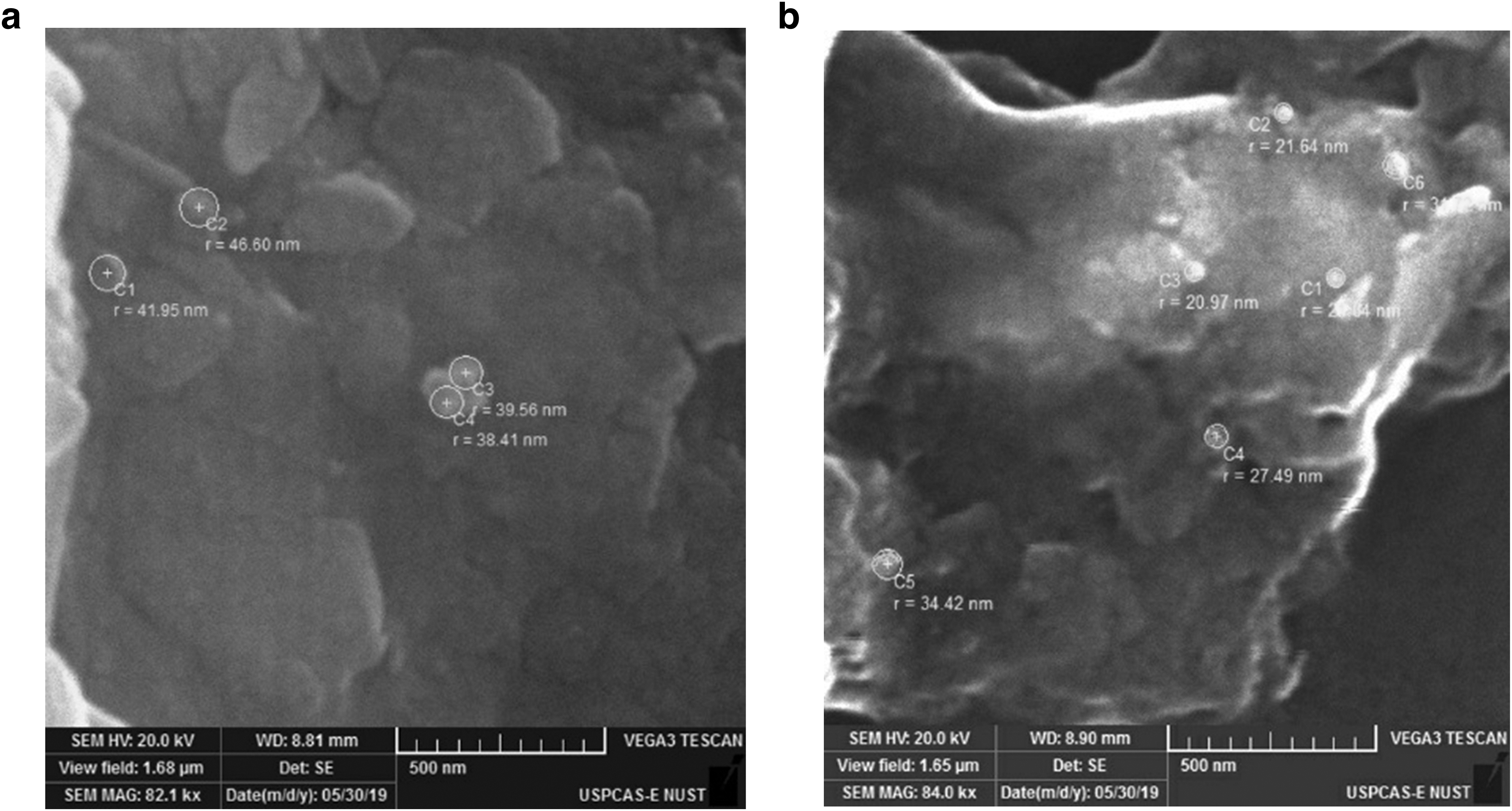
Detection of EVs through scanning electron microscopy, diameter size of EVs enriched from plasma.
Expression analysis of selected biomarkers
Transcriptional analysis of all the selected genes shows that in the EVs of HCC patients, the expression level of ANGPTL3, SH3BGRL3, and IFITM3 were significantly increased by 2.62-, 4.3-, and 9.03-folds, respectively (Fig. 3). Data were analyzed by applying ANOVA two-way. The mRNA expression of B-actin was used as an internal control. By considering the Ct values of the targeted genes and reference gene (B-actin), ΔCt values were calculated, which was used for fold change calculation by Pffafl formula.
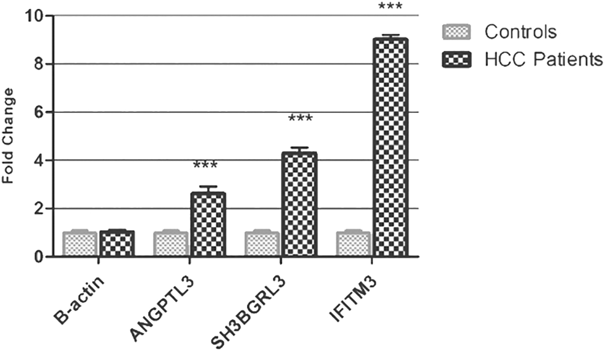
Expression analysis of ANGPTL3, SH3BGRL3, and IFITM3 between healthy controls and HCC patients. A collective representation of the selected biomarkers, where ANGPTL3, SH3BGRL3, and IFITM3 gene expressions have been in HCC patients, respectively. For statistical analysis, one-way ANOVA was used. Significance: ***p < 0.001.
Effects of the selected biomarkers on their downstream targets
Optimization of growth of Huh7 cells in EVs containing HP
In comparison to FBS containing complete medium as a control, Huh7 cells showed stunted growth in the presence of HIHP even after 24 h of subculturing. On the other hand, cells were 40–50% confluent after growing in HP (Fig. 4).
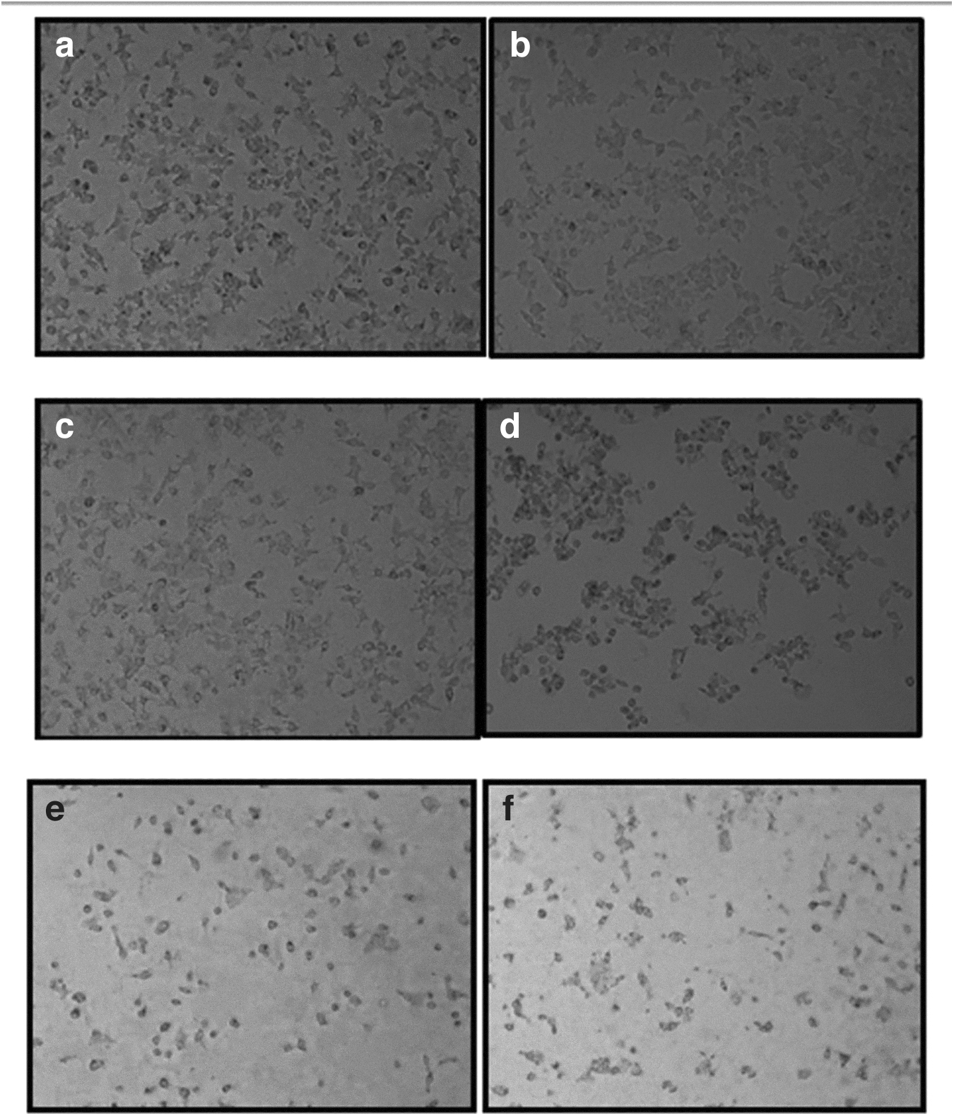
Growth of Huh7 cells in complete medium (DMEM supplemented with 10% FBS), DMEM +10% HP and DMEM +10% HIHP.
Expressional analysis of EVs signaling downstream targets
EVs signaling downstream target for each biomarker used in the current study showed a decrease in expression after 24 h. However, after 48 h, cells showed a significant upregulation in the downstream targets. Figure 5 shows comparative expressional analysis of EVs signaling downstream targets, that is, AKT, TNF-α, and MMP-9 after 24 and 48 h of EVs uptake.
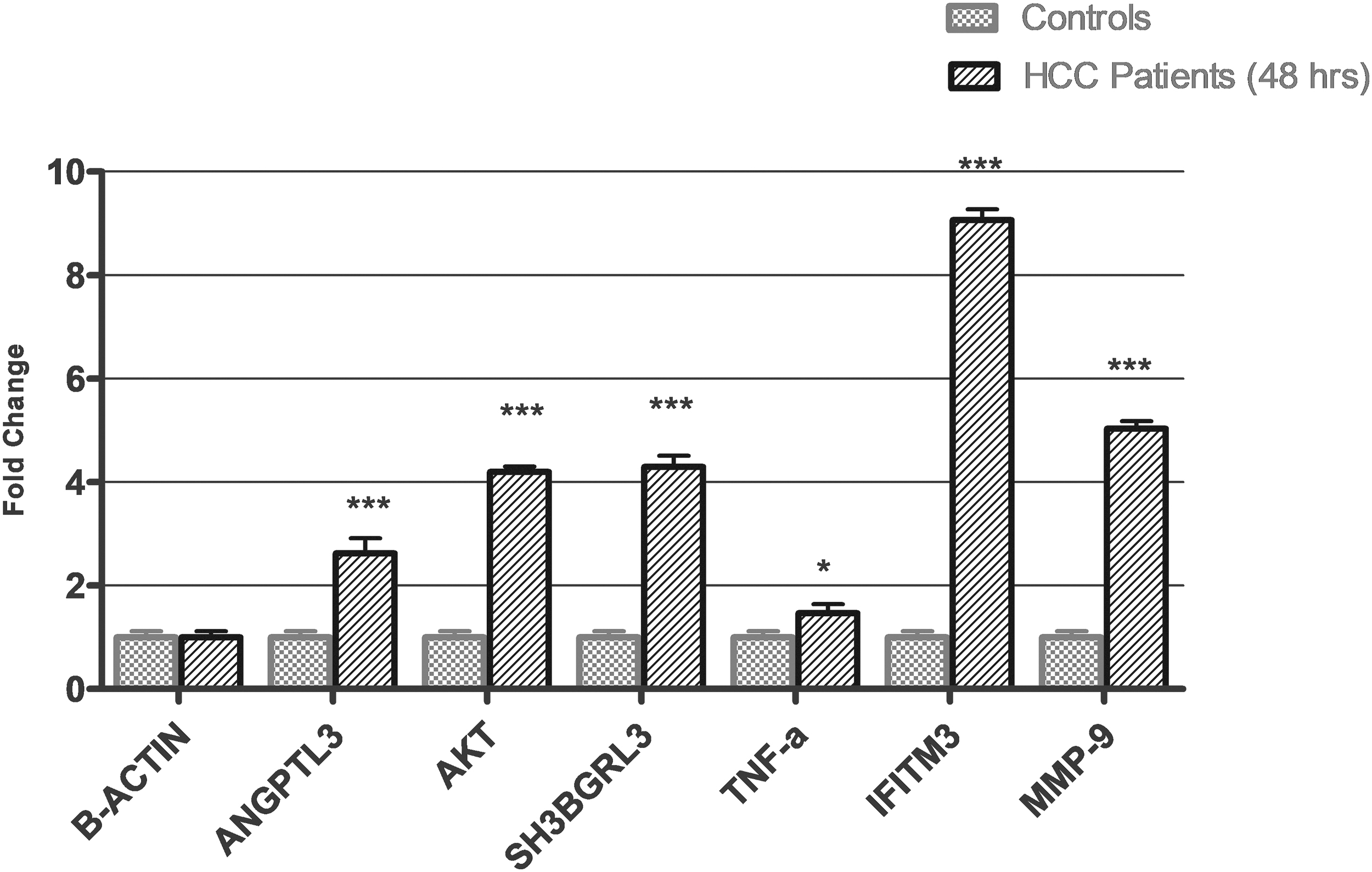
Comparative expression analysis of selected biomarkers with their downstream targets (after 48 h of EVs uptake). ANGPTL3 and AKT comparative expression, showed a significant increase of 2.62- and 4.1-folds, respectively. SH3BGRL3 was significantly increased by 4.3-folds, while TNF-α showed a fold change of 1.46 with a comparatively low significance. IFITM3 and MMP-9 showed a significant increase with a fold change of 9.03 and 5.05, respectively. Significance test: ANOVA two-way. Significance: *p < 0.05, ***p < 0.001.
Comparative expression analysis of selected biomarkers and their downstream target
In viral-induced HCC patient samples, ANGPTL3 showed an increase of 2.62-folds, while its downstream target AKT showed a fold change of 4.1. The biomarker for HCC as well as its target showed a significant increase in expression. SH3BGRL3 showed an increase of 4.3-folds in clinical samples, while its downstream target AKT showed a fold change of 1.46. In clinical samples, IFITM3 showed an increase of 9.03-folds, while its downstream target MMP-9 showed a fold change of 5.05 (Fig. 5).
Discussion
With the advancement of biomarker research, EVs, including exosomes and microvesicles, have drawn the attention of researchers globally. Studies show that EVs transfer extracellular RNA molecules from one cell to another and as a result contribute toward cancer development through angiogenesis, invasion, immune evasion, metastasis, and cancer progression (Redzic et al., 2014).
EVs are a means of communication between cells. It was suggested that the activation of MAPK and PI3K/AKT signaling pathways after the uptake of EVs by neighboring cells results in elevated levels of matrix metalloproteinases (including MMP-2 and MMP-9), resulting in cancer progression (Niland et al., 2022). In HCC, various signaling pathways and genes are involved in the crosstalk mediated by EVs. These genes can serve as a noninvasive biomarker in the diagnosis of viral HCC.
The current study focused on the identification of EVs-based biomarkers for viral-induced HCC using blood samples as liquid biopsy. The goal was to establish a noninvasive, cost-effective, and reliable way for early detection of HCC. For this purpose, biomarkers selection was done based on their role in HCC and their presence in the EVs. Transcriptional analysis of all the selected biomarkers showed a significant increase in their expression in HCC patients as compared to controls. The presence of ANGPTL3, SH3BGRL3, and IFITM3 was confirmed in EVs through Vesiclepedia (El-Shal et al., 2017; Min et al., 2018; Mollbrink et al., 2014).
Furthermore, the study confirmed the role of EVs in intercellular communication, where EVs from HP were incubated with human hepatocarcinoma cells (Huh7 cells). EVs signaling downstream targets for the selected biomarkers showed a significant change as well. All the selected biomarkers (ANGPTL3, SH3BGRL3, and IFITM3) and their downstream targets (AKT, TNF-α, and MMP-9) showed a significant change in the expression as shown in Figure 5.
Camenisch et al. (2002) and Koyama et al. (2015) also showed that ANGPTL3 can promote angiogenesis as well as cancer growth. El-Shal et al. (2017) also reported that the ANGPTL3 expression level is comparatively increased in HCC patients when compared to healthy controls. Many previous studies were done to assess the role of ANGPTL3 in angiogenesis through PI3K/AKT signaling (Camenisch et al., 2002; Karar and Maity, 2011). Our study focused on the involvement of ANGPTL3 in HCC angiogenesis via its downstream signaling target AKT, which also showed an increase in the expression corresponding to ANGPTL3. Hence, based on these findings, ANGPTL3 can be used as an efficient biomarker for viral HCC.
SH3 domain-binding glutamic acid-rich-like protein 3 (SH3BGRL3) was also evaluated as a biomarker with TNF-α as its downstream target. Several studies suggest the involvement of SHBGRL3 in redox-dependent processes where a significant upregulation of Glutaredoxins protein Grx5 has been reported in HCC. Also, oxidative stress aids in the development of liver cirrhosis and HCC, thus elevated level of Grx was observed in livers exposed to oxidative stress (Aravalli et al., 2013). Following these findings, our study revealed a similar change in the expression of SH3BGRL3 in viral-induced HCC patients (Min et al., 2018). Expression analysis of SH3BGRL3 strengthened its role as a biomarker for HCC. SH3BGRL3, also known as TNF inhibitory protein-B1 (TIP-B1), makes the cells TNF-resistant and inhibits TNF-induced apoptotic lysis (Berleth et al., 1999; Pompili et al., 2013; Wang and Lin, 2008). Similar to these findings, our study also demonstrates that TIP-B1 was inhibiting the lysis of cancerous cells ultimately aiding in carcinogenesis.
Another biomarker, IFITM3 was suggested to have its expression regulated by the downstream signaling target, MMP-9. Min et al. (2018) suggest the involvement of P38 MAPK signaling in the overexpression of IFITM3 in HCC. In accordance with our results, IFITM3 was found to be overexpressed in HCC, and the previous studies also showed that expression of the IFITM3 gene is frequently increased in HCC tissues as compared to normal tissues. Moreover, the regulation of MMP9 gene is reported as an essential factor involved in IFITM3-mediated HCC (Fig. 6) (Min et al., 2018).

Expression of selected biomarkers (ANGPTL3, SH3BGRL3, and IFITM3) and their effect on downstream targets (AKT, TNF-α, and MMP-9). As a result, aiding in the major hallmarks of HCC, that is, metastasis, inflammation, and progression. The findings of the current study showed that the selected biomarkers can serve as a promising liquid biopsy-based biomarker for the early detection of viral HCC. Moreover, EVs were up taken by Huh7 cell lines from the viral-induced HCC patients' plasma and the change in expression of the EVs signaling downstream targets revealed the involvement of EVs in intercellular communication.
Conclusion
The results of our study show that all the selected biomarkers can be used for the detection of HCC. The selected biomarkers (ANGPTL3, SH3BGRL3, and IFITM3) display a significant increase in their expression in HCC patients. Moreover, it can be suggested through in vitro data that EVs contribute to HCC pathogenesis. This could be concluded through the uptake of EVs by Huh7 cells, where the selected biomarkers affected the expression of their downstream targets (AKT, TNF-α, and MMP-9), encouraging metastasis, inflammation, and progression. The results of this study and other similar studies, to identify more biomarkers, can lead to the development of a simple, noninvasive, and reliable liquid biopsy-based test for diagnosis of viral-mediated HCC.
Footnotes
Acknowledgments
The authors are grateful to doctors and staff of Holy Family Hospital, Rawalpindi, Pakistan, and of Military Hospital, Rawalpindi, Pakistan, for their assistance in sample collection from patients.
Ethical Approval
For blood sampling, ethical approval was taken by internal review board (IRB) from the Ethical Committee Board of Rawalpindi Medical University (RMU).
Consent to Participate and Publish
Written informed consent to participate and publish was obtained from all individual participants included in the study.
Authors' Contributions
S.M. conceived and supervised the study. F.T. did the main experimental work and wrote the manuscript. M.A.M. assisted F.T. in experimental work. M.F. did the literature review and assisted F.T. in manuscript writing.
Author Disclosure Statement
The authors declare that the research was conducted in the absence of any commercial or financial relationships that could be construed as a potential conflict of interest.
Funding Information
Student research funds were used to conduct this research project.
Supplementary Material
Supplementary Data
References
Supplementary Material
Please find the following supplemental material available below.
For Open Access articles published under a Creative Commons License, all supplemental material carries the same license as the article it is associated with.
For non-Open Access articles published, all supplemental material carries a non-exclusive license, and permission requests for re-use of supplemental material or any part of supplemental material shall be sent directly to the copyright owner as specified in the copyright notice associated with the article.