
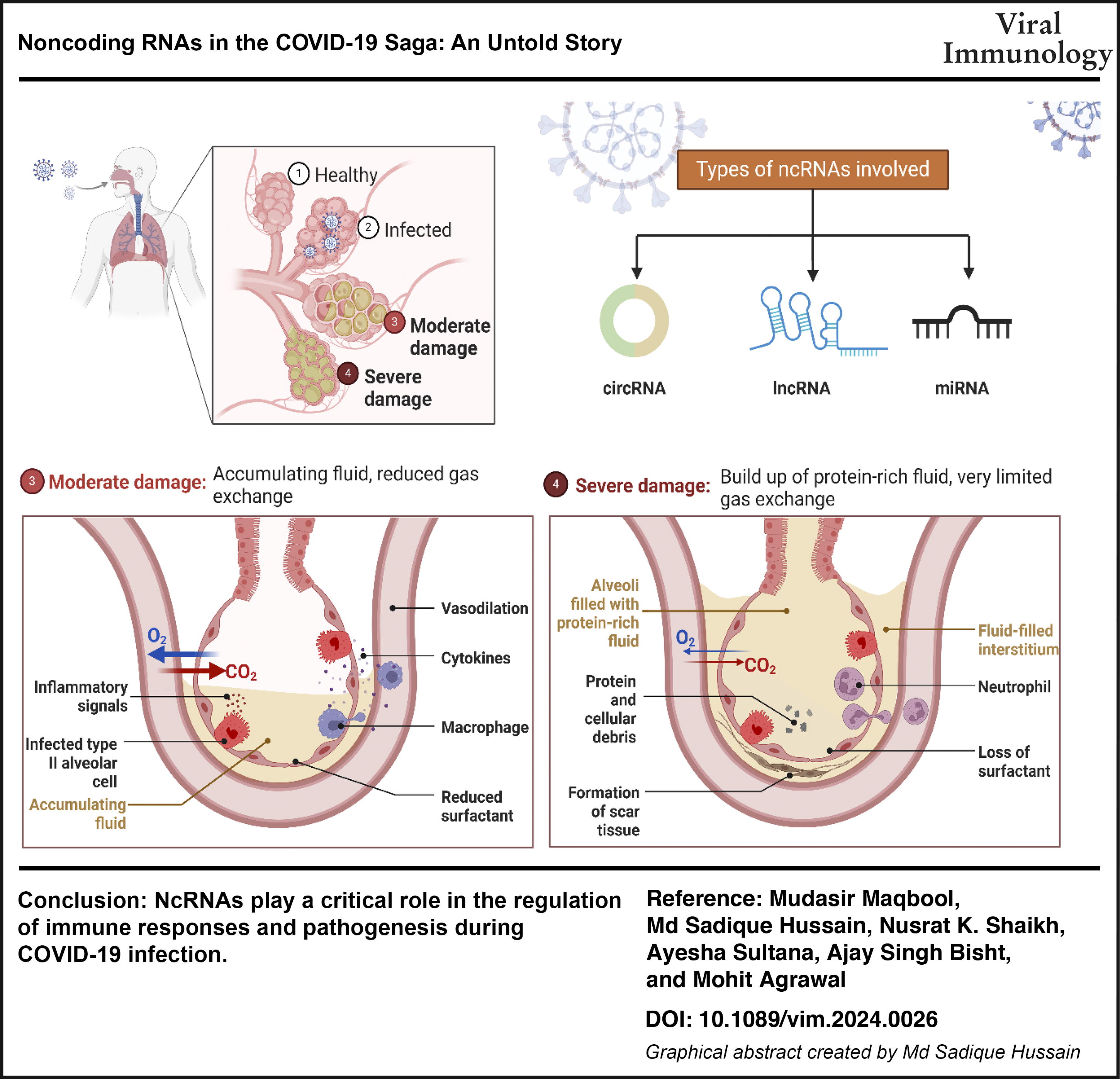
Abstract
In late 2019, the SARS-CoV-2 virus emerged as the cause of COVID-19, triggering a worldwide pandemic of unprecedented scale. A detailed comprehension is being established to elucidate the severe immunopathological condition in critical cases of COVID-19. Noncoding RNAs (ncRNAs) are the transcribed but untranslated part of the genome that used to be ignored or disregarded until recent times. The latest research has revealed the intricate role played by the immune system in responding to SARS-CoV-2 and the development of COVID-19, affecting important aspects such as cytokine storm syndrome, changes in blood clotting, attraction of immune cells, and regulation of blood vessels. Exploring the possibilities of host–virus RNA interactions and RNA-RBP interactions has garnered significant interest. Following SARS-CoV-2 infection, the levels of certain ncRNAs change to indirectly control the expression of antiviral genes and viral gene replication. Certain ncRNAs are utilized by SARS-CoV-2 to assist the virus in evading the immune system by reducing the production of type I interferon (IFN-1) and regulating cytokine levels.
Introduction
Since December 2019, there have been accounts of a cluster of instances of “unidentified viral pneumonia” linked to a neighboring Huanan seafood wholesale market in Wuhan, China. A new coronavirus, known as Severe Acute Respiratory Syndrome Coronavirus 2 (SARS-CoV-2), was identified as the cause of the mysterious pneumonia in a patient from Wuhan by researchers. Subsequently, the illness was formally designated as COVID-19 (Ciotti et al., 2020b; Hussain et al., 2021; Lone and Ahmad, 2020; Yang et al., 2020). The coronavirus (CoV) is a part of a broad family of positive-sense, single-stranded RNA viruses found within the Nidovirales order. The order consists of Roniviridae, Arteriviridae, and Coronaviridae families. Within the Coronaviridae family, there are two subfamilies as follows: Torovirinae and Coronavirinae. The classification of Coronavirinae includes alpha-, beta-, gamma-, and delta-CoVs. Grouping these virus subtypes is based on phylogenetic clustering. The length of their viral RNA genome varies from 26 to 32 kilobases. These can be extracted from various animal species. These consist of birds, livestock, and mammals like camels, bats, masked palm civets, mice, dogs, and cats (Malik and Maqbool, 2020; Shi et al., 2020; Yuki et al., 2020). Considering the widespread transmission and highly contagious nature of COVID-19, it is a significant pathogen. When viewed through an electron microscope, they display a crown-shaped structure due to the spike glycoproteins on their envelope. By examining genes, it has been found that bats and rodents are the origin of alpha-CoV and beta-CoV. Avian species are recognized as genetic reservoirs for delta-CoV and gamma-CoV (Ciotti et al., 2020a; Liu et al., 2020).
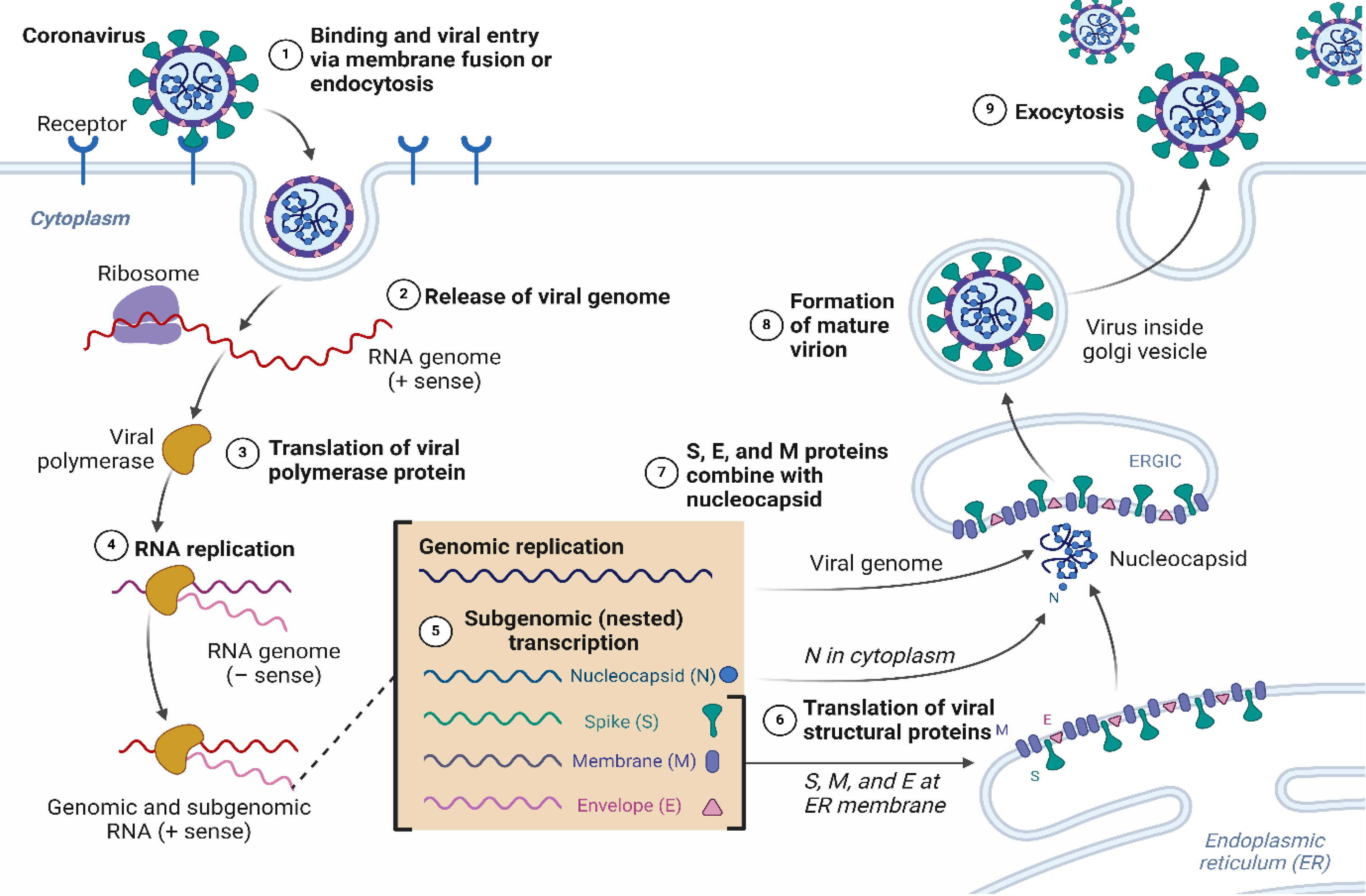
In individuals with a robust immune system, these CoVs usually present as moderate respiratory infections and common colds. In persons of advanced age and those with compromised immune systems, the infection can impact the lower respiratory tracts. MERS-CoV, SARS-CoV, and SARS-CoV-2, like other human coronaviruses, display both respiratory and nonrespiratory symptoms. Analysis of the genomic features of the new strain indicated that it shares 89% of its nucleotide sequence with bat SARS-like CoVZXC21. Furthermore, there is an 82% similarity in nucleotides between the SARS virus and the human SARS virus. Therefore, these findings establish the foundation for classifying the novel variant as SARS-CoV-2 (Esakandari et al., 2020; Hussain and Sharma, 2024; Wu et al., 2020). The genome length varies between 29,891 and 29,903 nucleotides. The virus exhibits susceptibility to UV radiation and high temperatures. The SARS-CoV-2 virus binds to certain cells by communicating with angiotensin-converting enzyme 2 (ACE2) (Hussain et al., 2023b; Husssain et al., 2021; Lan et al., 2020). Nevertheless, there has been an increase in clinical cases with a variety of exposure histories. This provides evidence that the virus can be transmitted between people. Hence, the primary mode of transmission is now recognized as being from person to person. People who show no symptoms can still spread the virus (Chilamakuri and Agarwal, 2021; Lee and Choi, 2021). Transmission occurs by the dissemination of respiratory droplets through coughing or sneezing. Studies suggest that transmission can also happen through direct touch between individuals in proximity. Figure 1 represents the replication process of CoV.
Noncoding RNAs (ncRNAs) are RNA molecules that are transcribed from DNA but do not undergo translation to produce proteins (Hussain et al., 2024e). Formerly thought to be DNA, ncRNAs are currently acknowledged for their pivotal functions in regulating gene expression and the three-dimensional structure of the genome. ncRNAs have a regulatory function in influencing gene expression both during transcription and after transcription has occurred. When looking at transcription, ncRNAs can attach to DNA or function as enhancers or inhibitors of transcription factors. These changes can affect gene expression levels without modifying the coding sequence. ncRNAs can regulate mRNA stability, translation efficiency, and splicing patterns by interacting with molecules like microRNA (miRNA) (Fernandes et al., 2019; Hussain et al., 2024f; Statello et al., 2021). Aside from controlling gene expression, ncRNAs are also involved in structuring the 3D configuration of the genome. ncRNAs can attach to particular locations on chromosomes and create loops that play a role in preserving chromosome structure. Ensuring accurate gene expression involves maintaining the proximity of genes for effective interaction. ncRNAs also play a role in organizing chromatin structure through interactions with histones or other epigenetic modifiers like DNA methylation or acetylation marks (Gil and Ulitsky, 2020; Zhang et al., 2019b). Depending on the specific miRNA, this can lead to either an increase or decrease in protein production levels. MiRNAs play crucial roles in various cellular pathways, including development, cell cycle progression, apoptosis, and immune responses. Long noncoding RNAs (lncRNAs), which represent another form of ncRNA, can engage with proteins, influencing their function or guiding them to particular locations on the DNA (Hussain et al., 2023a). In addition, lncRNAs play a role in various epigenetic processes like methylation and histone modification, which regulate gene expression levels (Soni and Biswas, 2021; Zhang et al., 2019b). Another category of ncRNA includes circular RNAs (circRNAs). When two exons from a pre-mRNA molecule are linked together, these molecules are formed through splicing. CircRNAs have a significant impact on gene expression by serving as reservoirs for miRNA molecules, preventing them from binding to other mRNA targets and blocking protein translation (Tachiwana et al., 2020). Exploring the involvement of ncRNAs in controlling gene expression and genome structure is gaining more attention. Small RNA molecules, which do not encode proteins, are crucial for gene regulation. They play roles as transcriptional regulators, epigenetic modifiers, and regulators of 3D chromosome architecture. ncRNAs play a role in regulating gene expression through various mechanisms. Binding to particular DNA sequences can prevent transcription factors from binding to their intended sites. This hinders their ability to activate or suppress gene expression (Coker et al., 2019; Hussain et al., 2024a). Table 1 explores the summary of ncRNA-mediated SARS-CoV-2 functions.
Investigation of ncRNAs concerning SARS-CoV-2 and Its Associated Functions
circRNA, Circular RNAs; NcRNAs, Noncoding RNAs; LncRNAs, long NcRNAs.
Investigating the function of ncRNAs in relation to COVID-19 provides insights into their probable involvement in the development and advancement of the disease. Although there has been extensive research on the immunology and virology of SARS-CoV-2, the investigation into the involvement of ncRNAs in COVID-19 has been incomplete. Delving into the influence of ncRNAs on different facets of the COVID-19 narrative, such as viral replication, host immune response, and disease severity, this review aims to address the existing knowledge gap. Through a thorough examination of current research and new findings, the goal is to offer understanding on the impact of ncRNAs on susceptibility to infection, disease outcomes, and potential treatment approaches. This mini-review covers various classes of ncRNAs such as miRNAs, lncRNAs, and circRNAs and their interaction among virus and the host cellular machinery, exploring the possible diagnostic and prognostic implications of ncRNAs as biomarkers for COVID-19 detection and patient stratification.
ncRNAs Implicated in COVID-19 Pathogenesis
Research indicates that a mere 2% of the genome is responsible for encoding protein-coding RNAs, whereas a substantial 70% comprises ncRNAs. Alongside circRNAs, ncRNAs are pivotal in various diseases such as cardiovascular ailments, metabolic disorders, and inflammatory conditions (Andreassi, 2018; Marinescu et al., 2022). During viral infections, ncRNAs are induced or repressed by viruses to modulate the infection process. Many viruses have evolved mechanisms that exploit ncRNAs to evade the host immune system. Understanding the regulatory roles of ncRNAs in host immunity during viral infections is crucial for the development of antiviral therapies (Li et al., 2024). With an average length of 21–25 nucleotides, microRNAs help break down RNA and inhibit translation inside the RNA-induced silencing complex, which regulates gene expression. MicroRNAs possess the capacity to selectively interact with diverse RNA categories, such as protein-coding RNAs, lncRNAs, and even proteins. The inherent flexibility of miRNAs enables them to exert their influence on a multitude of RNA molecules, hence impacting a diverse array of biological functions (Bartel, 2009; Jiang et al., 2019; Kalayinia et al., 2021; Kingston and Bartel, 2019; Natarelli and Weber, 2019; Natarelli et al., 2018; Nouri et al., 2022, Santovito et al., 2020). ncRNAs play diverse roles in influencing the outcome of viral infections. They can function as miRNAs or protein inhibitors (‘sponges’), regulate protein function, or act as templates for translation themselves. In addition, ncRNAs can modulate signaling pathways (Abbas et al., 2023). miRNAs can control the expression of genes involved in the immune response against viral infections. For instance, miR-200c has been identified as a regulator of immune responses to SARS-CoV-2 (Sodagar et al., 2022).
With lengths exceeding 200 nucleotides, lncRNAs are a diverse class of RNA molecules originating from various regions of the genome. This also applies to these circRNAs, which are produced by back splicing exons. These compounds function as transcription modulators, miRNA sponges, and epigenetic regulators in various clinical states, acting as biomarkers in each case. There have been observations of circRNA dysregulation in various diseases (Chen et al., 2018; Hussain et al., 2024c; Jiang et al., 2019; Ulitsky and Bartel, 2013). Figure 2 shows the ncRNAs involved in COVID-19 immune response. LncRNAs are particularly pertinent in the context of viral infections, acting as regulators of the cellular response during infection by single-stranded RNA viruses. Upregulation of lncRNAs has been observed in response to SARS-CoV-2 infection, and their specific roles in the infection process are under active investigation (Enguita et al., 2022). CircRNAs are a newly recognized class of ncRNAs involved in the regulation of viral infections. They can act as templates for translation and modulate signaling pathways. CircRNAs have been shown to be induced by respiratory syncytial virus (RSV) infection and have been found to inhibit RSV replication (Yao et al., 2021).
Coronavirus replication cycle.
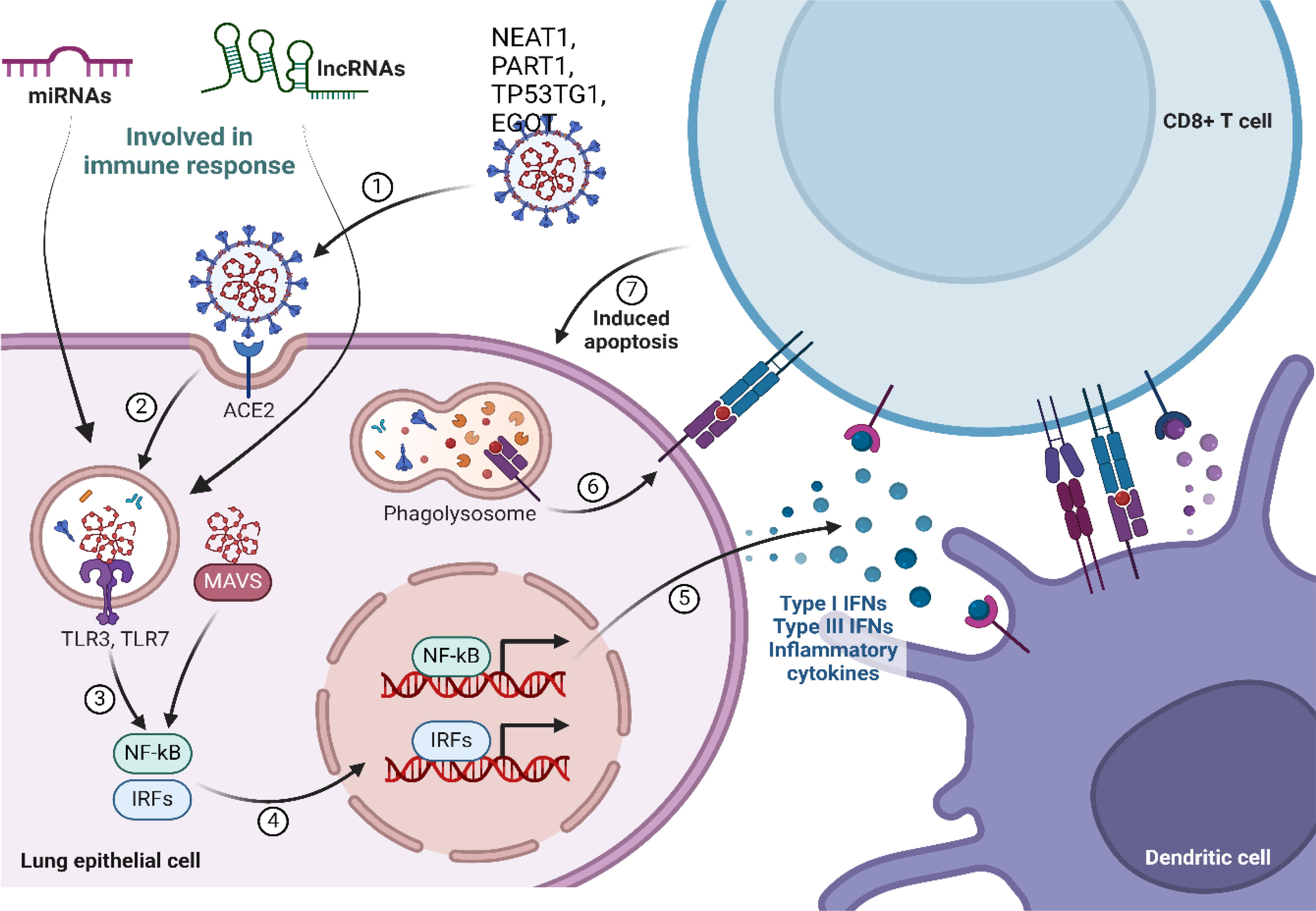
NcRNAs involved in SARS-CoV-2 immune response. NcRNAs, Noncoding RNAs.
ncRNAs in SARS-CoV-2 and Viral Infections
ncRNAs are essential for regulating a variety of cellular processes, as noted before. Both miRNAs and lncRNAs play a crucial role in the intricate interactions between host cells and viruses during infection (Jopling et al., 2005; Trobaugh et al., 2014; Wong et al., 2020). Studies have demonstrated that targeted miRNAs can impede viral infections by targeting certain untranslated regions, indicating potential for the advancement of RNA-based therapies. There may be a relationship between the transcripts and genome of the virus based on the observation of the regulation of human host miRNAs during SARS-CoV-2 infection (Natarelli et al., 2021b; Suleiman et al., 2024; Vierbuchen and Fitzgerald, 2021; Zhang et al., 2024).
miR-3661 directly influences the production of SARS-CoV-2 proteins in lung tissues, as illustrated in Table 2. Patients with COVID-19 exhibit reduced levels of miR-17-5p and miR-20b-5p in their blood. In addition, researchers have found that human miR-190a-5p and miR-184a-3p play a role in controlling SARS-CoV-2 mRNA, impacting the production of ORF8 and ORF6 proteins, which contribute to the body’s defense against viral multiplication. These findings have been well-documented in prior research (Khan et al., 2020; Saçar Demirci and Adan, 2020).
Impact of ncRNAs on SARS-CoV-2
NcRNAs, Noncoding RNAs.
Through a thorough examination of SARS-CoV-2 genomes globally, three miRNAs (miR-23b, miR-198, and miR-125a) were pinpointed for their notable antiviral properties in respiratory illnesses, potentially focusing on specific binding locations in the SARS-CoV-2 genome. Furthermore, several miRNAs, including let-7 family members, miR-200b/c, and miR-18, have been discovered as possible regulators of ACE2 expression in renal illness associated with COVID-19. Conversely, when miR-145 is overexpressed, it leads to a decrease in ACE protein levels. Furthermore, the miR-221/222 cluster has a role in regulating inflammation and the restructuring of blood vessels. It is also linked to the regulation of ACE2 expression in nephropathy associated with COVID-19 (Rai et al., 2024; Sardar et al., 2020; Widiasta et al., 2020).
Through the utilization of computational techniques and the examination of publicly available gene expression datasets from individuals infected with SARS-CoV, MERS, and SARS-CoV-2, plausible situation arises suggesting that the virus might depend on host ncRNAs for replication. The clinical implications of these discoveries have not been proven yet. Yousefi and colleagues have identified many miRNAs that play a role in the infection of host cells by SARS-CoV, affecting the regulation of the TGF-beta cascade. A number of miRNAs have been discovered, including hsa-miR-203a, miR-92b, miR-23b, miR-155, miR-145, miR-21, miR-125a, and the let-7 family (Yousefi et al., 2020).
There is currently a scarcity of research on the role of lncRNAs in viral infections. Only a limited number of lncRNAs have been identified to either facilitate or impede viral replication following infection. NEAT1, NRON, EGOT, and lncRNA-CMPK2 have been shown to be affected by hepatitis B virus (HBV) and hepatitis C virus (HCV). Furthermore, NEAT1 exhibits elevated levels in instances of influenza infections, encephalitis, and HIV (Zhang et al., 2013b; Zhang et al., 2013a). Computational analyses and RNA-seq data from COVID-19 patients worldwide suggest that the diverse roles of lncRNAs may provide a strong foundation for developing lncRNA-based vaccines against SARS-CoV-2 infection, notwithstanding the paucity of information regarding the role of lncRNAs in SARS-CoV-2 infection. This theory is supported by the discovery made by Moazzam-Jazi and colleagues of six lncRNAs with variable regulation in BALF and PBMC obtained from COVID-19 patients. Among these lncRNAs, the viral genome was significantly attracted to HOTAIR, lncRNA-PGCs, PVT1, and AL392172.1, whereas MALAT1 and NEAT1’s attachment may have an effect on inflammatory responses in infected cells (Moazzam-Jazi et al., 2021; Vishnubalaji et al., 2020).
Four lncRNAs—EGOT, ENSG00000271646, WAKMAR2, and EPB41L4A-AS1—exhibited increased expression in cells infected with SARS-CoV-2, as per a study by Mukherjee et al. While the precise functions of these lncRNAs remain unclear, EGOT and WAKMAR2 have been associated with the production of cytokines and may facilitate viral replication in the lungs. Furthermore, a computational examination of lncRNA interactions with the SARS-CoV-2 genome revealed that H19 can interact with the Spike transcript in addition to the SARS-CoV-2 genome (Mukherjee et al., 2021; Natarelli et al., 2021a; Rai et al., 2024; Vishnubalaji et al., 2020).
Studies suggest that RNA viruses like SARS-CoV-2 can impact the activity of cellular miRNAs, leading to changes in the host transcriptome that may benefit the virus. However, alterations in miRNA expression might hinder the replication and translation of viral genomes, thus enhancing the antiviral effects. The regulation of the entry and replication of SARS-CoV-2 can be influenced by microRNAs that control TMPRSS2 and ACE2 receptors. This indicates that targeting these microRNAs could be a potential treatment strategy (Natarelli et al., 2021a).
A study conducted by Fulzele et al. found that miR-16-2-3p has been shown to target specific regions or genes within the genome of SARS-CoV-2, rather than the entire genome. This suggests that miR-16-2-3p may play a crucial role in SARS-CoV-2 infection. miRNAs have a crucial role in modulating the host’s immune response to viruses. Researchers noted a notable rise in miR-155 levels in human cell lines infected with SARS-CoV-2, along with increased expression of ISGs and tissue-damaging cytokines such as CXCL10 or IL6 in comparison to SARS-CoV infection. By removing miR-155, a decrease in pulmonary damage was observed, suggesting a possible treatment target for addressing SARS-CoV-2 infection (Fulzele et al., 2020; Wyler et al., 2021).
Khan et al. have conducted a study and found that SARS-CoV-2 infection causes the disruption of several miRNAs. In addition, they proposed a possible association between these miRNAs and important immunological signaling pathways such as TRAF6, IFN, and TLR-signaling pathway suppression. There is a proposition suggesting that viruses have the ability to generate sequences that imitate miRNAs or viral miRNAs. These sequences may disrupt the expression of genes in the host, facilitating viral replication within the host cell (Barbu et al., 2020). Saini et al. identified two unique miRNAs, MD3-3p and MD241-3p, that target p53 and BMPR2, respectively, in relation to SARS-CoV-2. These findings are summarized in Table 2 (Bhat et al., 2022).
CircRNAs
Insufficient research has been conducted on the distinctively expressed (DE) circRNAs in COVID-19. However, this study has the potential to provide useful knowledge about the DEcircRNAs that are found after SARS-CoV-2 infection and their occurrence in different samples from persons with COVID-19. By conducting genome-wide dynamic screening, over five thousand circRNAs were identified in human lung epithelial cells infected with SARS-CoV-2, situated at various positions in the genome (Yang et al., 2021). In addition, DEcircRNAs were identified in the whole-blood sample of individuals experiencing recurrent COVID-19, in comparison to the normal control group (Wu et al., 2021). Previous studies indicate that infection with SARS-CoV-2 has the potential to interfere with the production of circRNAs in the blood. Furthermore, a distinct disruption in the functioning of circRNAs in the cerebrospinal fluid (CSF) has been noted. This is evidenced by a distinctive pattern of circRNA expression in the CSF of COVID-19 patients, which differs from that of both healthy individuals and individuals with neurological disorders (Reinhold et al., 2023). Further work is necessary to determine whether there are similarities or differences in the categories of DEcircRNAs between the brain and red blood cells. Gaining a comprehensive understanding of the broader ramifications of SARS-CoV-2 is essential for furthering our understanding. Techniques such as single-cell RNA sequencing and spatial transcriptomics sequencing are highly useful tools for accomplishing this objective.
LncRNAs
It has been observed that differentially expressed long non-coding RNAs (DElncRNAs) are present in both COVID-19 cases ranging in illness seriousness and people with differing disease seriousness compared with healthy individuals. This depicts that lncRNAs may serve a role in the pathophysiology of this condition (Cheng et al., 2021; Li et al., 2021). The lncRNA expression exhibited changes over time, either within a short period (e.g., from being admitted to 7 days) or a longer duration encompassing treatment, recovery, and rehabilitation (Rombauts et al., 2023; Zheng et al., 2020). Furthermore, DElncRNAs have been found in instances of recurrent COVID-19, as shown by the identification of almost one thousand DElncRNAs in recurrent COVID-19 individuals as opposed to healthy subjects (Wu et al., 2021). However, more studies are necessary to ascertain if there are disparities in the expression of lncRNAs among people with a solitary SARS-CoV-2 infection and those who experience recurrence This is important because multiple infections may heighten the possibility and stress of the illness, indicating the presence of conceivable fundamental variations (Bowe et al., 2022).
Despite the discovery of DElncRNAs at the cellular level, nothing is known about the expression properties of lncRNAs in COVID-19 patients. Single-cell RNA sequencing research demonstrated the presence of DElncRNAs in blood leukocytes from patients with severe COVID-19 compared with healthy persons. This finding suggests that lncRNAs may play a more complex role in the disease’s progression (Aznaourova et al., 2022).
One study established the association between aberrantly expressed lncRNAs and the activation of host responses through innate immune signaling during SARS-CoV infection. This finding raises the possibility of a similar correlation with SARS-CoV-2 infection. Researchers have made several efforts to understand the role of lncRNAs in modulating the interaction between SARS-CoV-2 and the host, aiming to utilize these findings as disease biomarkers or therapeutic interventions. For instance, a study analyzing RNA-seq data from SARS-CoV-2 infected primary normal human bronchial epithelial (NHBE) cells identified 21 differentially expressed lncRNAs and protein-coding genes. Among these, 9 lncRNAs were upregulated, and 12 were downregulated, as detailed in Table 3 (Rai et al., 2024; Turjya et al., 2020).
Differentially Expressed lncRNAs Identified from RNA Sequencing of SARS-CoV-2-Infected Primary Normal Human Bronchial Epithelial Cells
lncRNAs, long noncoding RNAs.
In addition, among the differentially expressed protein-coding genes, six were found to closely interact with these aberrantly expressed lncRNAs, with the interactions primarily being RNA–RNA and a few involving protein-RNA interactions. By constructing biological networks, researchers identified lncRNA-interacting differentially expressed genes that encode proteins. Moreover, the SARS-CoV-2 protein interactors were associated with pathways that facilitate cell survival, viral proliferation, and immune response. The study also suggested that these dysregulated lncRNAs might act as competing endogenous RNAs to regulate the levels of virus-induced microRNAs [75].
Furthermore, transcriptome profiling revealed significant enrichment of differentially expressed genes during SARS-CoV-2 infection. This study utilized primary NHBE cells and the A549 alveolar basal epithelial cell line to investigate the host response to SARS-CoV-2 infection, comparing it with other respiratory viruses. It highlighted the driving force behind the extreme inflammatory state observed during COVID-19. The study demonstrated that SARS-CoV-2 infection induced limited levels of type I and type III interferons, in contrast to the significantly elevated levels of pro-inflammatory cytokines and chemokines such as IL-6, resulting in a reduced innate immune response against the virus [76]. This finding helps explain why elderly individuals or those with compromised immune systems experience severe outcomes upon infection.
Similarly, systemic changes in the host response to SARS-CoV-2 infection were investigated by another research group focusing on lncRNA expression and function. This study utilized NHBE cells and lung tissue samples from SARS-CoV-2-infected subjects, as previously examined by Blanco-Melo et al. [77]. Using various computational methods, the researchers aimed to elucidate virus–host interactions mediated by lncRNAs. Pathway and functional analyses indicated an enhanced interferon-mediated response as the primary defense mechanism against the virus, evidenced by the upregulation of interferon-responsive gene targets, consistent with the findings of Blanco-Melo et al. Key genes such as IRF9, IFIT1, IFIT2, IFIT3, IFITM1, MX1, OAS2, OAS3, IFI44, and IFI44L were significantly elevated in NHBE cells compared with controls, underscoring the pivotal role of interferon signaling in the development of SARS-CoV-2 infection [77].
In addition, network reconstruction through computational analysis revealed biological networks that were both activated and repressed in the NHBE cell model and patient tissue samples. For example, genes involved in type I and III interferon signaling were activated to combat the virus, whereas genes related to p53 binding and cell cycle progression were repressed. The study also demonstrated the expression of several viral proteins both in vitro and ex vivo and identified numerous host-derived lncRNAs that were dysregulated in response to SARS-CoV-2 infection. RNA sequencing of SARS-CoV-2-infected NHBE cells revealed 155 upregulated and 195 downregulated lncRNAs compared with mock-infected cells. Some of these differentially expressed lncRNAs are listed in Table 4 [77]. However, the precise functions of these lncRNAs in modulating the host response to SARS-CoV-2 infection remain to be fully explored.
A Selection of Differentially Expressed lncRNAs Identified through RNA Sequencing of Primary Normal Human Bronchial Epithelial Cells Infected with SARS-CoV-2
LncRNAs, long noncoding RNAs.
These lncRNAs offer a new perspective on their role in modulating the antiviral immune response in the host by interacting with chromatin, RNA, or protein molecules [78]. Furthermore, previous studies have highlighted the roles of MALAT1 and NEAT1 in the context of HIV-1 infection and various carcinomas. Investigating their roles in SARS-CoV-2 infection would be valuable for understanding the mechanisms involved in disease pathogenesis and progression. Collectively, such mechanism-centric studies are crucial for determining the effects of dysregulated lncRNAs during SARS-CoV-2 infection and managing the excessive inflammation associated with COVID-19.
MiRNAs
Multiple studies have indicated that the behavior of specific miRNAs is altered in individuals with COVID-19 in comparison to the general population, suggesting a possible role for miRNAs in the development of COVID-19 (Fernández-Pato et al., 2022). Furthermore, the activity of individual miRNAs can be affected by the seriousness of the sickness. This highlights the essential role of miRNAs in differentiating the extent of COVID-19 by revealing the varied profile of miRNAs among individuals with different levels of infection seriousness (Parray et al., 2021). Moreover, the DEmiRNAs profile shows a clear time-dependent responsiveness in relation to COVID-19. The fast alterations become evident within a few days after the commencement of the illness, shifting from the early phase (within 3 days) to the second phase (>7 days), suggesting the ability to promptly anticipate emerging symptoms following SARS-CoV-2 infection (Donyavi et al., 2021; Duecker et al., 2021). When considering the overall timeline of therapy, recovery, and rehabilitative phases, the expression profile of certain miRNAs affected by SARS-CoV-2 also shows distinguishable characteristics (Zheng et al., 2020). In addition, exposure to SARS-CoV-2 could lead to a distinct human miRNA expression pattern different from those observed in people impacted by different pathogens. Three specific miRNAs were identified in the blood of individuals with severe COVID-19, as opposed to those with influenza-induced acute respiratory distress syndrome (ARDS). These small RNA molecules serve a significant part in unraveling the unique progression of SARS-CoV-2 (Garg et al., 2021). Some miRNAs that deviate from the norm have been confirmed by different methods in various scientific circles (Garg et al., 2021; Wilson et al., 2022). In addition, the abundant PCR or sequencing data allowed for a thorough examination of the molecular profile of DEmiRNAs in people affected by COVID-19. Multiple studies have consistently shown that miRNAs, including miR-106b-5p and miR-1246, have distinct levels of expression, suggesting that SARS-CoV-2 has a significant effect on the patient’s miRNA profile (Farr et al., 2021; Fernández-Pato et al., 2022; Gutmann et al., 2022).
miR-21: miR-21 has been shown to modulate the immune response by targeting genes involved in the TLR (Toll-like receptor) signaling pathway. It can downregulate the expression of pro-inflammatory cytokines, thereby preventing excessive inflammation and tissue damage during viral infections. This regulatory role is crucial for maintaining a balanced immune response (Aalaei-andabili and Rezaei, 2013; Qian and Cao, 2013).
miR-29: This miRNA targets the mRNA of IFN-γ (interferon-gamma), a key cytokine in antiviral immunity. By fine-tuning the levels of IFN-γ, miR-29 helps in controlling the extent of the immune response, preventing potential immunopathology due to an overactive immune response while still enabling effective viral clearance (Ma et al., 2011; Schmitt et al., 2013; Schmitt et al., 2012).
miR-155: miR-155 is upregulated in response to viral infections and is involved in the regulation of multiple components of the immune system, including macrophages and dendritic cells. It enhances the production of type I interferons and pro-inflammatory cytokines, crucial for the antiviral state and the activation of adaptive immunity (Mashima, 2015).
miR-223: This miRNA plays a role in modulating the innate immune response by targeting several key molecules involved in inflammation and antiviral defense, such as STAT3 and NLRP3 (Morales et al., 2022; Shi et al., 2023). By regulating these targets, miR-223 contributes to a controlled immune response, reducing the risk of cytokine storm and associated complications in viral infections (Morales et al., 2022; Roffel et al., 2020).
miR-146a: miR-146a is known for its role in the negative regulation of the NF-κB pathway, which is pivotal in the inflammatory response. By targeting TRAF6 and IRAK1, miR-146a reduces the production of pro-inflammatory cytokines, helping to modulate the immune response and prevent excessive inflammation during viral infections (Nahand et al., 2020).
miR-124: This miRNA is involved in the regulation of the JAK/STAT signaling pathway, which is essential for the antiviral response. By targeting STAT3, miR-124 can influence the production of interferons and other cytokines, thereby playing a role in the innate immune response to viral infections (Qin et al., 2016; Sonkoly et al., 2008).
By elucidating the specific mechanisms, we aim to provide a clearer understanding of how the referenced miRNAs contribute to antiviral immunity. Each miRNA discussed plays a distinct and crucial role in regulating the immune response, highlighting their relevance in the context of COVID-19 and other viral infections.
ncRNA-Based Biomarkers for COVID-19 Diagnosis and Prognosis
Certain ncRNAs have been identified as potential markers for each of these processes due to the role of host ncRNAs in various aspects such as replication, viral invasion, immune response, multiorgan damage, and the emergence of lengthy COVID produced by the SARS-CoV-2 virus. Although ncRNAs have shown promise as indicators for a variety of disorders, particularly cancer, their diagnostic utility for COVID-19 may be limited (Beermann et al., 2016; Slack and Chinnaiyan, 2019). Currently, there exist three primary diagnostic tests for COVID-19. These tests utilize molecular testing techniques that include taking samples from the nasopharynx or nasal cavity using swabs. The purpose of these tests is to detect the presence of viral RNA. In addition, antigen testing is used to identify viral proteins, whereas serology testing is used to detect host antibodies that are created as a reaction to infection. The initial two tests are efficacious in diagnosing acute infections (Peeling et al., 2022).
The ncRNAs derived from the host offer advantages in closely monitoring and assessing the progression of COVID-19. According to the criteria set by the World Health Organization (WHO), severe cases of COVID-19 are characterized by particular diseases such as severe pneumonia, low levels of oxygen in the blood, or evidence of significant difficulty in breathing. In contrast, critical cases of COVID-19 meet the criteria for conditions like ARDS or septic shock (Liu et al., 2023). By incorporating ncRNAs in disease management, it can improve the ease of tracking COVID-19 advancement, offering notable benefits in giving prompt treatment suggestions. Compared to other molecular biomarkers, ncRNAs have distinct characteristics such as cell-specificity, tissue-specificity, developmental stage-stability, and specificity (Chen et al., 2022).
New avenues for the use of ncRNAs as therapeutic targets have been made possible by targeted therapies that manipulate them. These methods often entail RNA- or DNA-level treatments meant to regulate the activation or suppression of ncRNA expression sites, making use of RNA interference knockdown, agomirs, CRISPR/Cas9 tools, antisense oligonucleotides, and mimics (Beermann et al., 2016). With the progress in gene editing technologies, there has been a surge in research studies investigating the therapeutic potential of ncRNA candidates in different diseases such as viral HCV, cardiovascular disease, liver cancer, and Alzheimer’s disease (Dube et al., 2019; Gebert et al., 2014; Hussain et al., 2024b; Lu and Thum, 2019; Xu et al., 2011). With this promising trend and their crucial involvement in the development of COVID-19, ncRNAs show potential as a feasible treatment option for tackling COVID-19, even in instances of long COVID.
Emerging Research Frontiers
Single-cell RNA sequencing revealing ncRNA dynamics
The importance of ncRNAs (ncRNAs) in diverse physiological processes should be acknowledged, despite the fact that protein-coding sequences have received a lot of attention in genome research. The regulation of the cell cycle, differentiation, imprinting, and other processes and pathways all depend on ncRNAs. The different kinds of ncRNA that have been researched include circRNA, miRNA, tRNA, snoRNA or sdRNA, and lncRNA. tRNAs are essential ncRNAs that are typically 70–100 nucleotides long and are involved in the translation of mRNA into proteins. Owing to their significant impact on translation, mutations in mitochondrial tRNA and issues with the enzymes involved in tRNA processing and modification have been linked to a variety of human illnesses, including cancer, neurological disorders, and mitochondrial diseases. There are variations in the way different diseases impact different tissues, may be due to distinctions in individual cells. Since tRNA expression varies across developing and proliferating cells and is intimately correlated with the translation requirements of distinct tissues, this fluctuation is frequently observed (Cardona-Alberich et al., 2021; Johnsson et al., 2022).
A role in cellular diversity may be suggested by the complex genomic architecture and varied expression patterns of RNA molecules, which impact the availability of particular anticodon-carrying molecules and, in turn, the rate of protein synthesis. These noncoding, small nucleolar RNAs (snoRNAs) range in length from 60 to 300 base pairs and are mainly responsible for adjusting and adjusting ribosomal RNA (rRNA) (Johnsson et al., 2020). Small nucleolar ribonucleoproteins (snoRNAs) are crucial components that play a role in post-transcriptional modifications of rRNA, such as 2′-O-methylation (2'-O-Methyl) and pseudouridylation. Through the production of proteins, ribosomes are essential for maintaining the functionality of cells. Studies have connected the proliferation of tumor cells to the expression of snoRNA, and several cancer types have been associated with genes that include intronic snoRNA. Different snoRNAs have been linked to neurological disorders such as Prader–Willi syndrome (Isakova et al., 2021; Li et al., 2020). It has been demonstrated that alterations in snoRNA expression lead to oxidative stress traits in cells. It is noteworthy that several findings indicate that snoRNA influences alternative splicing by interacting with lncRNAs. Previously, bulk RNA transcriptomes from several cells were averaged to determine the expression of ncRNA. Numerous ncRNAs are detected at low concentrations, show transient expression, or are linked to other regulating transcription processes. These features make bulk measures less sensitive in identifying the expression of ncRNAs. Examining the effects of ncRNA on differentiation can be challenging when working with expression measurements from distinct cell populations, especially since a small number of cells can result in significant genomic modifications (Zhang et al., 2021).
The evaluation of ncRNAs’ impact in individual cells has prompted recent research to focus on single-cell transcriptome sequencing. A single cell’s transcriptome, which normally consists of 0.2 pg of mRNA and 10 pg of total RNA, can be examined in its entirety using RNA amplification. The amplification of single-cell RNA is facilitated by two primary methods that depend on 3′ poly-A tails to start the process in one or more directions. In vitro transcription techniques and PCR-based approaches are included in these procedures. Being able to remove rRNA, which makes up most RNA, is one benefit of Poly-A priming. Standard single cell RNA sequencing can be used to identify several forms of ncRNAs, such as pri-miRNAs and poly-A tailed annotcRNAs. That being said, poly-A dependent methods cannot be used to study mature miRNAs. In recent times, many techniques have been devised to target single cells’ miRNAs or short RNAs (Huang et al., 2022). Researchers can analyze the transcriptomes of individual cells in a sample and carry out cell-type analysis of gene expression under particular circumstances using single-cell RNA sequencing (scRNA-seq). It has made significant discoveries in a number of disease research investigations. It has been quickly applied to study SARS-CoV-2 infection and immune response at the cellular and molecular levels since the spring of 2020. It is crucial to emphasize that scRNA-seq has effectively unveiled a number of COVID-19 features (Vishnubalaji et al., 2020). Researching the expression of SARS-CoV-2 receptors and infected cells, assessing alterations in cell subpopulations, scrutinizing transcriptomes and immune signaling pathways as the disease advances, comprehending the immune response, and other associated responsibilities have been essential. Finding differences between COVID-19 patients and control groups is the main goal of several research initiatives that use single-cell RNA sequencing for the virus. Further scRNA-seq investigations are needed to investigate the variations in the impact on immunity of the new SARS-CoV-2 variants. As viruses change over time, some strains have shown increased infectivity. Studying how the immune system adjusts in individuals affected by new variants is crucial since antibodies’ capacity to bind and neutralize various variants, such as B.1.351, B.1.617.2, and B.1.1.529 (which includes BA.1, BA.2, BA.3, BA.4, BA.5, and associated lineages), has decreased (Wahiduzzaman et al., 2022; Wang et al., 2020). It is imperative that present and future studies incorporate the most recent dominant variations to uncover their distinct immunological characteristics, as previous research has concentrated on patients infected by earlier variants. Taking into account patients who have not received vaccinations, as well as those who have, offers important insights into how vaccines impact immune cell populations and repertoires in response to novel SARS-CoV-2 variants. scRNA-seq has greatly advanced our ability to study the molecular processes of complex disorders such as COVID-19. Through the investigation of gene expression patterns at the individual cell level made possible by this technique, unique insights into cellular variety and temporal changes can be obtained (Zheng et al., 2020).
Amid the COVID-19 situation, scRNA-seq has played a crucial role in uncovering the intricacies of ncRNAs and their impact on the development of the disease and immune system reaction. One significant discovery in scRNA-seq research on COVID-19 involves the disruption of different types of ncRNAs, such as miRNAs, lncRNAs, and circRNAs, in various cell types. For instance, miRNAs can regulate the expression of genes related to viral entry, replication, and host immune response. Through ScRNA-seq analysis, specific miRNA signatures linked to COVID-19 severity and immune dysregulation have been identified, indicating their potential as diagnostic and prognostic biomarkers (Bhargava et al., 2023). lncRNAs have been linked to controlling immune cell functions and inflammatory responses in COVID-19. Through ScRNA-seq studies, cell type-specific expression patterns of lncRNAs in lung epithelial cells, immune cells, and endothelial cells have been uncovered, offering valuable insights into their functions in coordinating cellular responses to viral infection. In addition, circRNAs, a type of ncRNAs that have regulatory roles, have been demonstrated to impact gene expression through different mechanisms, such as miRNA sponging and protein binding. Analysis of single-cell RNA sequencing data has revealed disrupted circRNA networks linked to the development of COVID-19, indicating a role in influencing interactions between the host and the virus, as well as inflammatory responses (Emanuel et al., 2021). Furthermore, single-cell RNA sequencing allows for the analysis of immune cell populations and their functional states in individuals with COVID-19, providing insights into the molecular processes responsible for immune dysregulation and cytokine storm syndrome (Fig. 3). Through analyzing immune cells from peripheral blood, bronchoalveolar lavage fluid, and lung tissue, distinct subsets of T cells, B cells, natural killer cells, and myeloid cells with changed gene expression patterns have been discovered in reaction to SARS-CoV-2 infection. By combining scRNA-seq data with bulk RNA-seq and other omics datasets, researchers have been able to pinpoint important ncRNAs and signaling pathways that play a role in immune cell activation, exhaustion, and cytokine production in COVID-19 (Lin et al., 2022).
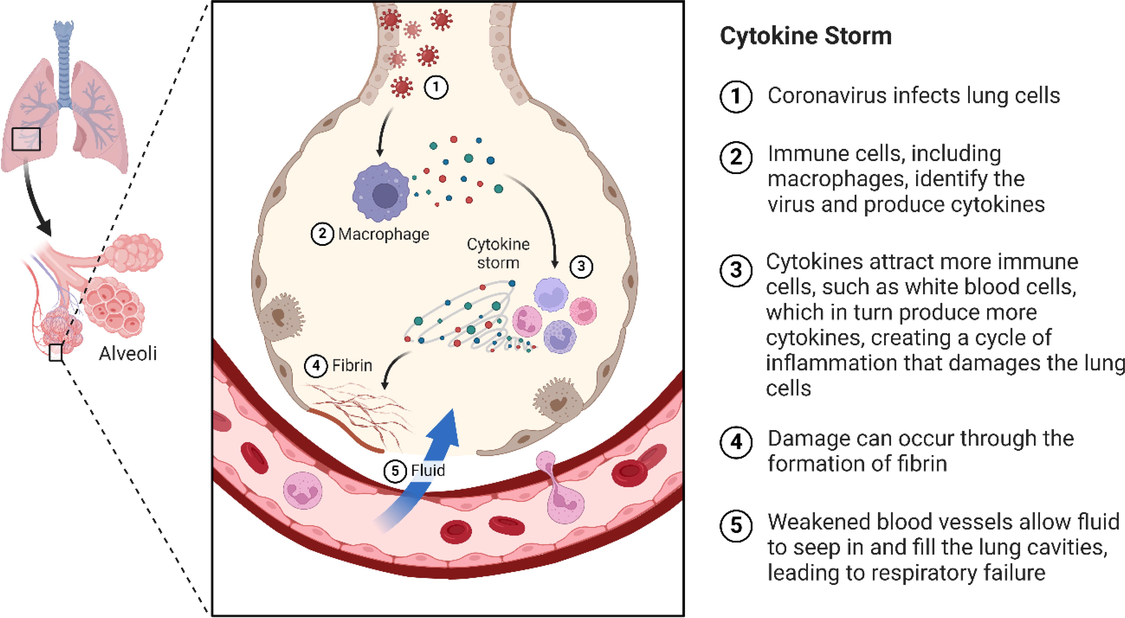
Visual representation of cytokine storm in COVID-19.
Single-cell RNA sequencing studies have revealed unique reactions of different cell types to antiviral medications and immunomodulatory drugs, offering valuable information on resistance to treatment and mechanisms of therapeutic resistance. Through profiling individual cells before and after treatment, scientists can pinpoint molecular signatures linked to treatment response and resistance, which helps in tailoring personalized therapeutic approaches for COVID-19 patients (Huang et al., 2021).
Epitranscriptomic modifications of ncRNAs
Studying chemical modifications of RNA molecules has revealed a new way to control protein expression, prompting a reevaluation of certain aspects of cellular biology. Studying epitranscriptomic modifications is crucial for understanding RNA metabolism, influencing various processes such as transcription, translation, splicing, subcellular localization, and stability. They have a significant influence on the transcriptome, offering a quick and efficient method to alter its composition and function in reaction to different stimuli. One method to achieve this decidable task is by modulating RNA interactions with RNA-binding proteins (RBPs) (Huang et al., 2021; Jusic et al., 2022). An approximate number of over 3,000 RBPs are present in human cells. Even though the purpose of most of them remains a mystery, it is evident that numerous RBPs are involved in various stages of post-transcriptional RNA processing. Three primary RBPs linked to RNA epitranscriptomic modifications have been identified as follows: enzymes responsible for depositing, removing, and recognizing specific chemical groups. Nevertheless, not all RNA chemical groups have designated erasers or readers. Therefore, there is a need for targeted proteomic methods to comprehensively identify and quantify epitranscriptomic regulators and effectors (Atlante et al., 2020; Yahaya et al., 2021). When RBPs interact with RNA targets, it can happen in two ways as follows: either directly by recognizing the RNA modification or indirectly by influencing the RNA’s secondary structure, which in turn affects RBPs. Both coding and ncRNAs interact regularly with different RBPs. Part of the 'house-keeping' category RBPs can be active all the time and everywhere, whereas some RBPs show specific expression patterns or their RBPs could be controlled. Natural RNAs frequently contain alterations to RNA, sometimes known as epitranscriptomic changes of RNA. It has been determined that approximately 140 post-transcriptional changes are critical for the structural variety and metabolism of RNAs. At the cellular level, messenger RNA (mRNA), rRNA, and tRNAs undergo chemical changes. Studies have indicated the importance of RNA epigenetic modifications in viral infection (Arzumanian et al., 2022; Atlante et al., 2020). A-to-I editing and pseudouridine are examples of noncanonical nucleotides that have significant modifications in the virus genome, along with methylation of adenine and cytidine residues like N6-methyladenosine (m6A), or 7-methylguanosine, as well as 2'-O-Methyl. These alterations in methylation are often caused by cellular enzymes, although virus-encoded methyltransferases have also been linked to distinct consequences. Determining the impact of epigenetic modifications on viral RNA metabolism or virus infection is challenging since these modifications are dynamic and reversible during viral infection and are aided by enzymes (Stamatopoulou and Zaravinos, 2021). Despite the rapid advancements in RNA research, scientists have identified numerous RNA modifications in various virus genomes that could influence virus infection. When a virus infects the body, the immune system’s initial response is usually to activate innate antiviral defenses. TLRs, PRRs, Myeloid Differentiation Factor-88, TIR-domain-containing adaptor protein inducing interferon-beta, and other cytoplasmic receptors/adapters are activated when they recognize external nucleic acids, such as virus-derived RNAs or DNAs. This process also activates TNF receptor-associated factors (TRAFs) (Song et al., 2023). Activation of TRAFs triggers the activation of IRF3/7 and NF-κB signaling pathways, leading to the production of type I interferons and pro-inflammatory cytokines. Apart from the TLR pathway, another family of Pattern recognition receptors (PRRs) known as the retinoic acid-inducible gene I (RIG-I)-like receptor (RLR) family has been found to be essential cytosolic sensors of viral nucleic acids. The RLR signaling pathway’s receptor protein is recognized as the mitochondrial antiviral-signaling protein, which can be found in the endoplasmic reticulum or mitochondria. When a virus infects, this route makes it possible for IFN-β to express itself quickly. Strong antiviral effects are shown by IFNs and pro-inflammatory cytokines (Hussain et al., 2024d; Jiang et al., 2023).
RNA alterations are becoming more and more important in controlling antiviral innate immune responses, according to recent study. Exploring modifications of ncRNAs has unveiled significant insights into the progression of COVID-19, exposing the intricate molecular mechanisms at play in viral infection, host immune response, and disease progression. Exploring epitranscriptomics entails analyzing the dynamic chemical modifications of RNA molecules that affect their structure, stability, and function. In the context of COVID-19, these modifications play a crucial role in controlling the interactions between the virus and host cells, as well as shaping the immune response to infection. One of the extensively studied epitranscriptomic modifications involves the addition of a methyl group to the adenosine nucleotide in RNA molecules, known as m6A (Courtney, 2021). Studies have demonstrated that m6A modifications are involved in regulating the stability, translation, and splicing of coding and ncRNAs. COVID-19 observations indicate that the m6A modification process is disturbed, leading to alterations in the expression of ncRNAs associated with viral replication, host immune response, and inflammatory signaling pathways. Modifications to viral RNA may influence its stability and translation efficiency, which could in turn impact viral replication and pathogenesis. Modifications in m6A patterns of host ncRNAs, such as miRNA and lncRNAs, could impact their connections with target mRNAs and regulatory proteins, ultimately altering gene expression patterns in response to viral infection (Ruiz Ramírez and Prado Montes de Oca, 2022). One of the epitranscriptomic changes linked to COVID-19 pathogenesis is 5-methylcytosine (m5C), where cytosine residues within RNA molecules are methylated. Modifications of m5C impact RNA stability, structure, and RBPs, thus altering RNA processing, translation, and function. There may be a disruption in m5C modification machinery in COVID-19, potentially causing changes in the expression and function of ncRNAs related to antiviral defense, immune regulation, and tissue balance. Furthermore, additional epitranscriptomic changes, like pseudouridylation and adenosine-to-inosine (A-to-I) editing, have been linked to COVID-19 pathogenesis (Zheng et al., 2024). Changing uridine to pseudouridine can affect RNA structure, stability, and interactions with proteins. When adenosine deaminases acting on RNA catalyze A-to-I editing, it causes nucleotide modifications in RNA molecules, which can impact RNA splicing, stability, and translation. When these epitranscriptomic modifications are disrupted, it can lead to abnormal expression and functioning of ncRNAs that play a role in viral–host interactions, immune evasion, and inflammatory signaling pathways.
Interaction networks between ncRNAs and viral genomes
Recent findings in virology indicate that ncRNAs are essential in various stages of viral infection, including controlling virus growth, replication, and cell death. Based on research findings, it is evident that viral ncRNAs can influence cellular and viral gene expression to create a favorable environment for the viral life cycle. Just as medical researchers have discovered, numerous cellular ncRNAs have the ability to impact viral replication and target viral genomes, either directly or indirectly. The complex functional interaction network is formed by the interaction of viral and cellular ncRNAs with their viral and cellular targets. Understanding intricate interactions is crucial for comprehending viral infection and creating novel antiviral treatments (Enguita et al., 2022; Suga et al., 2023). Severe viral infections are caused by intricate interactions between two organisms, where the infectious agent relies on the infected cell’s molecular and metabolic functions for both growth and replication. Viral components and cellular structures establish crucial molecular linkages as the virus penetrates the host’s cellular machinery. These interactions are necessary to alter cellular metabolism, help viruses enter cells more easily, and circumvent certain defense mechanisms. Studying the molecular components of cells and viruses during infection primarily centers on proteins. Studying viral polypeptides that facilitate infection or evade the immune system, along with their cellular counterparts, is a key aspect of this research (Enguita et al., 2022; Liu et al., 2019).
It is critical to understand the role that viral and cellular RNAs play as critical components during an infection. The past 10 years have seen the beginning of research into the roles of lncRNAs in viral infections. Research has demonstrated the critical function of lncRNA mediators in controlling the inflammatory and immunological reactions to viral infections. These particular instances show how some lncRNAs control the spread of RNA viruses. The ability of the human norovirus to strongly activate a lncRNA-mediated response in host cells that regulates the interferon reaction is one important element in the development of viral gastroenteritis. By lowering lncPINT expression, human HCV strains can withstand interferon defense and the innate immune response, allowing them to survive for prolonged periods of time (Serpeloni et al., 2021). The recently identified lncRNA AP000253 illustrates how HBV might go unreported in the host for extended periods of time using a similar technique. Research from experimental models has connected a complicated system of RBPs with lncRNA regulators of infection by looking at various cases. A respiratory RNA(+) virus known as SARS-CoV-2 quickly spread throughout the world, causing the pandemic that began in late 2019. Acknowledged as a member of the coronaviridae family, SARS-CoV-2 enters cells by attaching itself particularly to the host’s ACE2 receptor. Once inside the cell, infection causes alterations in the pattern of gene and protein expression, exhibiting an increase in genes linked to the synthesis of interleukin and the interferon response (Cheng et al., 2022; Serpeloni et al., 2021).
In severe instances of SARS-CoV-2 infection, there is typically a significant acute inflammatory reaction associated with an overproduction of cytokines, referred to as a “cytokine storm.” Researchers discovered that certain proteins are engaged by genomic SARS-CoV-2 RNA and its RNA transcripts, influencing cellular responses to the infection, through high-throughput proteomic analysis (Zhang et al., 2019a). Studying the intricate networks between ncRNAs and viral genomes reveals a multifaceted relationship that impacts different facets of viral infection, replication, and pathogenesis. It is essential to comprehend these networks to uncover the molecular mechanisms behind viral–host interactions and pinpoint potential therapeutic targets for fighting viral diseases like COVID-19. One of the key connections between ncRNAs and viral genomes is through miRNAs (Wang, 2018; Zhang et al., 2019a). Exploiting host cellular machinery, viruses can encode viral miRNAs that regulate both viral and host gene expression. Examining viral miRNAs reveals their ability to impact different phases of the viral life cycle, such as viral entry, replication, and avoidance of host immune reactions (Lanjanian et al., 2021). In contrast, cellular miRNAs from the host can target viral genomes, either by directly stopping viral replication or by adjusting host immune responses to control viral spread. For instance, within the realm of COVID-19, researchers have pinpointed various host miRNAs that could potentially control SARS-CoV-2 replication and pathogenesis. These miRNAs focus on viral RNA sequences or host factors related to viral entry and replication, like transmembrane protease serine 2 (TMPRSS2) and ACE2, which serve a crucial role in viral entry into host cells. Moreover, viral-encoded miRNAs play a role in controlling host immune responses and adjusting viral gene expression to enhance viral persistence and pathogenesis. lncRNAs play a role in interaction networks with viral genomes, influencing viral replication, host immune responses, and inflammatory processes (Arora et al., 2020; Lanjanian et al., 2021). lncRNAs have the ability to function as molecular scaffolds, decoys, or competitive endogenous RNAs to regulate the expression of viral and host genes. Through interactions with viral RNA or proteins, lncRNAs have the ability to impact viral replication and assembly, along with host antiviral defense mechanisms. When it comes to COVID-19, the disruption of host lncRNAs has been linked to changes in immune responses, cytokine production, and disease severity. For instance, specific lncRNAs have been demonstrated to control the expression of pro-inflammatory cytokines and chemokines, which play a role in the cytokine storm syndrome seen in severe instances of COVID-19. Furthermore, alterations in lncRNA expression profiles caused by viruses can help viruses evade the immune system and remain in host cells for longer periods (Madhry et al., 2021; Withers et al., 2019). CircRNAs have also been linked to interaction networks with viral genomes. These circRNAs can act as molecular sponges for miRNA or RBPs, influencing gene expression and signaling pathways related to viral replication and host immune responses. Changes in circRNA expression profiles caused by viral infection can disrupt cellular processes and immune evasion mechanisms (Pan et al., 2021).
During the COVID-19 situation, increasing evidence suggests the important role circRNAs play in regulating viral replication and host immune responses. One illustration is how circRNAs are involved in regulating the expression of crucial host factors related to viral entry and replication, along with inflammatory signaling pathways linked to COVID-19 development. In addition, circRNAs encoded by viruses can function as competitive inhibitors of host circRNAs or miRNAs, interfering with cellular processes and enhancing viral survival. Studying the dynamic and context-dependent interaction networks between ncRNAs and viral genomes involves considering factors like viral strain, host cell type, and immune status. To comprehend these intricate networks, a combination of high-throughput sequencing technologies, bioinformatics analyses, and experimental validation is essential. Through understanding the molecular mechanisms involved in viral–host interactions, scientists can discover new targets for antiviral treatments and create plans to fight against viral diseases like COVID-19 (Jafarinejad-Farsangi et al., 2020; Pan et al., 2021; Sun and Chen, 2020).
Challenges and Future Perspectives
Our current understanding of COVID-19, particularly concerning ncRNAs, is continuously evolving and faces several challenges. These challenges primarily stem from the intricate nature of the virus, its dynamic interactions with the host, and the pressing need for more precise and comprehensive diagnostic tools. A significant hurdle is the identification of specific ncRNAs uniquely associated with COVID-19. Although several ncRNAs, such as NEAT1 and MALAT1, have been identified as differentially expressed in COVID-19 patients, there is still a need for more systematic and comprehensive approaches to uncover the full spectrum of ncRNAs involved in the disease (Baj et al., 2020; Genena et al., 2023; Huang et al., 2022).
Another limitation lies in the lack of standardization in methods used for ncRNA detection and quantification. This variability can lead to inconsistent findings across studies, making it challenging to compare results. Therefore, there is a critical need for the development of standardized protocols and reference materials for the detection and quantification of ncRNAs in COVID-19 (Ruivinho and Gama-Carvalho, 2023). Furthermore, our understanding of the roles that ncRNAs play in the pathogenesis of COVID-19 remains incomplete. While some studies suggest that certain ncRNAs may regulate the host immune response or virus replication, further research is necessary to fully elucidate their functions and the underlying mechanisms contributing to the disease (Huang et al., 2022; Ruivinho and Gama-Carvalho, 2023).
In addition, the potential of ncRNAs as diagnostic biomarkers for COVID-19 remains largely unexplored. Although specific ncRNAs have been detected in patient samples in some studies, more research is required to determine their sensitivity, specificity, and clinical utility as diagnostic tools. Finally, the development of therapeutic strategies targeting ncRNAs is still in its early stages. While some studies have identified ncRNAs as potential therapeutic targets, more research is needed to fully understand their mechanisms of action and develop effective therapeutic strategies (Ruivinho and Gama-Carvalho, 2023; Wu et al., 2022).
Conclusion
ncRNAs have become important factors in the complex realm of COVID-19 pathogenesis and interactions between host and virus. Using different mechanisms, such as miRNA, lncRNAs, and circRNAs, ncRNAs have significant effects on viral replication, host immune responses, and disease advancement. The disruption of ncRNAs during COVID-19 impacts a wide range of cellular processes. MiRNAs, for example, have the ability to directly target viral genomes or regulate host gene expression, impacting viral entry, replication, and evasion of immune surveillance. In a manner akin to a medical researcher, lncRNAs function as molecular scaffolds or competitive inhibitors, influencing host–virus interactions and controlling inflammatory responses. Functioning as miRNA sponges or regulators of gene expression, circRNAs impact viral replication and host immune signaling pathways. Furthermore, ncRNAs are essential in the disrupted immune responses seen in severe cases of COVID-19. By regulating the expression of pro-inflammatory cytokines and chemokines, they play a role in the cytokine storm syndrome and hyperinflammation seen in severe cases. Through their impact on immune cell activation, differentiation, and function, ncRNAs play a crucial role in shaping the host’s immune response to viral infection, ultimately affecting disease severity and clinical outcomes. ncRNAs are valuable as diagnostic and prognostic biomarkers for COVID-19. Studying the varied expression of ncRNAs during viral infection offers important insights into the advancement of diseases and categorization of patients. Studying ncRNA expression patterns in clinical samples helps discover new biomarkers for early detection, prognosis, and treatment response monitoring in COVID-19 patients. Overall, ncRNAs show great potential as targets for therapeutic intervention in COVID-19. Approaches focused on altering ncRNA expression or function may help reduce viral replication, dampen inflammatory reactions, and enhance patient outcomes. More investigation is necessary to clarify the exact functions of certain ncRNAs in COVID-19 development and to create specific treatment strategies. ncRNAs play a vital role in the COVID-19 pandemic but have not been thoroughly investigated. Delving deep into viral–host interactions, immune responses, and disease pathogenesis highlights their importance as potential diagnostic biomarkers and therapeutic targets. Exploring the intricate functions of ncRNAs in COVID-19 is crucial for enhancing our comprehension of the disease mechanisms and creating successful approaches to address the current global health emergency.
Footnotes
Acknowledgments
The authors are thankful to their institutions for providing support in carrying out the study.
Authors’ Contributions
M.M.: Data curation and Writing—original draft. M.S.H.: Formal analysis, Software, Supervision, and Writing—review and editing. N.K.S.: Formal analysis, Validation, and Writing—original draft. A.S.: Software, Supervision, and Visualization. A.S.B.: Data analysis and Writing—review and editing. M.A.: Data acquisition and Writing—review and editing.
AI-Assisted Work Statement
During the preparation of this work, author(s) used the ChatGPT for language and grammar corrections. After using the tool, author(s) reviewed and edited the content as needed and take(s) full responsibility for the content of the publication.
Author Disclosure Statement
No competing financial interests exist.
Funding Information
Our work received no particular grant from any funding agency in the public, commercial, or not-for-profit sectors.