
Abstract
The widespread use of efficient vaccines against infectious diseases is regarded as one of the most significant advancements in public health and techniques for preventing and protecting against infectious diseases and cancer. Because the purpose of vaccination is to elicit an appropriate, powerful, and long-lasting immune response against the pathogen, compounds such as adjuvants must be used to enhance these responses. Adjuvants have been widely used since their discovery to boost immune responses, prevent diseases, and activate protective immunity. Today, several types of adjuvants with varying properties are available for specific applications. Adjuvants are supramolecular substances or complexes that strengthen and prolong the immune response to antigens. These compounds have long-term immunological effects and are low in toxicity. They also lower the amount of antigen or the number of immunogenic reactions needed to improve vaccine efficacy and are used in specific populations. This article provides an overview of the adjuvants commonly used in the vaccination industry, their respective mechanisms of action, and discusses how they function to stimulate the immune system. Understanding the mechanisms of action of adjuvants is crucial for the development of effective and safe vaccines.
Introduction
In recent decades, the increase in the incidence of infectious diseases has prompted researchers to try to provide effective solutions for the control and prevention of these diseases, and in this regard, many vaccines have been successfully presented to deal with these diseases (Excler et al., 2021, Shen and Cooke, 2019, Zarei Taher et al., 2019). As per the World Health Organization’s data, vaccination prevents the deaths of five individuals each minute, resulting in an estimated total of 25 million lives saved between 2011 and 2020 (Ozawa et al., 2011, Tahamtan et al., 2016). Primary vaccines are formulated using attenuated or inactivated microorganisms or their constituents. Currently, it is understood that vaccines consist not just of antigens obtained from diseases but also contain immunostimulating ligands that promote the initiation of an immune response (Hoebe et al., 2004, Zarei Taher et al., 2019). The purpose of vaccination is to induce protective immunity and create a long-term response against infectious agents, which can strengthen the immune system by adding adjuvants to the vaccine (Jiang et al., 2021, Tahamtan et al., 2016). One of the issues raised in connection with vaccines is the effect of vaccine adjuvants on growth indicators. In other words, sometimes an adjuvant causes improvement in efficacy of a vaccine and enhancement of survival when exposed to infectious agent (Sarkar et al., 2019, Soltani and Shafiei, 2022). The term “adjuvant” originates from the Latin word “adiuvare” and pertains to any substance that enhances the cellular or humoral response to an antigen (Aiyer-Harini et al., 2013, Fazeli et al., 2006). Adjuvants are a varied collection of specific molecules or more intricate compositions that have been used since the early 20th century to enhance immune responses. Adjuvants are necessary because numerous vaccinations alone provoke a weak immune response (Aiyer-Harini et al., 2013, Bajpai and Susan, 2016). When selecting these compounds in addition to the vaccination, it is important to examine certain factors such the antigen type, target species, immune response type, and length of the immune response. The utilization of adjuvants to enhance and stimulate the immune response to vaccine antigens is varied. Adjuvants have the effect of augmenting the immunogenicity of weak antigens and contribute to accelerating and prolonging their immunological response. Furthermore, it enhances the binding capacity of antibodies to antigens. Adjuvants are efficacious in enhancing the immune response in individuals of all age groups, hence diminishing the required dosage of antigen incorporated in the vaccine formulation, resulting in cost savings (Kaur et al., 2022, Parvizpour et al., 2020, Tahamtan et al., 2016, Thomas and Prendergast, 2016).
The Necessity of Using Adjuvants in the Vaccine Industry
Occasionally, the inclusion of a specific molecule in the primary composition of vaccines is required to augment the immune response. Previously, antibodies were generated as a reaction to proteins, carbohydrates, intricate lipids, and nucleic acids that were extracted from natural origins. Modern chemical, biosynthetic, and recombinant DNA (rDNA) methods produce antigens today. Most of them are ineffective at stimulating the immune system because of the absence of an innate immunological response. In order to become effective immunogens, small polypeptides and nonprotein antigens need to be chemically linked to a larger immunocarrier protein. Because of this, it is suggested to use them with an adjuvant to ensure a better antibody reaction that is amplified by memory (Petrovsky and Aguilar, 2004, Song and Hu, 2009).
Mechanism of Effect of Adjuvants
Adjuvants have the ability to enhance the immune response to antigens through several mechanisms. Some of the methods include enhancing the circulation of antigens in the bloodstream, facilitating the proliferation of antigen-presenting cells (APCs), stimulating macrophages and lymphocytes, and promoting the production of cytokines (Zarei Taher et al., 2019).
Formation storage at the vaccine injection site
The most widely recognized mechanism of action for adjuvants is the development of storage at the injection site. The depot effect is widely recognized as a fundamental mechanism of action for numerous adjuvants. The antigen persists within the granulomas induced by alumina gel for a duration of 2–3 weeks, progressively diminishing over time. Alum readily absorbs antigens, with the binding process being attributed to robust electrostatic interactions between the antigen and alum. These interactions promote the uptake and presentation of antigens by APCs, as depicted in Figure 1. Other adjuvants, such as water-in-oil emulsions, can enhance the duration and stability of antibody titers by creating a depot effect. The AS04 adjuvant, which is made up of alum and monophosphoryl lipid A (MPL), worked best when it was given along with the antigen. Multiple investigations have demonstrated that the immune responses were unaffected when the antigen-alum depot was surgically removed 14 days following immunization (Awate et al., 2013). Novel microparticle adjuvants have the ability to create durable reservoirs and can gradually release a specific quantity of antigen over an extended period of time. Additional adjuvants can potentially achieve efficacy by fully saturating Kupffer cells in the liver and, therefore, decreasing the uptake of antigen by the liver. These adjuvants have the potential to enhance the quantity of antigen that is accessible to APCs (Shanmugham et al., 2006). Some polysaccharide derivative adjuvants, like high-molecular-weight sulfated dextrans and diethylaminoethyl (DEAE), have been suggested as ways to use this method (Zarei Taher et al., 2019).
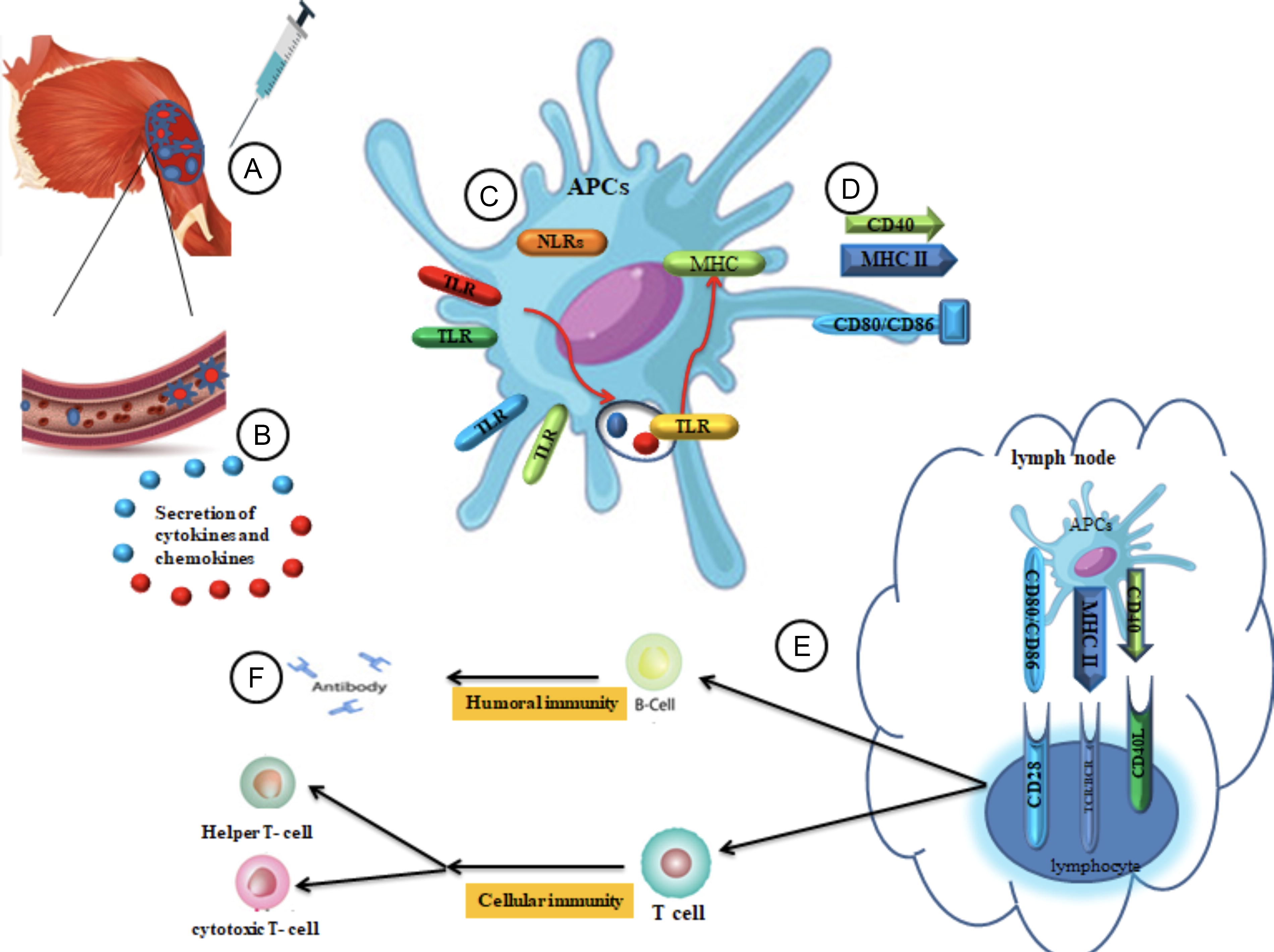
Mechanism of action of adjuvants.
Increasing cellular uptake at the injection site by regulating cytokines and chemokines
Adjuvant particles induce a localized state of inflammation to attract immune cells. Genome-wide research reveals that at the injection site, Alum, MF59, and CpG-59 affect a cluster of genes responsible for encoding cytokines, chemokines, innate immune receptors, interferon-induced genes, and adjuvant core response genes. MF59 exhibits a greater capacity than alum and CpG oligodeoxynucleotides (ODN) to modulate the expression of genes involved in the core response of adjuvants. Chemokines, which have a crucial function in the targeted movement of immune cells in tissues, have also been demonstrated to be controlled by adjuvants at the site of injection (Mosca et al., 2008, Seubert et al., 2008). MF59 exerts a substantial influence on the expression of CCR2, a receptor responsible for binding to CCL2 and facilitating the infiltration of monocytes. In addition, MF59 induces the secretion of chemotactic factors such as CCL2, CCL3, CCL3, and CXCL8. Adjuvant System 03 (AS03) is an extra oil-in-water emulsion that makes colony-stimulating factor 3 (CSF3) and IL-6 when mixed with antigen. It also attracts the chemokines CCL2, CCL3, and CCL5 to the site of injection (Di Pasquale et al., 2015, Dupuis et al., 2001, Morel et al., 2011, O’Hagan et al., 2013).
Antigen presentation
The immune response to antigens consists of two main phases: an innate, nonspecific response and an adaptive, specific response. Adjuvants interact with the innate immune response and exhibit specific expressions of the natural response to pathogens (Spickler and Roth, 2003). For an adaptive immune response to happen, it is very important that antigens are presented correctly by major histocompatibility complexes (MHCs) on APCs. Alum, oil-based emulsions, and microparticles are some of the adjuvants that work to expose antigens to APC. This makes it easier for MHC molecules to present antigens. Alum has demonstrated the ability to enhance the absorption of antigens by dendritic cells (DCs) and modify the quantity and duration of antigen presentation. The process of adsorbing the antigen onto alum results in an augmentation of antigen internalization. Recent research indicates that alum does not directly enter DC but instead transfers antigens through a process called abortive phagocytosis. As alum interacts with membrane lipids on DC, it sorts the lipids, brings in molecules that contain ITAM and Syk, and turns on PI3. Last but not least, these actions cause the antigen to bind to the alum, which then activates DCs and makes major histocompatibility complex class II (Awate et al., 2013).
Activation and maturation of DCs
DC activation is essential for initiating adaptive immune responses. Enhanced MHC class II expression, activation of CD86, and maturation of CD83 result in a heightened capacity of APC to stimulate T lymphocyte activation and differentiation. It has been shown that Freund’s complete adjuvant, lipopolysaccharide (LPS), liposomes, CpG ODN, MF59, AS04, and α-galactosylceramide (α-GAL) can all help DCs mature, which improves the adaptive immune response (Copland et al., 2003, Kool et al., 2008, Shah et al., 2003). Studies have shown that injecting OVA and alum adjuvants into the peritoneal cavity results in the absorption of antigens and the development of mature DCs.
Nevertheless, experiments conducted on human cells invitro have demonstrated that alum and MF59 do not have the ability to directly activate DC. However, they do enhance the presence of MHC class II and costimulatorymolecules (CD83 and CD86) on monocytes, macrophages, and granulocytes, which leads to increased proliferation of T cells (Awate et al., 2013, Seubert et al., 2008, Sokolovska et al., 2007). Evidence demonstrates that AS04 stimulates DCs via Toll-like receptor 4 (TLR4), subsequently directing their migration to the nearby lymph nodes where they activate antigen-specific T cells. In addition, CpG enhances the control of CD40, CD54, CD80, CD86, MHC class II molecules, and the process of antigen processing and presentation in plasmacytoid DCs (pDCs) (Didierlaurent et al., 2009, Kerkmann et al., 2003).
Activation of inflammation
Innate immune cells possess a variety of pathogen recognition receptors (PRRs) that enable them to identify infectious pathogens. Novel types of PRRs have been discovered in recent years. The mentioned receptors encompass Toll-like receptors (TLRs), C-type lectin-like receptors, nucleotide oligomerization domain (NOD)-like receptors, and retinoic acid-inducible gene 1 -like receptors. Several immunological adjuvants function by either signaling through PRRs or acting as ligands for receptors in the innate immune system (Awate et al., 2013).
Classification of Adjuvants
Adjuvants are commonly categorized into the following groups: mineral substances, bacterial products, oil-based emulsions, immunological adjuvants, mucosal adjuvants, and nanoparticle (NP)-based adjuvants. Out of these options, aluminum-based inorganic compounds are the most prevalent and favored by humans (Fig. 2).
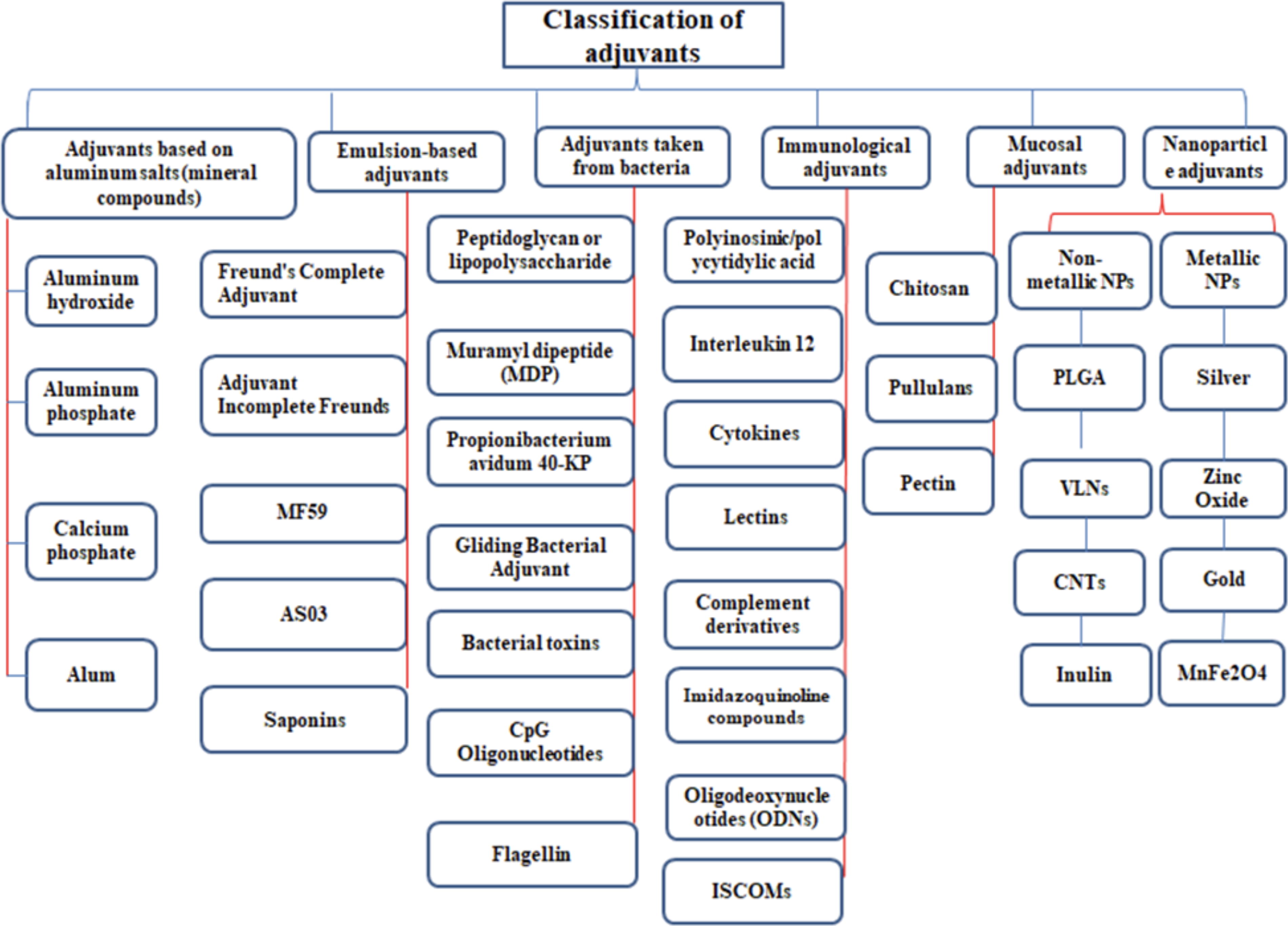
Classification of adjuvants.
Adjuvants Based on Aluminum Salts
Aluminum salts are highly effective at stimulating T helper cells (Th2)-type immunological responses in mice. Through human studies, it was discovered that aluminum salts elicit a mild T cell response. This could be attributed to the insufficient activation of the innate immune system in comparison to TLR stimulation. Animal studies have shown that aluminum salts can cause inflammation, with the injection site and the particular type of aluminum salts used influencing how much inflammation occurs. The mechanisms underlying the intrinsic stimulation caused by aluminum salts are currently the subject of ongoing discussion. It is clear that aluminum salts do not stimulate the immune system through TLR-dependent signaling because the antibody responses to T cell-dependent antigens stayed the same when alum adjuvant antigens were given to mice that did not have the MyD88 and TRIF genes (Gavin et al., 2006).Various investigations indicate that NOD-like receptor protein 3 (NLRP3) is the molecular target of alum. Recently, there has been a hypothesis suggesting that the activation of NLRP3 could be triggered by the phagocytosis of aluminum crystals. This occurs when the phagolysosomes, which contain the aluminum crystals, enlarge and burst, causing the release of cathepsin B into the cytosol. Cathepsin B then activates caspase 1, leading to the release of IL-1β. In order to evaluate these concepts on actual individuals, a group of physically fit participants were administered canakinumab, a human monoclonal antibody (mAb) targeting IL-1β, followed by immunization with a conjugate vaccine containing meningococci, which was further enhanced by hydroxide aluminum. The antibody response to the immunization was indistinguishable from that found in the control group. Research on gene expression in mice shows that additional adjuvants like MF59 and CpG share inflammatory pathways, including IL-1. In general, as compared with other substances that enhance the immune response, aluminum salts did not cause any noticeable increase in cytokine levels in the peripheral blood. Aluminum salts have the capability to enhance the uptake of antigens by APCs, but they do not have the ability to directly activate DC. In addition, research has shown that even in the absence of alum, local inflammation in living organisms can indirectly trigger the activation of APCs. This finding could potentially elucidate the reason why aluminum salts do not effectively stimulate cellular immunity (Del Giudice et al., 2018, Jones et al., 2005).
Aluminum hydroxide
Aluminum, which was initially used as an adjuvant in 1920, has now been entirely substituted by aluminum hydroxide and aluminum phosphate. Vaccines that use this adjuvant use insoluble aluminum salts, typically in gel form, to display vaccination antigens on their surface. These antigens are then released gradually upon injection. At the injection site, an aseptic inflammatory response takes place, causing the infiltration of inflammatory cells and macrophages, which subsequently facilitates the presentation of antigens to the immune system. Ultimately, it impacts Th2 and induces the creation of IL4 and IL5 cytokines, thereby promoting the activation of B lymphocytes and enhancing the synthesis of immunoglobulins, particularly immunoglobulin G (IgG) (He et al., 2015, Li et al., 2014).
Other mineral salt adjuvants
Calcium, iron, and zirconium salts have all been used to absorb antigens, albeit not as effectively as alum salts. For triple immunizations (diphtheria, tetanus, and pertussis), calcium phosphate has proven very useful. Calcium phosphate has the same qualities as alum salts, but it is a natural substance that the human body tolerates well. Calcium phosphate absorbs antigens effectively and induces a high amount of IgG antibodies, but it does not boost IgG synthesis (Foumani et al., 2012).
Emulsion-Based Adjuvants
Oil emulsion adjuvants contain a combination of oil in the aqueous phase that is stabilized by the surfactant. Oil adjuvants are classified into three types: water-in-oil emulsions, oil-in-water emulsions, and water-in-oil-in-water emulsions (Fig. 3). One type of water-in-oil emulsion is incomplete Freund’s adjuvant (IFA), which contains fine droplets of the aqueous phase in oil. IFA has been used in animal vaccinations such as foot and mouth disease, equine influenza, swine fever, rabies, para-influenza, Newcastle disease, and infectious canine hepatitis (Aguilar and Rodriguez, 2007, Petrovsky and Aguilar, 2004).
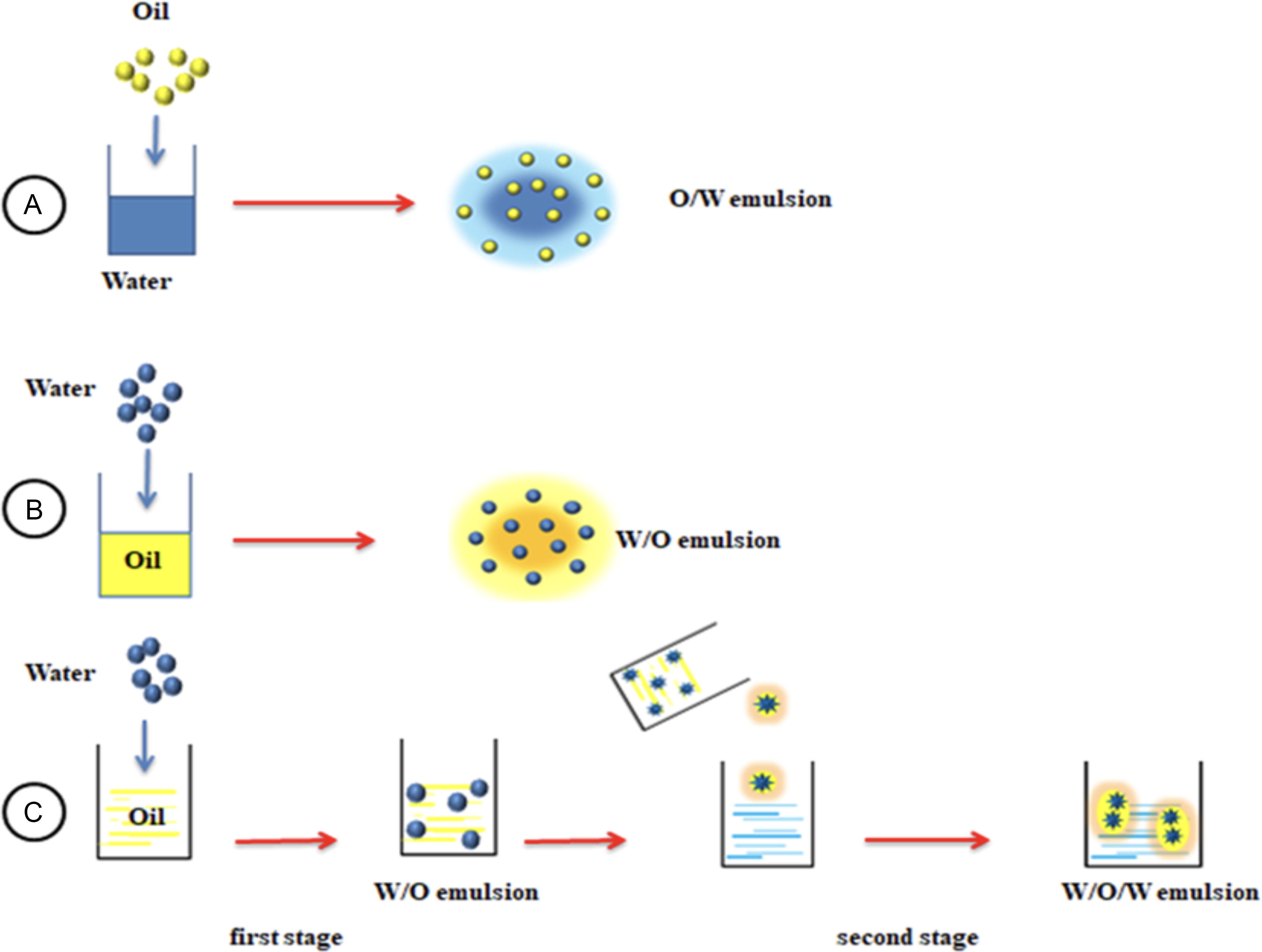
Emulsion adjuvants may be prepared in different ways.
These adjuvants are among the most potent immune system stimulants and boosters, with Freund’s adjuvant, including Freund’s complete and Freund’s incomplete adjuvant, as well as MF59, AS03, and Saponin, being the most well-known (Foumani et al., 2012).
Freund’s complete adjuvant
These adjuvants are crucial and contain a water-in-oil emulsion with mycobacterium that has been destroyed through heat. Typically, these adjuvants are used to assess the ability of antigens to provoke an immune response in mice and to produce autoimmune disorders such encephalomyelitis. These adjuvants can exert their influence by impacting DCs. It is essential to activate DCs in order to initiate certain immunological responses. This activation is achieved through upregulating the expression of particular markers, such as MHC class II, on the surface of DCs. This ultimately enhances the capacity of DCs to induce stimulation and differentiate T lymphocytes, resulting in targeted immune responses (Tahamtan et al., 2016).
One of the most important concerns related to the use of complete adjuvant is the induction of persistent local inflammation, which can cause wounds at the injection site (Zurbriggen et al., 2000) (Zurbriggen et al., 2000). The evidence shows that in order to induce autoimmunity, the components of mycobacterium cause the differentiation of T lymphocytes to Th1 and, as a result, create a delayed hypersensitivity response (Felnerova et al., 2004, Zurbriggen et al., 2000).
Freund’s incomplete adjuvant
These adjuvants include a water-in-oil emulsion, which does not contain mycobacteria. This adjuvant exerts its effect by gradually releasing the antigen from the fat droplets, increasing the half-life of the antigen, and stimulating local innate immunity, which increases phagocytosis, the secretion of lymphocytes and cytokines. During studies conducted by WHO, it has been shown that Freund’s incomplete adjuvant has severe side effects in humans (Gao et al., 2020).
MF59 and AS03
Emulsions have been used as vaccine adjuvants for an extended period. However, the mineral oils used in the initial generation of vaccines were nonmetabolizable, leading to the occurrence of aseptic abscesses and hindered absorption, despite their potent ability to enhance the antibody response. Fortunately, this issue has been resolved over time (Chen et al., 2021). The challenge has been resolved by the creation of oil-in-water emulsions, such as MF59, and emulsion-based adjuvant systems, such as AS03, which use metabolizable oils. This advancement has enabled the production of enhanced seasonal inactivated influenza vaccines, and subsequently, avian influenza vaccines. The vaccines mentioned, such as the adjuvanted MF59 seasonal inactivated vaccine for the elderly in Europe and the United States, the adjuvanted MF59 and AS03 pandemic vaccinations in Europe, and the MF59- and H5N1 avian influenza vaccines with AS03 for storage in the United States, have been licensed for use in these regions (Garçon et al., 2012, Wilkins et al., 2017). Emulsion vaccine adjuvants have been extensively used by millions of individuals worldwide, and their benefits and safety profile are now firmly established. Squalene, a metabolizable lipid produced by the human body during cholesterol synthesis, is a frequently used ingredient in oil-in-water emulsions such as MF59, AS03, and AF03. Furthermore, one of these adjuvants, known as AS03, includes an immunostimulant called α-tocopherol, which is a kind of vitamin E. As a result, it is referred to as an adjuvant system (Del Giudice and Rappuoli, 2015).
Oil-in-water emulsions have greater adjuvant potency and operate through distinct processes as compared with aluminum salts. Nevertheless, they share two commonalities: (1) Their method of action does not involve binding to TLR receptors and (2) their development, similar to alum, was experimental and relied on an older technology for formulating chemicals, without a full comprehension of the mechanisms responsible for their immunostimulatory qualities. MF59 in mice triggers the activation of cells at the injection site and the absorption of antigen by DC without causing a depot effect. This results in the recruitment of CD11b and F4/80 + mononuclear cells. MF59 elicits a more potent inflammatory reaction compared with CpG and aluminum salts. This reaction is linked to the activation of genes involved in the innate immune response, including IL-1b, caspase-1, and Ccr2, as well as their corresponding ligands (Ccl2, Ccl7, and Ccl8). Recent results indicate that MF59, in contrast to adjuvants like aluminum hydroxide or calcium phosphate, stimulates the release of extracellular ATP from muscle, which could potentially function as an endogenous warning signal. In summary, this indicates that the detection of lipid droplets in emulsions initiates the release of internal stress signals and the stimulation of innate immune pathways. In addition, MF59 enhances the swift infiltration of CD11b + cells into the muscle in comparison to other adjuvants (Jones et al., 2005, Vono et al., 2013).
MF59 can induce the formation of a localized immune-stimulating milieu marked by the production of several cytokines. In addition, it is feasible to indirectly activate DCs through processes that do not rely on TLRs. AS03 has been found to produce comparable outcomes. Thus, it appears that cells produced from monocytes, rather than genuine DCs, have a significant impact on the functioning of the emulsion. Emulsions not only facilitate the absorption and activation of cells at the injection site but also enhance the uptake of antigens by APCs and their transportation to the draining lymph nodes. The inclusion of immunostimulatory alpha-tocopherol in AS03 was linked to enhanced absorption of antigens by monocytes, as well as heightened production of CCL2, CCL3, IL-6, CSF3, and CXCL1, resulting in a stronger antibody response (Calabro et al., 2011, Morel et al., 2011). Finally, emulsions’ superiority over aluminum salts in increasing the antibody response may be due to their capacity to produce a high Th response. Indeed, MF59 stimulates a significant Th response, which regulates the breadth of the germinal center (GC) B cell response. In humans, influenza vaccinations adjuvanted with MF59 or AS03 have repeatedly shown higher immunogenicity than unadjuvanted vaccines (both avian and pandemic). Furthermore, both MF59 and AS03 adjuvants were more effective against hospitalization for influenza and influenza-related illness, and both helped to increase efficacy against seasonal or pandemic influenza in young children. According to the findings, comparing two simple H5N1 vaccines and an H5N1 vaccine with an alum adjuvant revealed that the presence of oil adjuvants in water induces the secretion of IgG antibodies, which are primarily directed against the HA1 region of the spherical head of the HA protein, which contains the binding site (Del Giudice et al., 2018). Pre-vaccination antibodies, as well as those generated by plain or adjuvant vaccinations, primarily target the stem region (HA2) of HA. A study of monoclonal antibodies produced from participants inoculated with the MF59-adjuvanted pandemic H1N1 vaccination found that these antibodies were more capable of recognizing wild-type viruses than antibodies evoked by the nonadjuvanted vaccine. This implies that oil-in-water adjuvanted vaccinations outperform non-adjuvanted vaccines (Khurana et al., 2010, Raymond et al., 2016, Vesikari et al., 2011).
Saponins
Saponins are glycosides, either steroidal or triterpenoid, that are present in several plant species, including both wild and cultivated varieties. Triterpenoid saponins are often the main type of saponins found in crops, although steroidal saponins are frequently present in plants that are used for medical purposes or have health benefits. Saponin-based adjuvants have a distinct capacity to enhance cellular immunity and augment antibody synthesis (Aiyer-Harini et al., 2013, Marciani, 2018).
Bacteria-Derived Adjuvants
Cell wall peptidoglycan or lipopolysaccharide of gram-negative bacteria
These bacterial constituents enhance the immune response to the antigens that are co-distributed with them. The efficacy of this adjuvant is influenced by the activation of receptors known as TLRs through the start of danger signals in the host’s immune defense system. Lipid A is the primary component in the structure of LPS, and it is accountable for its adjuvant activity. Under situations of low acidity, lipid A can undergo hydrolysis to become MPL. MPL is a molecule that preserves the adjuvant properties of lipid A while reducing its toxicity. Lipid A and MPL are immunomodulators that have the ability to induce robust type 1 immune responses (Melander et al., 2023, Shimoyama and Fukase, 2021).
Propionibacterium avidum 40-KP
These adjuvants have the ability to enhance the immune response to the antigen that is co-administered with them. In order to use bacterial products in new vaccines, it is necessary to subject them to additional purification and frequently detoxification (Buket and DİKMEN, 2019).
Muramyl dipeptide (MDP)
They are part of mycobacteria’s peptidoglycans, which have a function in immunological modulation. Muramyl dipeptide has serious adverse effects such as fever, arthritis, and iris inflammation; however, it also forms fewer toxic derivatives. Hydrophilic derivatives (including threonyl-MDP, muramitide, nor-MDP, N-acetylglucosaminyl-MDP, and murabiotide) primarily elicit type 2 reactions. Hydrophobic derivatives, such as muramyltripeptide phosphatidyl or ethanolamine (PE-MTP), are frequently used in liposomes, water-in-oil, and oil-in-water emulsions to promote type 1 responses and high cellular immunity (Khan et al., 2021, Reddy et al., 2022).
Bacterial toxins
Some toxins that act through adenosine diphosphate (ADP)-ribosylation are being investigated for mucosal or skin use. Two of these toxins that have been widely investigated are the cholera toxin and the heat-sensitive exotoxin (LT) of Escherichia coli. In addition to strong humoral responses, these two also stimulate Cytotoxic T Lymphocytes (CTLs) (Vilander and Dean, 2019).
Gliding bacterial adjuvant
The compound is a substantial polymer of aminoglycoside that is obtained from the Cytophaga bacterium. This polysaccharide has the ability to induce the release of cytokines in cats, mice, and humans. Gliding bacterial adjuvant appears to be most efficacious when used in conjunction with other adjuvants, such as alum or oil emulsion (Foumani et al., 2012).
CpG oligonucleotides
CpG oligonucleotides are adjuvants that are similar to the bacterial DNA motif. This motif is six long chains of DNA deoxynucleotides with central CpG dinucleotides and is found in bacterial and viral DNA 3–20 times more often than in mammalian DNA. CpG oligonucleotides are immune system modulators that can stimulate antibodies and especially seem to be effective in switching the immune pathway toward type 1 responses (Foumani et al., 2012, Tahamtan et al., 2016, Zarei Taher et al., 2019).
Flagellin
Flagellin, the primary component of the flagellum of gram-positive and gram-negative bacteria, can be detected by cell surface receptors, leading to the production of TNF-α. The adjuvant has a considerable effect on CD4 + T cells. Furthermore, studies demonstrate that the use of flagellin adjuvant might cause a high titer of antibodies in the host’s body (Khim et al., 2021).
Immunological Adjuvants
Immunological adjuvants have the ability to indirectly stimulate innate immunity by interacting with pattern recognition receptors or directly affecting cytokines. Immune cells possess a diverse array of pattern recognition receptors on their surface that detect infectious pathogens, with one prominent example being TLRs. TLR ligands, which imitate pathogen-associated molecular patterns and stimulate immune cells via TLRs, are currently being formulated for human use as therapeutic agents against various diseases and as vaccine adjuvants. These drugs, such as imiquimod and resiquimod (R-848), are imidazoquinoline compounds that specifically interact with TLR7 and TLR8. In addition, CpG ODN specifically bind to TLR9 (Liu et al., 2020).
Synthetic oxynucleotide-oligodes (ODNs)
These adjuvants consist of unmethylated CpG motifs, which function as agonists for the 9-TLR receptor. This adjuvant has the ability to enhance the immune response to recombinant protein antigens and enhances the functioning of specialized APCs. Consequently, the activation of innate immunity enables the production of heightened antibody responses and cell-mediated responses, specifically T + CD4 Th1 cells and CTL responses (Zarei Taher et al., 2019).
Research has demonstrated that both bacterial DNA and synthetic ODN that contain CpG motifs (CpG) can effectively activate or boost the activation of many types of immune cells. The stimuli encompass the following: (1) direct stimulation of murine and human B cells, resulting in the production of immunoglobulins, IL-6, and IL-10 (MHC II), as well as resistance to programmed cell death; (2) direct activation of macrophages and DCs, leading to independent maturation characterized by cluster designation (CD), followed by the release of proinflammatory chemokines and cytokines, including IL-12, upregulation of MHC class II molecules, expression of B7s, and expression of CD40. (3) Stimulation of natural killer (NK) cells resulting in swift initiation of interferon (IFN) generation and lytic abilities (Weeratna et al., 2005).
Imidazoquinoline compounds
Imidazoquinoline compounds, including imiquimod and risquimod (R-848), have demonstrated their ability to modulate the local immune response. They have also been found to stimulate the production of IFN-α, IFN-gamma, TNF-α, and IL-12 in cultured human blood mononuclear cells. In addition, they possess antiviral and anticancer characteristics. Recent research has demonstrated that in mice, R-848 is a powerful adjuvant that strongly favors Th1 immune responses. Similar to CpG, R-848 can also enhance Th2 immune responses that have already been triggered by immunization with the same antigen and alum (Miller et al., 2020, Weeratna et al., 2005).
Polyinosinic/polycytidylic acid
These adjuvants are artificial double-stranded RNAs that imitate viral RNAs and stimulate 3-TLR receptors found within the endosome. Administering this adjuvant stimulates DCs, leading to rapid activation of 12-IL and type I IFN. Both of these molecules are crucial in promoting Th1 reactions. This adjuvant is a highly significant 3-TLR antagonist that has been extensively evaluated for its effectiveness against diseases like HIV, dengue fever, malaria, and cancer (Nakano et al., 2020).
Interleukin 12
Interleukin 12, also known as a NK cell stimulating factor, has the ability to induce the secretion of gamma interferon (IFN-γ) from T lymphocytes and NK cells. Research indicates that multiple infections have the capability to trigger a Th1 immune response through the stimulation of IL12 production (Tahamtan et al., 2016).
Cytokines
Vaccine adjuvants encompass cytokine proteins and their corresponding genes. The cytokines included in this group are IL-2, IL-1, INF-γ, granulocyte-macrophage colony-stimulating factor (GM-CSF), and IL-12. INF-γ is a versatile cytokine that can augment cellular immune responses through multiple methods. GM-CSF improves the first immune response by stimulating and trapping cells that present antigens (Foumani et al., 2012, Zarei Taher et al., 2019).
Lectins
Lectins are proteins that attach to carbohydrates and function as receptors for different glycoproteins on the surface of cells. The presence of immunomodulatory properties in plant lectins encourages their screening for potential medicinal applications, including adjuvant development. One of the important features of plant lectins is their ability to communicate with the mucosal epithelium and transfer it inside the intestine, which can be used in vaccine formulation to create mucosal and systemic immunity. In the last few years, lectins from Jackfruit integrifolia Artocarpus, ArtinM, and Jacalin have been used as potential adjuvants in vaccines against protozoan parasites. Another recently isolated plant lectin is a
Complement derivatives
The constituents of the mammalian complement system can serve as potent adjuvants to provoke antibody responses. Segments of this protein selectively attach to external antigens and designate them for recognition by antibodies and immune cells. According to a study, the attachment of three C3D molecules to an antigen resulted in a 1000-fold increase in its ability to provoke an immune response. The antigens that have been effectively altered by C3D encompass influenza virus hemagglutinin, anti-idiotype antibodies, and capsular polysaccharides. In addition, the C3b component has demonstrated adjuvant action (Solairaja et al., 2021).
Liposomes
Liposomes are cholesterol and phospholipid vesicles that serve as both adjuvants and delivery routes for antigens and adjuvants. Allison and Gregoriadis were the first to describe the capacity of liposomes to stimulate an immunological response to antigens that are either encapsulated within the liposome or attached to its surface. Subsequently, there has been ongoing progress in the field of utilizing liposomes and lipid NPs for the creation of vaccines to prevent and treat infectious and oncological diseases. Commercially accessible liposomal vaccines include Inflexal® for influenza, Epaxal® for hepatitis A viruses, Mosquirix® for malaria, and Shingrix® for varicella zoster virus. Several liposomal formulations are currently being tested in clinical trials as prophylactic and therapeutic vaccines for malaria, influenza, TB, HIV, dengue, and other diseases (Tretiakova and Vodovozova, 2022). If liposomes are used as an adjuvant, the antigen is placed on the surface or between the layers of these compounds, and by creating a physical connection between the antigen and this lipid structure, the immunogenic agent is presented to the immune cells (Foumani et al., 2012). After absorption, liposomes are broken down by macrophages in the liver and spleen, and their antigens are presented to T cells. These compounds are able to increase cellular immunity in addition to stimulating humoral immunity and antibody production (Zarei Taher et al., 2019).
Immune stimulating complexes
Utilizing adjuvants composed of various particles, ranging from microscopic virus-like particles (VLP) to bigger liposomes and emulsion droplets, is an effective method to induce targeted immune responses by vaccination. Immune stimulatory complexes (ISCOMs) are categorized as diminutive lipid-based particles measuring 40–60 nm. These particles have demonstrated promise as adjuvants and carriers of antigens for the purpose of preventive or therapeutic immunization. Reports indicate that immunization with ISCOM antigens and adjuvants can elicit both cellular and humoral immune responses (Lövgren Bengtsson et al., 2011). The antigen in the vaccine is presented to the immune system by entering the fatty part of ISCOMs, which is actually the cholesterol contained in saponin, and then by the MHC class I molecule, they stimulate cytotoxic T lymphocytes. Of course, these adjuvants also control the MHC class II pathway by releasing gamma interferon. Due to their nontoxicity, these compounds can be used orally in addition to the injection form, and they also stimulate the production of immunoglobulin A in the case of mucosal administration (Foumani et al., 2012, Lövgren Bengtsson et al., 2011). ISCOMs are versatile and flexible delivery systems that can deliver antigen to B cells and increase antigen uptake by APC (Lövgren Bengtsson et al., 2011).
Mucosal Adjuvants
As the interest in intranasal or oral vaccinations grows, it becomes necessary to develop adjuvants that enhance their efficacy on mucosal surfaces. Mucous surfaces serve as a physical barrier to hinder the infiltration of foreign substances, hence eliminating antigens and vaccination adjuvants. Nevertheless, these surfaces possess specialized cells that are responsible for sampling antigens, such as M cells and intraepithelial DCs. M cells have the ability to internalize antigens and subsequently transmit them to APCs. Goblet cells can also internalize some antigens. The mucosal epithelium expresses several innate immune receptors, such as TLRs. Therefore, mucosal locations are targeted by pathogen-associated molecular patterns (PAMPs) such as muramyl dipeptide, poly I:C, flagellin, and CpG oligonucleotides (Srivastava et al., 2015, Tizard, 2021). Some bacterial PAMPs, such as cholera toxin and E. coli heat-sensitive enterotoxin, can also act as mucosal adjuvants by stimulating DCs. Cyclic guanosine monophosphate dimer induces both Th1 and Th17 responses on mucosal surfaces. Damage-associated molecular patterns (DAMP) adjuvants can also affect the mucosa and cause cellular stress or damage. These include cyclodextrins and some oleic acid derivatives. They may work simply by causing enough inflammation and mild injury to allow antigen entry. Some compounds added to intranasal adjuvants may increase their half-life at mucosal surfaces. Pectin, which forms a gel on mucosal surfaces, increases the antigenicity of the intranasal influenza vaccine by increasing its contact time with the mucosa. Other complex carbohydrates such as pullulans and mannans may have a similar effect (Srivastava et al., 2015, Tizard, 2021). Chitosan is a biopolymer obtained from glucosamine in the outer shell of crustaceans and is widely used as a mucosal adjuvant in intranasal applications. Chitosan adjuvant can facilitate antigen phagocytosis process. Studies have shown that diphtheria toxin intranasal vaccine with chitosan adjuvant increases Th2 responses and interferon gamma production, compared with previous vaccines (Nakano et al., 2020). In addition, the successful use of Helicobacter pylori vaccine with chitosan adjuvant in laboratory conditions in mice showed that this adjuvant has the same functional capacity as the adjuvant of previous vaccines used for this pathogen, and the mice that were protected by chitosan had a lower density of this bacterium in the stomach mucosa (de Jong et al., 2007).
Nanoparticles
Nanotechnology has emerged as a very promising scientific field with profound impacts on diverse aspects of human existence, particularly in the realms of pharmacology and medicine, since the late 21st century. NPs are typically solid colloidal particles with a size below 1000 nm, exhibiting distinct variations in terms of size, shape, physical characteristics, and chemical properties. NPs are categorized into many types based on their content, including polymeric agents, VLP, liposomes, emulsions, inorganic components, self-assembled proteins, and immunostimulating complexes (Badiee et al., 2013). Self-assembled peptide NPs (SAPNs), which are primarily made up of helical or beta-sheet secondary structures, have recently gained popularity as vaccine adjuvants due to their nontoxicity, biodegradability, and self-adjuvanticity. NPs are used for medicine delivery and diagnostic purposes. Today, NP engineering is a growing subject in vaccine science, with scientists hoping that NP adjuvants may usher in a new immunization method. The first NPs for medicine administration and immunization were produced in the late 1960s (Badiee et al., 2013, Li et al., 2018). Nanomaterials are being extensively researched as vaccine adjuvants in the fields of infectious illnesses and malignancies. Nanomaterials can effectively shield antigens from degradation and enable controlled and prolonged release. Consequently, APCs can uptake them more effectively (Fig. 4). NP vaccine adjuvants consist of vaccine adjuvants that have been reduced to small particle sizes, typically ranging from 1 to 1000 nm. As an illustration, MF59 is a nanoadjuvant that measures 165 nm in diameter. Additional nanoadjuvants now being studied in clinical trials include PLGA (poly lactide-hemoglycolide) NPs with a size range of 100–200 nm, virus-like NPs measuring 15–30 nm, cationic liposomes, nanoemulsion W805EC with a size of 400 nm, and nanogels containing cholesterol with a size range of 30–40 nm. Metal NPs (MeNPs) are highly significant nanomaterials that have garnered considerable interest in the domains of microbial immunization and cancer immunotherapy (Behzadi et al., 2021, Jin et al., 2019). They are relatively nonbiodegradable, have strong structures, and are generally simple to synthesis. MeNPs are frequently used for diagnostic and therapeutic purposes, as well as vaccine carriers. Various investigations have shown that MeNPs can trigger immunological responses and act as vaccine adjuvants, either directly or indirectly (Fig. 2). Some MeNPs have powerful adjuvants. They can also increase APC absorption, activate cytokines and chemokines, and elicit humoral and/or cellular immunological responses. However, the molecular processes underlying the immunological response triggered by NPs, particularly MeNPs, remain unclear. Gold and iron oxides are the most commonly used vaccine adjuvants among MeNPs (Behzadi et al., 2021, Jin et al., 2019).

Schematic images of the interaction of metal-based nanoparticles used in adjuvants with immune cells.
Adjuvants in mRNA Vaccines
Typically, these mRNA vaccines incorporate specific adjuvants to achieve the intended immune response. Vaccine adjuvants are substances that traditionally stimulate the immune system by binding to PRR found on innate immune cells. This binding increases the strength or effectiveness of immunological responses, thereby improving the effectiveness of vaccines. Adjuvants are crucial constituents of effective vaccinations. Based on the existing literature, adjuvants in mRNA vaccines can be categorized into three main groups: (1) RNA with intrinsic self-amplifying capabilities; (2) components of the delivery mechanism; and (3) external immunostimulants (Xie et al., 2023; Arunachalam et al., 2021). Adjuvants play a crucial role in determining the specific immune response triggered by mRNA vaccines. Therefore, the selection of suitable adjuvants is vital in the development of mRNA vaccines for the treatment of different diseases. An optimal adjuvant for mRNA vaccines should augment vaccine effectiveness by eliciting a robust immune response and mitigating the limitations of mRNA, such as its low stability and translation efficiency. The presence of ionizable cationic lipids in the LNP system can significantly impact the effectiveness of mRNA vaccines. Furthermore, the utilization of mRNA to encode precise functional immunity proteins and the evaluation of innate immune receptor agonists as suitable adjuvants are additional encouraging methods for the development of adjuvants in mRNA vaccines. Exploring proficient adjuvants will enhance comprehension of mRNA vaccines and facilitate the creation of safer and more efficacious mRNA vaccines for many diseases (Xie et al., 2023; Arunachalam et al., 2021; Karam and Daoud, 2022).
Systems Analysis of Adjuvants in Licensed Human Vaccines and Clinical Development
Alum is the primary and most extensively used adjuvant in vaccines for humans. The process by which alum adjuvant works is believed to involve increasing the absorption of antigens and triggering danger or harm signals, which in turn attract various types of immune cells. MF59 was the first adjuvant used in approved human vaccines, following alum. MF59 is a type of emulsion that consists of squalene, Tween 80, and Span 85. It has the capability to stimulate powerful antibody responses to co-antigens. Studies have demonstrated that MF59 enhances the range and strength of the immune response produced by influenza vaccination in adult humans. (Del Giudice et al., 2018; Harandi, 2018) AS03 and AS04 adjuvant systems are used in a limited number of authorized human vaccinations. AS03 is a squalene-based oil-in-water emulsion that includes α-tocopherol (a variant of vitamin E) and polysorbate 80. AS04 consists of alum and MPL, which is a TLR-4 agonist. Both adjuvants were demonstrated to activate the transcription factor NF-κB. Systems analysis was used to examine the immunological and transcriptional responses in individuals who were given the malaria vaccine candidate RTSS, S/AS01, in a sequential manner. The RTSS-S/AS01 vaccine candidate comprises the circumsprozoite protein (CSP), which is the primary constituent of Plasmodium falciparum sporozoite. Encapsulation involves the formation of VLP using AS01, a liposome-based adjuvant, a TLR4 ligand called QS-21, and a saponin. Recent studies have examined numerous potential vaccination adjuvants in animal models and have shown promising outcomes. Nevertheless, only a small number of these tools for finding new things have advanced to the phase of testing on humans to determine their effectiveness. The adjuvants used in licensed human vaccines include 2% alhydrogel (Alum) and MF59. In addition, there are three adjuvants now in clinical development: ribosylation cationic adjuvant formulations (CAF01), IC31, and glucopyranosyl lipid adjuvant stable emulsion (GLA-SE). CAF01 is a liposomal adjuvant made up of two components (Del Giudice et al., 2018; Harandi, 2018; Villarreal and Casale, 2020). It consists of cationic liposomes that contain a surfactant called dimethyldioctadecylammonium, which is stabilized with trehalose dihenate. Trehalose dihenate is a synthetic version of a compound found in mycobacteria called trehalose dimycolate. CAF01 works by interacting with a type of protein called C-type lectin. IC31 is a type of adjuvant that has two components: the cationic peptide KLK and the TLR-9-stimulating oligodeoxynucleotide ODN1a, which are fused together. GLA-SE is a man-made substance that activates the TLR4 and is formulated as an emulsion consisting of squalene-based oil dispersed in water (Del Giudice et al., 2018; Harandi, 2018; Villarreal and Casale, 2020).
Conclusion
The primary objective of vaccination is to confer immunity against infections. Effective immunization against various infections necessitates distinct immune responses that can be stimulated by using suitable vaccination adjuvants. Hence, a precise understanding of the mechanisms by which adjuvants function is crucial in the logical development of vaccines. Appropriate adjuvants that stimulate and enhance immune responses, when mixed with specific vaccine antigens, expedite, extend, and intensify immune reactions against related antigens. Desirable adjuvants should possess attributes such as a prolonged half-life, significant biodegradability, economical synthesis, and a strong ability to stimulate immunological responses. Recently, there has been notable advancement in comprehending the processes by which different adjuvants function, particularly in terms of stimulating innate immunity through diverse routes. A diverse range of chemical compounds with distinct modes of action and varying side effects are being developed for the manufacture of novel adjuvants. The objective of adjuvant research is to identify adjuvants that are more potent while causing fewer adverse effects. Over the past 20 years, there has been significant advancement in the creation of novel vaccine adjuvants. These adjuvants are currently being used in clinical practice and have proven highly effective in preventing infectious diseases like influenza and cervical cancer caused by Human papilloma virus (HPV) in real-world scenarios. In addition, they have demonstrated promising results in clinical settings for diseases such as malaria and herpes zoster. According to the information provided in the study and other reports, numerous adjuvants have been proposed. However, when considering factors such as safety, effectiveness, and feasibility of mass production, only a few of the adjuvants mentioned in this study are suitable for use in vaccines. However, because most commercial vaccines have similar structures in terms of their primary component, which is the antigen, vaccine production centers strive to discover appropriate compounds and blend different types of materials to create a unique formulation for their vaccines. This is done to enhance the effectiveness of the vaccines and maintain their superiority in the industry. Hence, it is necessary to possess knowledge regarding the suitable adjuvants, their attributes, and their mechanism of action in order to formulate and produce vaccines. Despite recent advancements in comprehending the role of adjuvants and the workings of the human immune system, only a limited number of vaccination adjuvants have obtained the required licensing for medical application. Hence, it is imperative to do thorough study to ascertain appropriate adjuvants for specialized vaccinations that offer optimal efficacy and safety in the future.
Footnotes
Acknowledgment
The authors are grateful to all the researchers who have done worthy research in this field and were our guide in writing this article.
Data Availability Statement
No data was used for the research described in the article.
Author Disclosure Statement
Dr. Shams and Dr. Jaydari are from Lorestan University (Khorramabad, Iran); Dr. Najafi and Dr. Pouladi are from the University of Tehran (Tehran, Iran); Ms. Hataminejad is from Mazandaran University of Medical Sciences (Sari, Iran), and Dr. Khanizadeh are from Lorestan University of Medical Sciences (Khorramabad, Iran), all where education and research are the primary functions.
Funding Information
The authors reported that there is no funding associated with the work featured in this article.