
Abstract
The plausible effects of SARS-CoV-2 infection on the expression of anti/proapoptotic molecules have been suspected. This cohort study examined the expression of p53, Bcl-2, Bid, Bak, and Bax molecules, the genes associated with induction or inhibition of apoptosis, in the SARS-CoV-2-infected patients with severe and mild symptoms in an Iranian population. In this 6-month cohort study, the expression of p53, Bcl-2, Bid, Bak, and Bax molecules was evaluated at onset of diagnosis, 24 h after symptom onset, and 6 months later in the nasopharyngeal cells of SARS-CoV-2-infected hospitalized patients and outpatients in comparison with healthy controls using the real-time PCR technique. At the onset of the study, the relative expression of p53, Bcl-2, Bid, Bak, and Bax significantly increased in the SARS-CoV-2-infected hospitalized patients and decreased after 6 months. The healthy controls showed potential positive correlations among the molecules, but the patients did not show these correlations. Since SARS-CoV-2 needs host cell survival, it appears that the virus induces the expression of Bcl-2 as an antiapoptotic molecule, and the host cells upregulate the proapoptotic molecules to neutralize the effects. Dysregulation of correlation expression of the molecules among the patients proved that SARS-CoV-2 affects the expression of the molecules involved in apoptosis. SARS-CoV-2 could be considered an important factor that regulates the expression of several molecules participating in cancer pathogenesis.
Introduction
Coronavirus (SARS-CoV-2) has spread as an epidemic in Iran (Fattahi et al., 2022). Studies have shown that this virus infects people through the respiratory system and enters the cells of the respiratory tract and causes different symptoms in various ethnicities (Lamers and Haagmans, 2022). There is a possibility that the virus, which causes viremia, can penetrate into some tissues, leading to both acute and chronic inflammation (Lamers and Haagmans, 2022). Accordingly, scientists hypothesized that SARS-CoV-2 infection could be considered a risk factor for inducing and progressing some cancers in the infected patients (Jahankhani et al., 2023). In agreement with the hypothesis, bioinformatic studies consider the possibility of the reaction of some proteins of SARS-CoV-2 with some tumor-related proteins, such as suppressor molecules (Alpalhão et al., 2020; Saini et al., 2020). Therefore, assuming that it is possible that infection with SARS-CoV-2 may lead to cancer.
Previous investigations revealed that p53, Bcl-2 (B-cell lymphoma 2), Bid, Bcl-2 homologous antagonist/killer (Bak), and Bax play key roles in the pathogenesis of several cancers (Kirkin et al., 2004). p53 is a main suppressor protein that induces apoptosis in several cell systems (Ozaki and Nakagawara, 2011, Voskarides and Giannopoulou, 2023). Accordingly, the downregulation of p53 can be associated with the progression of several tumors (Aubrey et al., 2018). Bax, which is known as Bcl-2-like protein 4, Bid, and Bak are other proapoptotic molecules whose expressions are defective in some cancers (Goldar et al., 2015). The molecules normally act on the mitochondrial membrane and promote permeabilization, followed by the release of cytochrome c and ROS (Savitskaya and Onishchenko, 2015). The last molecules are important signals for inducing apoptosis (Savitskaya and Onishchenko, 2015). However, Bcl-2 is a main antiapoptotic molecule and can induce the survival of various cell systems (Chipuk and Green, 2008). Therefore, environmental factors that affect the expression of the molecules can be associated with an increased risk of cancer in mammals (Goldar et al., 2015; Hanahan and Weinberg, 2011; Morana et al., 2022).
Cohort studies are important and valid investigations to clarify environmental factors’ roles in the pathogenesis of human disorders. In this 6-month cohort study, the expression levels of p53, Bcl-2, Bid, Bak, and Bax in the cells of the nasopharyngeal cells were explored in an Iranian population with different statuses of clinical symptoms of SARS-CoV-2 infection.
Materials and Methods
Subjects
This cohort study included 40 healthy controls, 40 hospitalized patients, and 40 outpatients infected with SARS-CoV-2. The patients were entered into the study, and the samples were taken at the onset of the diseases, 24 h after symptom onset, and also were under any treatment. The study’s entry criteria required patients referred to Kerman province health centers in Kerman, Iran, to have COVID-19 symptoms and a positive PCR test, whereas the control group consisted of healthy individuals not infected with SARS-CoV-2, PCR negative, and symptomless. The study excluded patients who suffered from other respiratory infections, chronic diseases such as cancer or allergies, were on immunosuppressive drugs, or had systemic diseases such as diabetes and autoimmune diseases.
Following the diagnosis of the disease and prior to any treatment, we collected nasopharyngeal cells from both patients and healthy controls. Six months later, we sampled both patients and healthy individuals. The nasopharyngeal cells were collected in the virus-transforming media (Karmania Pars Gene Company, Rafsanjan, Iran) and transferred to the laboratory for the purification of genomic and viral RNA and evaluation of the expression of p53, Bcl-2, Bax, Bid, and Bak.
RNA purification and cDNA synthesis
Total RNA was purified from the nasopharyngeal cells using a commercial kit from Karmania Pars Gene Company, Rafsanjan, Iran, and according to the manufacturer’s guidelines. The purified RNA was used for either evaluation of the SARS-CoV-2 infection or expression of p53, Bcl-2, Bid, Bak, and Bax. Accordingly, the purified RNA was directly used for one-step real-time PCR for diagnosis of SARS-CoV-2 and cDNA synthesis for evaluation of p53, Bcl-2, Bid, Bak, and Bax expressions. We used a smart commercial kit from Karmaina Pars Gene Company, Rafsanjan, Iran, for the synthesis of cDNA.
Detection of SARS-CoV-2
To diagnose SARS-CoV-2, 5 µL of purified RNA was used directly in a one-step commercial kit from Pishtaz-Teb Company, Tehran, Iran. The kit detected three SARS-CoV-2 genes simultaneously and RNase P as an internal control.
Real-time PCR for evaluation of P53, Bcl-2, Bid, Bak, and Bax expressions
For the evaluation of p53, Bcl-2, Bid, Bak, and Bax mRNA expressions, the real-time PCR technique was used. Accordingly, relative expressions of the molecules were carried out using SYBR Green master mix (Biosystem Company, England) in a Rotor-Gene Q instrument using the following program: 95°C for 2 min (one cycle) and then 40 cycles with 95°C/15 sec and 60°C/35 sec, followed by a melting curve ranging from 55°C to 95°C (acquiring fluorescence data every 0.5°C). The specific primers (Table 1) were designed using Primer 3 software. Finally, the raw data from the evaluation of p53, Bcl-2, Bid, Bak, and Bax in parallel with beta-actin, as a housekeeping gene, were analyzed using the 2−ΔΔCt formula.
Primer Sequences of p53, Bcl-2, Bax, Bid, Bak, and Beta-Actin
Statistical analysis
Data analysis was performed using SPSS software version 18. One-sample Kolmogorov–Smirnov test was used to check the data distribution, and due to the nonnormality of the data distribution, nonparametric tests, such as the Kruskal–Wallis test, were used to check the quantitative variables of patients with coronavirus in the hospitalized patient and outpatient groups in comparison with healthy controls, and the Wilcoxon signed ranks test was used to examine quantitative variables in hospitalized and outpatient coronary patients in the first round compared with the same patients in the second round. Mann–Whitney test was used to analyze the differences between men and women in each group. The Spearman test was used to examine the correlations among the molecules. Quantitative data were reported as mean rank and statistical descriptive quartile, and significance was considered lower than 0.05 in all tests.
Results
The participants in the three groups were similar regarding sex (p > 0.05) and age (p > 0.05). Table 2 shows the demographic data of the participants. At the start of the disease, the relative expression of p53 was 96.13 (4.2700; 1.3325–13.7250) in hospitalized patients, 22.83 (0.0027; 0.0011–0.0070) in outpatients, and 62.55 (0.2660; 0.1184–0.8037) in healthy controls. The statistical analysis revealed that the differences among the groups were significant (p < 0.001, Fig. 1A). The results demonstrated that p53 relative expression after 6 months from the onset of the disease was 53.72 [0.0403 (0.0167–0.2356)] in hospitalized patients, 32.29 [0.0168 (0.0039–0.0453)] in outpatients, and 75.11 [0.2660 (0.1184–0.8037)] in healthy control. The statistical analysis revealed that the differences among the groups were significant (p < 0.001, Fig. 1B). The results demonstrated that p53 relative expression in the hospitalized patients at the onset of the disease was 17.61 [4.2700 (1.3325–13.7250)], and after 6 months, it was 8.75 [0.0403 (0.0167–0.2356)], which was significantly different (p < 0.001, Fig. 1C). The p53 relative expression in the outpatients at the onset of the disease was 17.83 [0.0027 (0.0011–0.0070)], and after 6 months, it was 18.63 [0.0168 (0.0039–0.0453)], which was significantly different (p < 0.001, Fig. 1D).

The relative expression of p53 among the groups at the onset of the study
Demographic Data of Patients and Healthy Controls
Data analysis revealed that the participants in all three groups were similar regarding age and sex.
Relative expression of Bak at the onset of the disease in hospitalized patients, outpatients, and healthy controls was 87.88 [4.4900 (2.032–15.0200)], 56.30 [1.2426 (0.4852–3.2305)], and 37.33 [0.3800 (0.1600–1.3475)], respectively, and the differences among the groups were significant (p < 0.001). The Bak relative expressions after 6 months from the onset of the disease among the groups were also significantly different (p < 0.001). However, Bak relative expression in the hospitalized patients at the onset of the disease was not different when compared with the Bak relative expression after 6 months in this group (p = 0.675). The Bak relative expression in the outpatients after 6 months significantly increased when compared with the onset of the disease (p = 0.010).
Figure 2 illustrates the relative expression of Bak among the groups at the onset of the study (A), after 6 months (B), as well as in hospitalized patients (C) and outpatients (D), comparing first sampling versus secondary sampling.
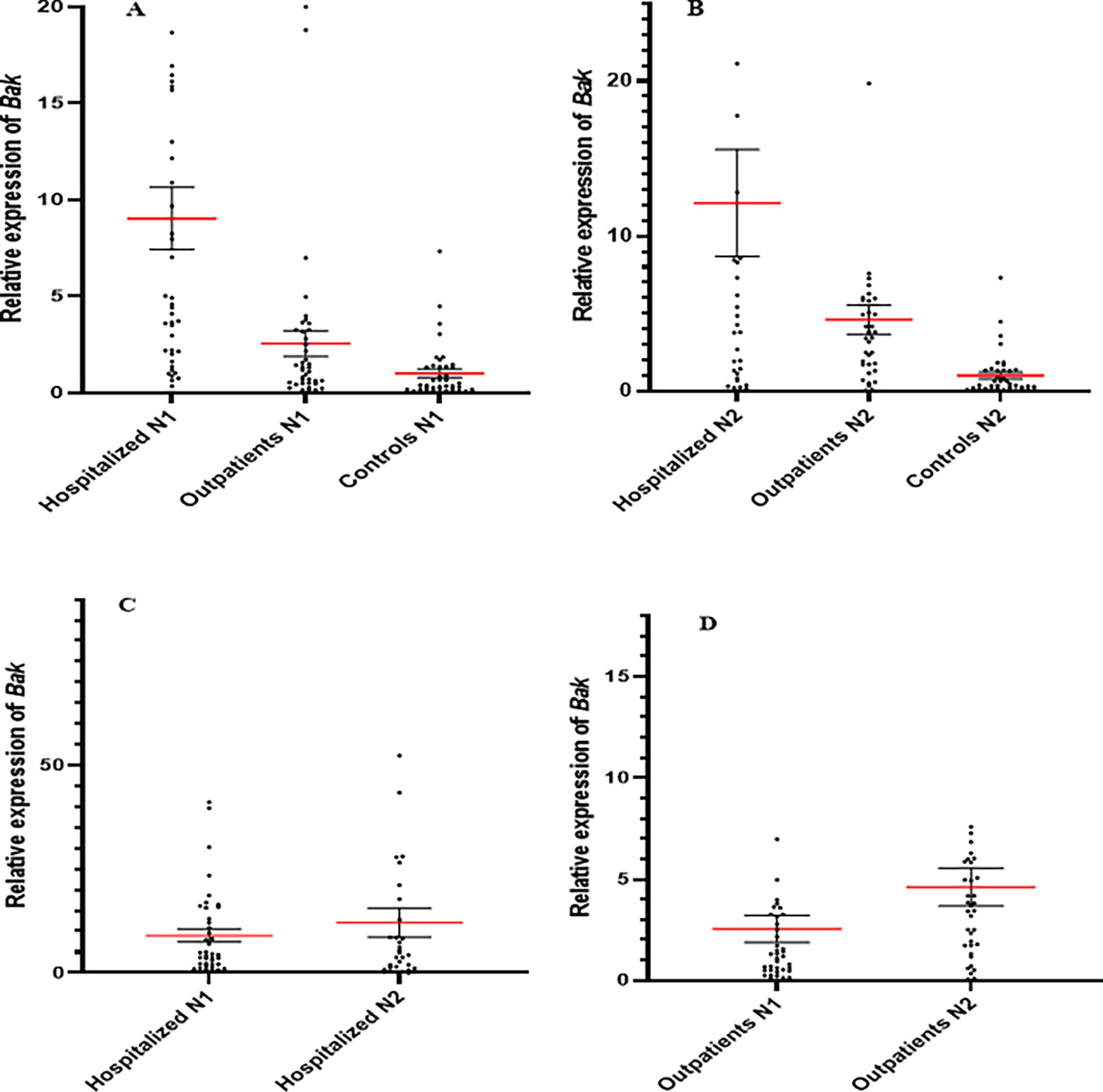
The relative expression of Bak among the groups at the onset of the study
As illustrated in Figure 3, Bcl-2 relative expressions at both onset and 6 months after the onset of the disease were significantly higher in hospitalized patients when compared with either outpatients or healthy controls (p < 0.001). The results demonstrated that Bcl-2 relative expressions in both hospitalized patients (p = 0.172) and outpatients (p = 0.379) were not significantly different when compared with its expression at the onset of the study to 6 months after disease onset. The results demonstrated that Bid relative expression at the onset of the disease was 88.45 [5.1100 (1.7562–31.0725)] in hospitalized patients, 51.51 [0.9025 (0.2302–2.0780)] in outpatients, and 41.54 [0.4550 (0.2025–1.3777)] in healthy control. The statistical analysis revealed that the differences among the groups were significant (p < 0.001, Fig. 4A). The results demonstrated that Bid relative expression after 6 months from onset of the disease was 72.45 [6.0321 (1.5406–10.6910)] in hospitalized patients, 63.42 [2.5392 (1.0193–5.5816)] in outpatients, and 33.03 [0.4561 (0.2031–1.3783)] in healthy control. The statistical analysis revealed that the differences among the groups were significant (p < 0.001, Fig. 4B). Bid relative expressions in the hospitalized patients at the onset of the disease and after 6 months were not significantly different (p = 0.235, Fig. 4C), but in the outpatients they were significantly increased (p = 0.028, Fig. 4D).
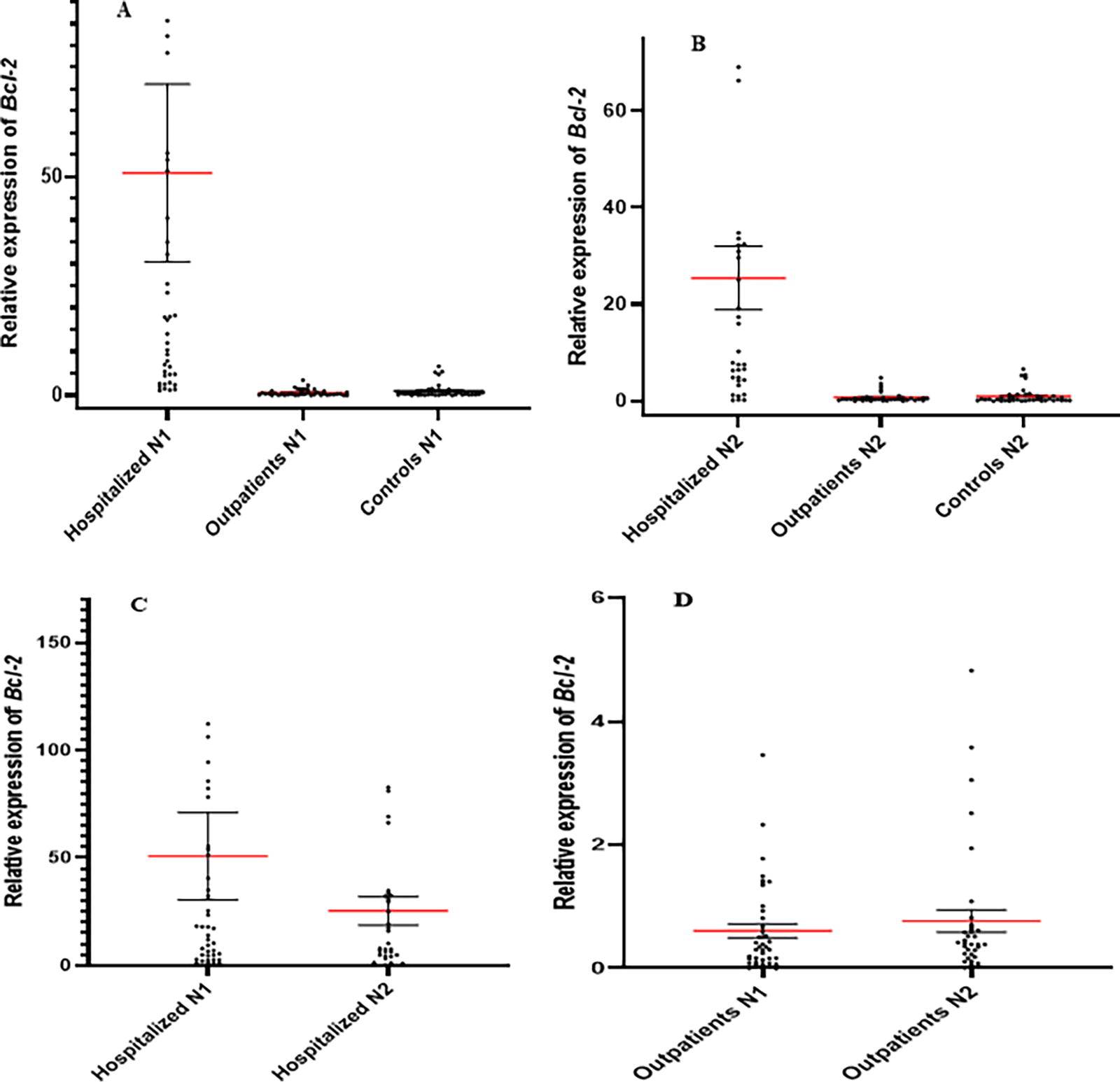
The relative expression of Bcl-2 among the groups at the onset of the study
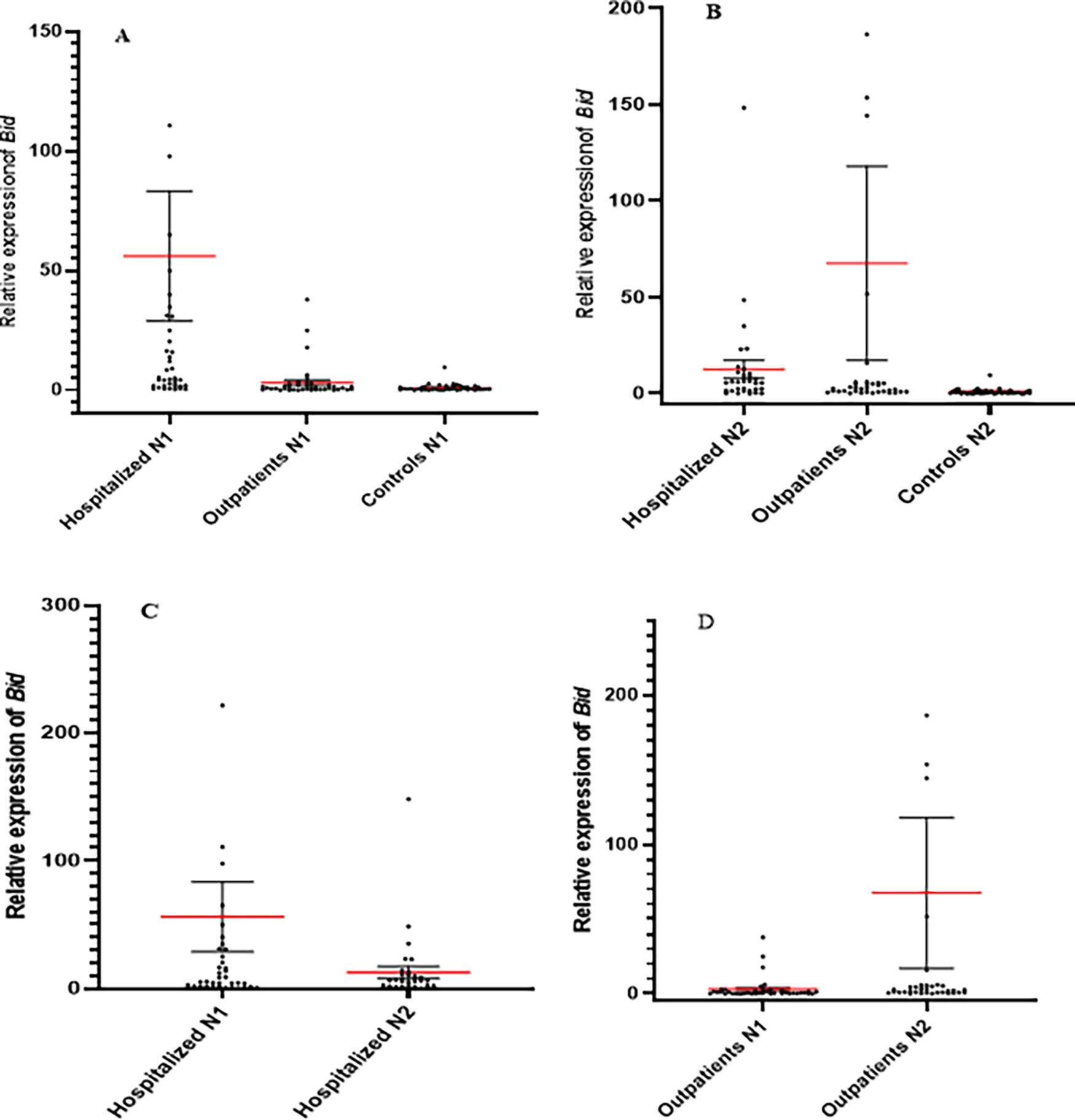
The relative expression of Bid among the groups at the onset of the study
As illustrated in Figure 5, Bax relative expressions were significantly increased in either onset or after 6 months in hospitalized patients when compared with both healthy controls and outpatients (p < 0.001). However, the relative expressions of Bax were not significantly different in the hospitalized patients (p = 0.477) and outpatients (p = 0.051) when comparing the participants at the onset of the study with 6 months after the study.

The relative expression of Bax among the groups at the onset of the study
The Spearman test showed that there were powerful correlations among all the molecules involved in the apoptosis in healthy controls, whereas the correlations were not seen among all molecules in either hospitalized or outpatients (Tables 3–7). However, it was shown that there was a significant negative correlation between age and Bax in outpatients at the start of infection (Table 5) and a positive correlation between Bid and age in outpatients 6 months after infection (Table 6).
Correlations Among the Examined Molecules, Age, and Virus Load in the Hospitalized Patients at the Start of Study
The Spearman test showed that there were significant positive correlations between Bid with P53, Bak, and Bcl-2. There was a positive correlation between Bak and Bcl-2.
P-value lower than 0.05 was considered significant.
Correlations Among the Examined Molecules and Age in Hospitalized Patients After 6 Months
The Spearman test showed that there were significant positive correlations between Bid with P53, Bak, and Bcl-2. There were positive correlations between Bak with P53, Bcl-2, and Bax. Bax had also significant positive correlations with P53 and Bcl-2.
P-value lower than 0.05 was considered significant.
Correlations Among the Examined Molecules, Age, and Virus Load in Outpatients at the Start of Study
The Spearman test showed that there were significant positive correlations between Bid with Bak and Bcl-2. There were positive correlations between Bak with Bcl-2, Bid, and Bax. Bax had a significant negative correlation with age.
P-value lower than 0.05 was considered significant.
Correlations Among the Examined Molecules and Age in Outpatients After 6 Months
The Spearman test showed that there were significant positive correlations between Bak with Bcl-2 and Bid, and between Bid and age.
P-value lower than 0.05 was considered significant.
Correlations Among the Examined Molecules and Age in Healthy Controls
The Spearman test showed that there were significant positive correlations between all the molecules, while there were no correlations between age and the molecules.
P-value lower than 0.05 was considered significant.
Using Mann–Whitney test to analyze the differences of relative expression of p53, Bcl-2, Bid, Bak, and Bax showed that there were no differences between men and women in each group, separately (Fig. 6).

The relative expression of examined molecules (P53, Bak, Bid, Bcl-2, and Bax) at the start of the study and 6 months in the male and female participants. The figure illustrates that there were no differences between male and female in the hospitalized, outpatients, and healthy controls, separately. CG men, control group men; CG women, control group women; HP men, hospitalized patient men; HP women, hospitalized patient women; OP men, outpatient men; OP women, outpatient patient women.
Discussion
The roles played by SARS-CoV-2 infection in the induction and progression of cancers and their related molecules, including p53, Bcl-2, Bid, Bak, and Bax, are yet to be clarified (Alpalhão et al., 2020; Yuan et al., 2023). In descriptive studies, investigators evaluated the relative expressions of the molecules following SARS-CoV-2 infection. We designed our project as a 6-month cohort study to achieve validated results.
The results demonstrated that p53 relative expression was significantly increased in hospitalized patients at the onset of the infection with SARS-CoV-2. The expression was also significantly decreased after 6 months. However, its expression was lower after 6 months in outpatients in comparison with both healthy controls and hospitalized patients. Due to the fact that p53 is a key inducer of apoptosis (Aubrey et al., 2018; Kubra Acikalin et al., 2022), it appears that increased molecule in the epithelial cells of hospitalized patients may be associated with acute inflammation and elimination of the virus reservoir (Parasher, 2021). The results were repeated in the Bak expression at both hospitalized patients and outpatients, which showed increased Bak as a proapoptotic molecule (Konopleva et al., 1999, Parasher, 2021). However, the expression of the molecule was increased after 6 months in the outpatients but not in the hospitalized patients. Therefore, it seems that induction of apoptosis is the main response of epithelial cells to reduce the reservoir of SARS-CoV-2 (Cizmecioglu et al., 2021; Donia and Bokhari, 2021). The similar patterns were also repeated in the expression of Bid and Bax as other members of the proapoptotic molecule (Korsmeyer et al., 2000). Additionally, the results demonstrated that Bcl-2 as the antiapoptotic molecule shows similar patterns as the proapoptotic molecules and revealed a significant elevation in hospitalized patients at the onset and after 6 months after infection and also decreased after 6 months in hospitalized patients. Collectively, it seems that hospitalized patients suffer from a huge invasion of SARS-CoV-2 to the epithelial cells, and the virus induces Bcl-2 to survive the virus reservoir. In response, the host’s epithelial cells may naturally release molecules that help cells die, which will reduce the number of SARS-CoV-2 reservoirs (Nelli et al., 2021). Consequently, the outpatients did not exhibit the pattern, and the relative expression of Bcl-2 remained unchanged compared with healthy controls, even after 6 months. Therefore, we may hypothesize that the induction of antiapoptotic molecules may be associated with SARS-CoV-2 infection with severe symptoms and cytokine storm (Bader et al., 2022). However, after 6 months, its expression was decreased, and it may be related to a nonchronic infection (Mukund et al., 2021). In agreement with our results, a study by Inde et al. revealed that the expressions of proapoptotic molecules, such as BIM (BCL-2-interacting mediator of cell death), were significantly increased in the patients with active COVID-19 (Inde et al., 2021).
Given the association between chronic infections and cell exhaustion (Rha and Shin, 2021), it is plausible to hypothesize that SARS-CoV-2 could potentially induce cancerous molecules in a chronic format, making it a potential carcinogen virus (Alahdal and Elkord, 2022). In agreement with our results, Ma-Lauer et al. showed that SARS-CoV-2 uses a method to escape from the host’s immune system, in which it increases the replication of the virus by destroying the p53 protein, which has the role of inhibiting viral infections by inhibiting the cell cycle (Ma-Lauer et al., 2016). Upregulation of p53 in the lymphocytes of the SARS-CoV-2-infected patients has also been demonstrated by Xiong et al. (2020) and Bordoni et al. (2021). Icard et al. also reported that infection with COVID-19 can lead to the Warburg effect in the cells of the respiratory tract, and this will probably be associated with the inactivation of the p53 molecule, although its expression was increased (Icard et al., 2021). In agreement with this study, other studies showed that probably the NSP3 and S2 proteins of the SARS-CoV-2 increased the survival rate of the host cell by inducing antiapoptotic and inhibition of proapoptotic molecules, which may be associated with increased possibility of cancer in the epithelial cells (Cardozo and Hainaut, 2021; Ramaiah, 2020; Singh and Bharara Singh, 2020). Another study confirmed the hypothesis, revealing that herbal drugs that induce p53 expression can be effective in treating SARS-CoV-2 infection (Khanal et al., 2022). Collectively, due to the results of the current study and in comparison with other investigations, it may be concluded that SARS-CoV-2 may be considered a factor for affecting expression of the molecules involved in the pathogenesis of tumors, and chronic infections with the virus may enhance the chance of cancer by deregulating apoptosis and antiapoptotic molecules.
The results also demonstrated that there were no significant differences regarding the expression levels of p53, Bcl-2, Bid, Bak, and Bax in men when compared with women in each group. Our previous investigations on various populations with different infections confirmed the results and did not show significant differences between men and women regarding several molecules in the Iranian population (Arababadi et al., 2010; Ayoobi et al., 2013; Bahramabadi et al., 2017; Karimi-Googheri et al., 2015). Therefore, it appears that gender cannot affect the expression of immune system-related molecules in Iranian population.
The results also confirmed the presence of powerful positive correlations among p53, Bcl-2, Bid, Bak, and Bax in healthy controls that were not seen in this format in either hospitalized or outpatients. The positive correlation between apoptosis-related molecules is a natural cell response to induce apoptosis in healthy cells, and disruption of the natural responses in epithelial cells of hospitalized and outpatients demonstrated abnormal responses. Therefore, it seems that the disrupted correlation can be a marker for the cancerous properties of SARS-CoV-2. Additionally, there were negative and positive correlations between age with Bax and Bid in outpatients. The negative correlation happened at the start of infection, whereas the positive correlation was seen 6 months after the infection. Thus, it appears that age is a risk factor for the development of cancers during infection with SARS-CoV-2, which needs to be explored by further investigations.
Conclusion
Due to the data presented here, it appears that SARS-CoV-2 needs host cell survival and, hence, the virus upregulates Bcl-2, an antiapoptotic molecule, for proliferation. Accordingly, it was significantly increased in the hospitalized patients. Additionally, it seems that the host cells upregulate the proapoptotic molecules as feedback responses to neutralize the effects. Age can also intensify the negative effects of SARS-CoV-2. Collectively, due to the potential effects of SARS-CoV-2 on the expression of apoptosis-related molecules, it may be hypothesized that SARS-CoV-2 could be considered a factor for deregulation of the molecules that participate in the pathogenesis of tumors.
Ethical Approval Statement
The Research Ethics Committee of Iran University Medical Sciences, Tehran, Iran (Ethical code: IR.IUMS.REC.1400.983) approved the relevant guidelines and regulations for all sampling and experiments. We collected all the samples from the patients who gave their consent.
Footnotes
Authors’ Contributions
M.G. and M.K.A. conceived and designed the study. Z.S. visited and selected the patients. M.K.-G. and M.K.A. performed the experiments. M.K.-G., M.G., and M.K.A. analyzed the data and interpreted the results. M.K.-G., Z.S., M.G., and M.K.A. prepared the article. M.G., M.K.A., Z.M., and J.K. reviewed and revised the article during preparation and finalization.
Author Disclosure Statement
The authors have no conflicts of interest to declare.
Funding Information