
Abstract
Objectives:
The aim of this study was to determine the feasibility of a portable electrospinning device for the application of wound dressings.
Approach:
Four polymer nanofibers dressings were applied on superficial partial thickness wounds to a porcine model and compared with a traditional paraffin tulle gras dressing. The polymer nanofibrous dressings were applied using a handheld portable electrospinning device activated at a short distance from the wound. The partial thickness donor sites were evaluated on day 2, 7, and 14 when dressings were removed and tissue samples were taken for histological examination.
Results:
No significant difference was detected between the different electrospun nanofibrous dressings and traditional paraffin tulle gras. Desirable characteristics of the electrospun nanofiber dressing group included nontouch technique, ease of application, adherence and reduction in wound edema and inflammation. There was no delayed wound healing or signs of infection reported in both the electrospun nanofiber and traditional tulle gras dressings.
Innovation:
Used on partial thickness wounds, polymer electrospun nanofiber dressings provide excellent surface topography and are a nontouch, feasible, and safe method to promote wound healing with the potential to reduce wound infections. Such custom-made nanofibrous dressings have implications for the reduction of pain and trauma, number of dressing changes, scarring, and an added cost benefit.
Conclusion:
We have demonstrated that this portable handheld electrospinning device can be utilized for different formulations and materials and customized according to the characteristics of the target wound at the various stages of wound healing.

Introduction
W
Electrospinning technology is a method for the creation and utilization of nanofibers and has received much attention in peer-reviewed literature. 4 –8 Electrospun nanofibers dressings have in the past received positive attention as an ideal dressing due to their unique architectural features that mimic the extracellular matrix and indeed provide an ideal wound environment for wound healing. 9,10 Nanofibers provide a large surface area-to-volume ratio, are highly permeable, and have small pore size suitable for biomedical applications such as wound healing. 11 –13 In particular, polymer nanofibers have attracted much interest in the domain of wound care providing a unique architectural structure and environment facilitating wound healing. 1,14,15 Furthermore, the high porosity and surface area facilitate the migration of keratinocytes accelerating the wound healing trajectory. 15
Currently, wound care dressings include products like hydrogels, hydrocolloids, alginates, hydrofibers, silicones, and foams that have the capacity to provide a moist wound healing environment, which have been traditionally used. Furthermore, advances in complex dressing technologies have seen the development of epidermal, dermal, and extracellular or scaffold-like replacement products such as TransCyte™, Biobrane™, and Epicel® cultured epidermal autograft and Suprathel. However, the main disadvantages of these dermal replacement templates are the high cost associated with these products as well as the availability, storage, and limited shelf life. 15 Subsequently, the use of electrospun biopolymer nanofibrous dressings allows a biocompatible, simple, and cost-effective alternative to wound management and has gained considerable interest to wound care clinicians. 11,16,17 Electrospun polymer nanofibrous dressings have shown to facilitate cell migration and proliferation of the wound bed, provide hemostasis, gaseous exchange, and management of wound exudate.
Clinical Problem Addressed
Despite a plethora of literature surrounding the efficacy of nanofiber technology, and from the authors' knowledge, there is currently no self-contained handheld portable device that uses electrospinning technology to form a polymer nanofibrous dressing for commercial use. Therefore, the aim of this study was to determine the feasibility of a new portable electrospinning device for the application of dressings to wounds.
Materials and Methods
Materials and scaffold fabrication
Four formulations of biocompatible medical grade polyester, polycarbonate, and polyurethane polymers were used (Corbion Purac, Lenexa, KS and AdvaceSource, Wilmington, MA) (Table 1). The blended nanofibrous dressings were prepared by electrospinning technique using a new portable handheld device manufactured and supplied by Nicast Ltd., Lod, Israel (Fig. 1) and tested for safety and Electro Magnetic Compatibility. All sterile solutions were prepared and supplied by Nicast Ltd., Lod, Israel, manufactured in the company's aseptic line. Briefly, the solutions were electrospun using the electrospinning device with 25 kV applied voltage, at 20 cm distance from the nozzle to the wound bed at a flow rate of 4.5 mL/h and the diameter of the needle tip 0.7 mm. The device used in this study is a portable handheld device. The clinician uses both hands to activate and create the dressing while aiming the nozzle toward the wound area. The clinician moves the device in a uniform motion to ensure full coverage of the wound bed and the surrounding skin (See Supplementary Video SV1; Supplementary Data are available online at
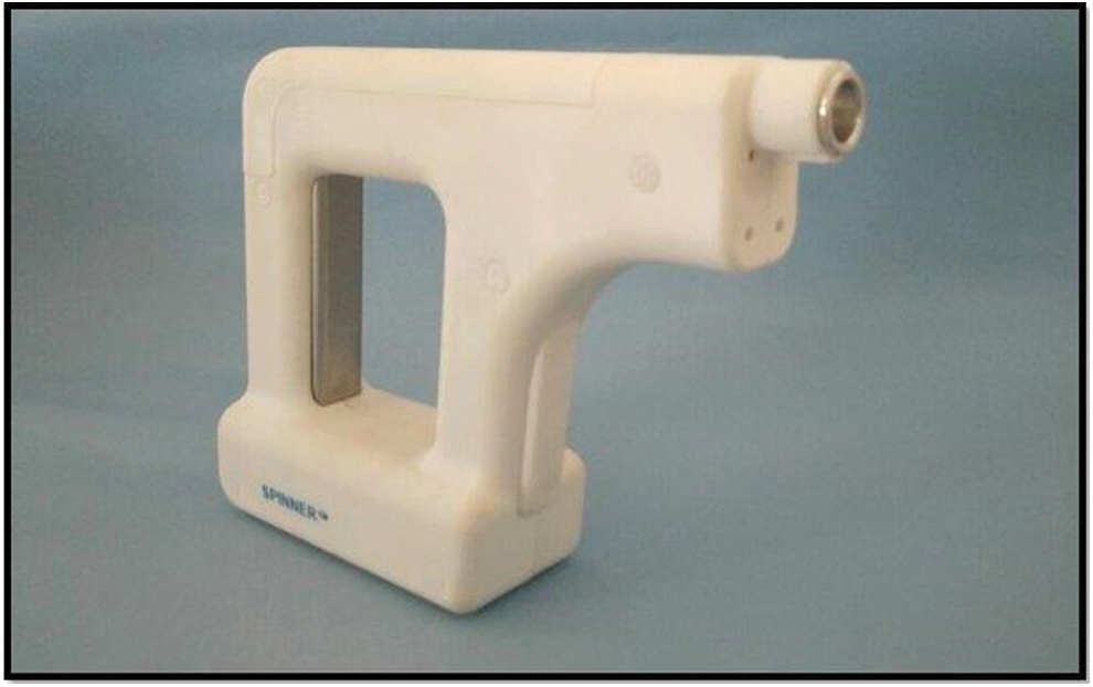
Handheld electrospinning device.
Polymer formulations and variables used for electrospinning nanaofibers
Histology slides were evaluated by light microscopy in an Olympus BX43 microscope. Photographs were taken with an Olympus DP21 camera and Olympus CellSens Entry software.
In vivo study
This study was approved by the Local Ethics Committee at Lahav Research Institute (746/12/ANIM), which is a recognized institute for performing animal experiments by the Israeli National Ethics Committee for Animal Experiments of the Israeli Ministry of Health. Three white Landrace Sus scrofa domestica pigs weighting an average of 28 kg were housed under controlled conditions and used as a porcine dermal tissue model. Upon arrival at the laboratory, the pigs were acclimated for a period of 1 week and housed in double-sized cages during the acclimation period and individually housed during the study. Sunlight was provided during the day time and the temperature and humidity were monitored and recorded daily. Standard diet was provided consisting of dry sow mix sourced from Ambar mixture institute (Beit Kama, Israel) available to the pigs twice a day with water availability ad libitum.
Pigs were induced with Isoflurane 5% via face mask and anesthetized with Ketamine Hydrochloride 10 mg/kg+Xylazine 2 mg/kg by intramuscular injection, followed by an intravenous administration of Diazepam 5 mg. After intubation, Isoflurane 1.5–2% was delivered via positive pressure ventilation utilizing 100% oxygen (2 L/min) for each procedure including wound formation, dressing application, and follow-up observations. Fifteen donor site wounds were created on each animal, which lasted for a longer period of time. This required the use of an induction of intravenous anesthesia and the additional inhalation anesthesia.
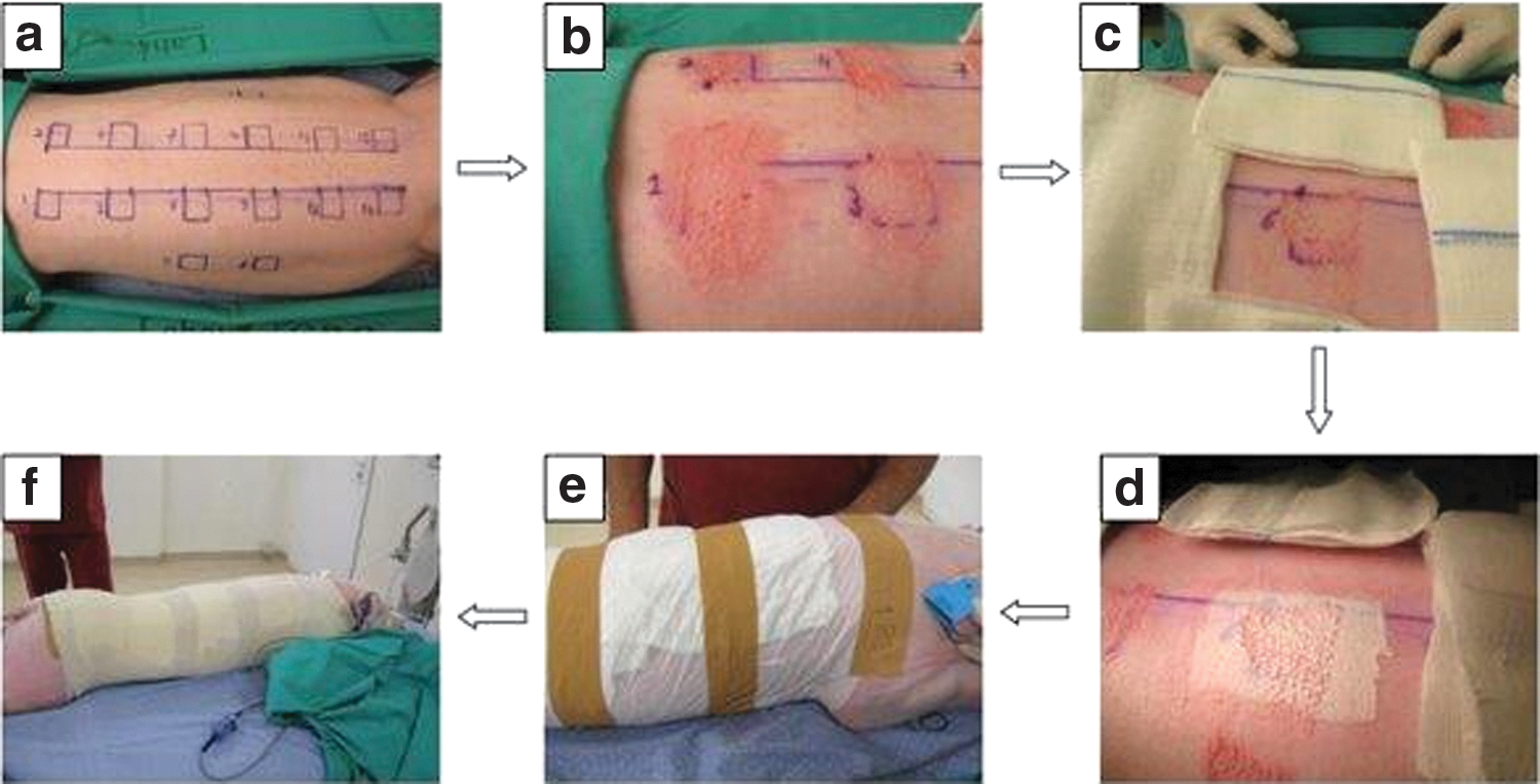
On day 1, hair on the back of the pigs was clipped with standard animal clippers and removal ointment. A marker was used for labeling the treatment areas. The skin on the back and sides of the animals, and surrounding area including the application area, was prepared by washing with Septal Scrub and sterile water. Initially, 15 superficial partial thickness wounds were formed on the dorsal aspect of the pigs using a dermatome. The surface area of each wound was 20 × 20 mm and 0.254 mm in depth and immediately thereafter dressing were applied. Each of the four formulations was designated to 3 of the 15 wounds formed on each pig. The remaining three wounds served as controls and were treated with commercially available paraffin nonmedicated tulle gras dressing (Jelonet, Smith & Nephew), gauze, and an abdominal pad was added and secured by stockinette (Fig. 2).
After 48 h, initial general observation and tolerability assessment was performed on one pig (no. 0894). One week later, a clinical assessment of the dressing and healing status was performed on the same pig. Finally, 14 days later, the final observation and evaluation was performed on all the pigs. Macroscopic investigations were performed to evaluate the different types of dressings: location and adherence of dressing; ease of application and removal; wound exudate including color and odor; eschar formation; wound closure (% wound surface area); time to complete healing; histology evaluation of the treated sites; wound bacteriology and mycology analysis; and any adverse reaction using a Draize scale. Histological assessment was performed at day 14 on one donor site from each nanofiber polymer formulation and pig. The skin samples consisted of two pieces of 0.5 × 1 cm rectangle longitudinal sections from each site. Tissue specimens were placed in formalin and histological processing was performed by Patho-Vet Diagnostics Ltd., Nes Ziona, Israel (
In this study, no animal experienced mortality due to infection or other complications. All pigs were euthanized as mandated by the Ethics Committee due to the number of cuts, their area and length, and the improbable closure of the wounds after the excisions.
Results
In vivo macroscopic results
Macroscopic evaluations on the donor site wound were performed at day 2, 7, and 14. The reported areas at each point in time are presented as the mean of the measurements.
Dressing application
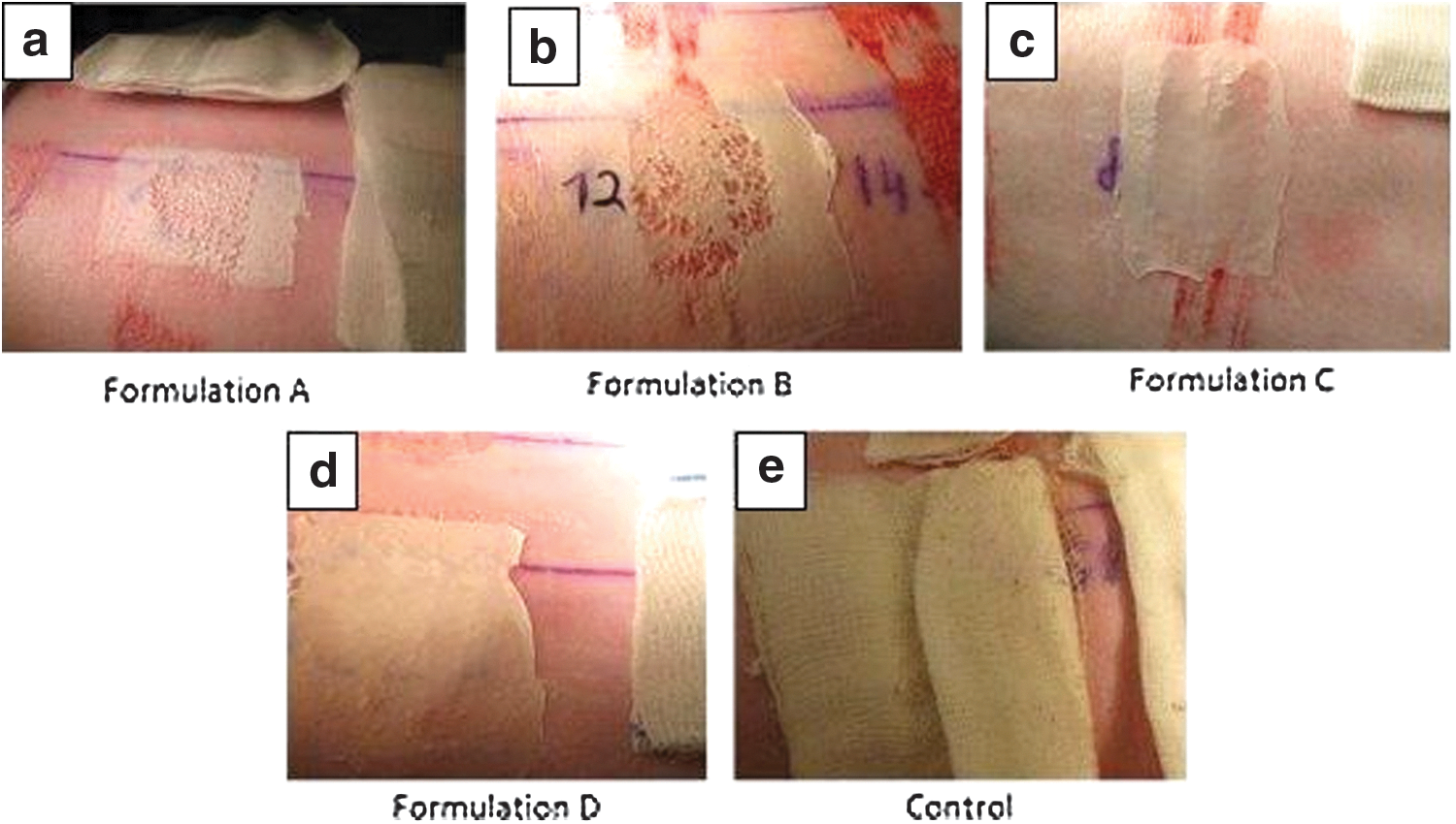
The ease of application was assessed immediately after application of the nanofibrous electrospun dressings. A simple scoring system was utilized reflecting the level of ease experienced during application: 1 = easy; 2 = moderate; and 3 = hard. The application of all nanofibrous dressings to the porcine model obtained a score of 1 (easy) indicating the ease the operator experienced during the application (Fig. 3).
Follow-up observations and evaluation day 2 and day 7
Follow-up observations were conducted on day 2 and day 7 for pig no. 0894 to evaluate five key areas of the donor site including adherence, wound exudate, presence of eschar, and any adverse skin reaction for each nanofibrous dressing formulation (Table 2). The scoring for adherence was ranked as: 0 for no adherence, 1 for partial adherence, and 2 for total adherence. All formulations (A–D) had total adherence to the wound bed at day 2 and day 7. The control had total adherence at day 2 and partial to no adherence on day 7. No wound exudate was evident across the nanofibrous groups and the control for day 2 and day 7 and no eschar was evident. Erythema across all nanofibrous dressings and including the control was scored as 4 (severe) on day 2 and 1 (very slight) on day 7.
Evaluation of dressing qualities (formulation A, B, D, and control) at day 2 and day 7 (pig no. 0894)
Adherence score: 0 = no adherence, 1 = partial, 2 = total. Odor score: 0 = no odor, 1 = mild, 2 = malodor. Skin reaction score: Erythema: 0 = no erythema, 1 = very slight, 2 = well defined, 3 = moderate to severe, 4 = severe. Edema: 0 = no edema, 1 = very slight, 2 = slight, 3 = moderate, 4 = severe.
Follow-up observations and evaluation day 14
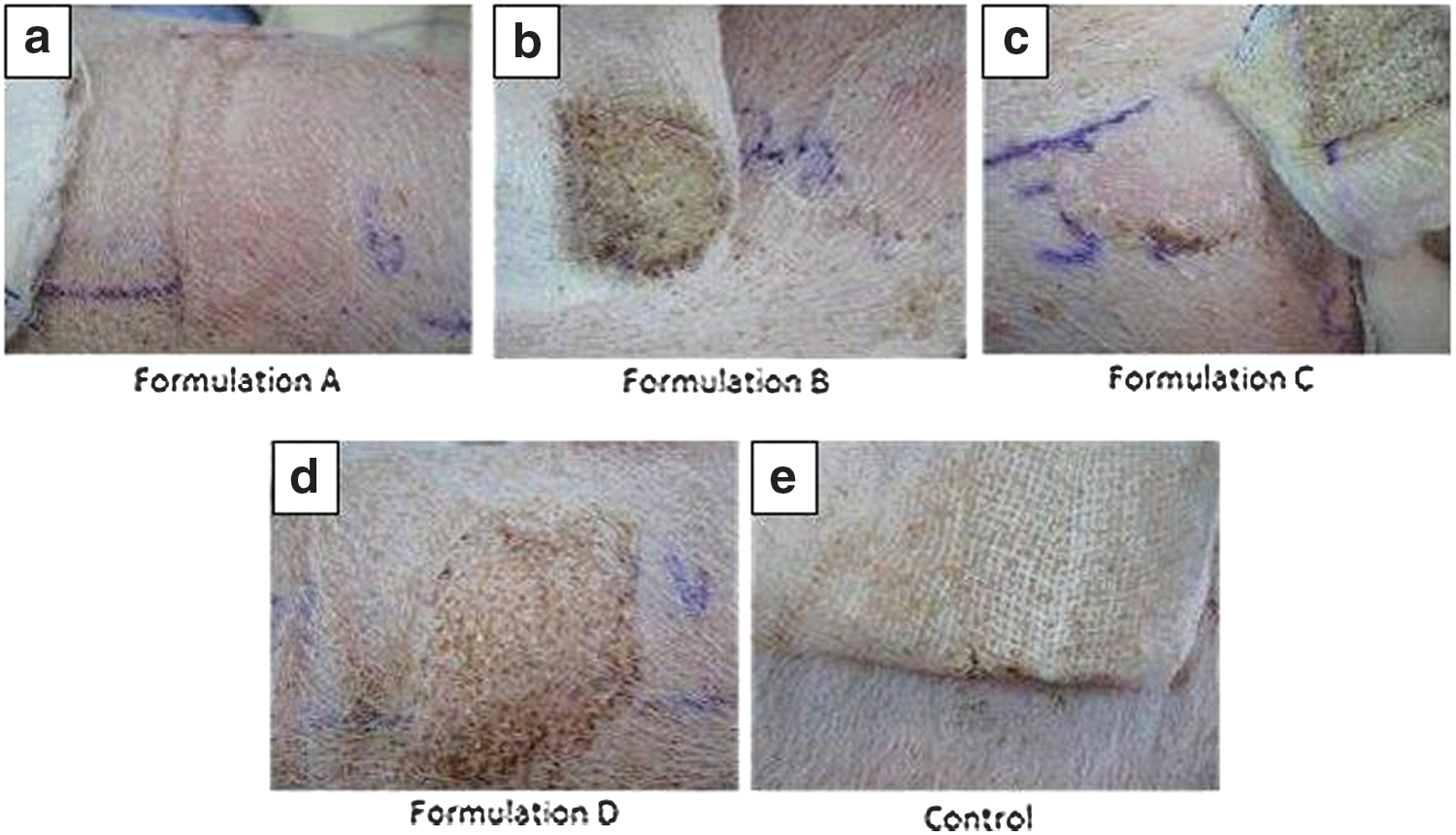
Fourteen days following the experimental porcine procedure, the nanofibrous dressings were removed from the donor site. In addition to adherence, wound exudate, and presence of eschar and erythema/edema, we also evaluated wound closure surface area, ease of dressing removal, and time to complete healing (Table 3). At day 14, adherence ranged from 1.9 to 2.0 for the nanofibrous dressing of all four formulas demonstrating good adherence rates with the control adherence situated at 1.1 with partial adherence. No wound exudate or eschar was evident across all groups including the control. By day 14, erythema had reduced ranging from 0.2 to 0.6 and edema was rated between 0.3 and 1.1 indicating scant erythema and edema across all samples including the control. The time to complete wound healing was 2 weeks across all nanofibrous dressing groups and the control. The surface area healed ranged from 98% to 100% for the nanofibrous dressings and 100% for the control indicating that the nanofibrous dressings were as effective as a standard paraffin tulle gras dressing. Of importance was the ease of removal, ranging from 1.0 to 1.2 indicating that removal was easy for all groups. Figure 4 clearly shows the donor sites following treatment with the four different nanofibrous electrospun polymer formulation dressings and the control post removal on day 14.
Evaluation of dressing qualities at day 14 for all pigs
The mean result is presented for each category. Adherence score: 0 = no adherence, 1 = partial, 2 = total. Removal score: 1 = easy, 2 = moderate, 3 = hard. Odor score: 0 = no odor, 1 = mild, 2 = malodor. Wound closure: % of donor site area. Skin reaction score: erythema: 0 = no erythema, 1 = very slight, 2 = well defined, 3 = moderate to severe, 4 = severe. Edema: 0 = no edema, 1 = very slight, 2 = slight, 3 = moderate, 4 = severe.
N/A, not applicable.
Histology assessment and bacteriology and mycology analysis
Histology from one donor site from each nanofibrous polymer formulation and each pig was analyzed (Fig. 5). For each sample, six domains of wound healing were analyzed: hyperkeratosis, crusting, epidermal hyperplasia, dermal fibrosis, clefting of the dermo-epidermal junction, and dermal inflammation (Table 4). A semiquantitative score (0–4) was used to provide an evaluation of the quality of the lesions observed. Where “0” indicated no lesion observed; “4” indicated severe wound observed. Overall, the histological findings were very similar in all samples with some variations in intensity: mild to moderate epidermal hyperplasia, minimal hyperkeratosis, occasional serocellular crusting, and a superficial band of dermal fibrosis that varies between 0.4 and 1 mm in thickness. The area of fibrosis was mildly edematous with a minimal lymphocytic infiltrate, presence of few eosinophils, (which are common background changes in pigs), and rare clefting of the dermo-epidermal junction. Findings are slightly more severe in samples from pig 2 (no. 0907). However, the control dressing of paraffin tulle gras received the highest wound total severity score mean of 7.3 ± 0.6 (Table 4). A specimen was taken from one donor site of each nanofibrous polymer formulation and control for each pig for culturing testing for both aerobic and anaerobic pathogens and mycology. No pathogenic aerobic or anaerobic bacteria were found in all samples analyzed. Furthermore, no fungal growth was evident in specimen examined.

Histology assessment results summary following dressing removal at day 14
Semiquantitative 0–4 score was used: 0 = normal or no lesion, 1 = minimal, 2 = mild, 3 = moderate, 4 = marked or severe.
Discussion
Electrospinning is reported to be the most frequently used method to fabricate nanofibers. 5,18 The further benefits of a handheld portable electrospinning device for the purposes of wound care are beginning to emerge and reported within the peer-reviewed literature. 19 –24 The development of biocompatible electrospun nanofibrous dressings speaks to the versatility, simplicity, adaptability and importantly cost effectiveness. The aim of this study was to determine the feasibility of a handheld electrospinning nanofibrous device for the application of dressings on donor sites. We have demonstrated that the electrospun nanofibrous dressings presented in this study have the capacity to be a biocompatible, safe, and effective alternative for wound healing and dressing of choice concurring with other in vivo animal studies. 15 –17
Electrospun nanofibers properties used for wound management including hydrophilicity, flexibility and strength, biocompatibility, and specific cell interactions are largely determined by the chemical composition of the materials used. 25 Formulation A was biodegradable polyester based requiring only 1 minute for creation of a dressing in a desirable and workable thickness and texture in comparison with 1.5 min for formulations B and C and 2 min for formulation D. Moreover, in formulations B and C the smell of the solvents was evident in the first minute of device operation, while in formulation A and D it was not noticeable, which is an important element in occupational health and safety for any associated risks from exposure to these materials. 26 Other considerations including raw material availability at pharmaceutical grade, formulation composition, electrospinning ability and stability, and in vitro bacterial penetration testing results will serve as input to further development of formulations for clinical use.
Overall, our study shows no significant differences detected between the formulations in all the parameters of the donor sites evaluated. The dressing adherence score showed a positive trend to the nanofibrous polymer dressing compared to the control paraffin tulle gras dressing. However, an advantage of the application of the dressing from a distance (without touching the wound) reduces the risk for infection as hands are the main source of transferring infection. 27 Furthermore, dressing large wounds with difficult geometries and locations presents issues with movement such as the back and over joints therefore, the application of dressing with existing dressing products often becomes cumbersome and difficult. Others such as Dong et al. 1 reported similar findings with rats treated with a simple gauze dressing as used in this study. Dong et al. 1 also found the electrospun nanofibrous dressing to be conformable, flexible, and easier to work with compared to the conventional gauze dressing concurring with our findings. Wound exudate was not an issue with the wounds presented in our study, however, Dong et al. 1 found that the rats treated with electrospun nanofibrous membranes demonstrated better exudate management compared to those treated with the gauze dressing.
The dressings described in this study were shown to be semi-permeable, conformable, and easily removed without inflicting unnecessary trauma to the wound bed. The handheld device was shown to be easy to use and effective in producing in situ nanofibrous dressings, applied at a distance from the wound, having the potential for reducing infection and cross-contamination rates. Furthermore, it is known that ineffective and inappropriate choice of dressings can result in poor wound healing, increased infection rate, and subsequent scarring. 28 Although not shown in this study, Liu et al. 28 demonstrated less prominent dermal scarring on a rat model with the use of electrospun polymer nanofibrous dressings.
Innovation
Electrospun nanofibers dressings are an ideal dressing with unique architectural features that mimic the extracellular matrix and indeed provide an ideal wound environment for wound healing. These formulations can be customized to the characteristics of the target wound and the various stages of wound healing. Such custom-made nanofibrous dressings have the potential to reduced pain, trauma, scarring, number of dressing changes, and added cost benefit. Used on partial thickness wounds, polymer electrospun nanofiber dressings provide excellent surface topography and are a non-touch, feasible, and safe method to promote wound healing with the potential to reduce wound infections.
• Electrospun nanofibrous dressings have the capacity to be a biocompatible, safe, and effective alternative for wound healing and dressing of choice.
• Electrospun nanofibrous dressings were shown to be semi-permeable, conformable, and easily removed without inflicting unnecessary trauma to the wound bed.
• The technology offered by this handheld portable electrospinning device can be used at the bedside in the application of non-touch dressings and can be utilized for different formulations and materials.
• Although additional studies are necessary to determine the safety on humans, there were no delayed wound healing or signs of infection reported with the use of the electrospun nanofiber dressing.
Footnotes
Acknowledgments and Funding Sources
This study was funded by Nicast Ltd, Lod, Israel.
Author Disclosure and Ghostwriting
J.H. is a Medical Advisor for Nicast Ltd. R.K., B.B., and M.H. have no disclosures. The content of this article was expressly written by the authors listed. No ghostwriters were used to write this article.
About the Authors
References
Supplementary Material
Please find the following supplemental material available below.
For Open Access articles published under a Creative Commons License, all supplemental material carries the same license as the article it is associated with.
For non-Open Access articles published, all supplemental material carries a non-exclusive license, and permission requests for re-use of supplemental material or any part of supplemental material shall be sent directly to the copyright owner as specified in the copyright notice associated with the article.