




Abstract
Objective:
At present, there is an urgent need to develop a novel and practical therapeutic approach to accelerate the healing of acute wounds. Mesenchymal stem cell (MSC)–based therapy is emerging as a promising therapeutic approach for acute skin wounds. However, there are still challenges in clinical application of this strategy, such as low survivability, low retention time, and less engraftment in skin wounds.
Approach:
Wharton's jelly mesenchymal stem cells (WJMSCs) were seeded into three-dimensional (3D) gelatin microspheres (GMs) to identify the biocompatibility of GMs. WJMSCs were embedded in GMs and then encapsulated with Pluronic F-127 (PF-127) and sodium ascorbyl phosphate (SAP) combination to transplant onto acute full-thickness skin wound in mice. Histology, immunohistochemistry, and immunofluorescence assay were used to investigate the skin wound healing, dermis regeneration, collagen deposition, cell proliferation, and neovascularization.
Results:
Three-dimensional GM had strong biocompatibility, compared with two-dimensional adherent culturing, GM loading increased the cell viability and proliferation ability of WJMSCs. WJMSCs+GM+PF-127+SAP transplantation increased skin wound healing rate, dermis regeneration, and type III collagen deposition through improving macrophage polarization, cell proliferation, neovascularization, cell retention, and engraftment at skin wound site.
Innovation:
The effective 3D encapsulation technology for WJMSCs solved the main problems of cell activity and residence time during MSC transplantation. WJMSCs+GM+PF-127+SAP transplantation will be a new and effective MSC biomaterials–based therapeutic strategy for acute skin traumatic wounds.
Conclusion:
WJMSCs+GM+PF-127+SAP transplantation facilitated acute full-thickness skin wound healing and regeneration and might be a new and effective therapy for acute skin traumatic wounds.


INTRODUCTION
Various types of triggers including trauma, burns, diabetic ulcers, atopic dermatitis (AD), aging, and oxidative stress, are easy to cause acute or chronic skin wounds, which brings burden on the patients both physically and psychologically. 1 Approximately 8.2 million people suffered acute and chronic skin wounds in 2018. The annual Medicare cost for the treatment of skin wounds in United States was estimated to be $28.1 billion to $96.8 billion. 2 Trauma is the leading cause of mortality in Europe and United States. Besides, millions of acute surgical wounds are created each year during routine medical procedures in the United States and Europe. 3 Traditional therapies for acute skin wound treatment still have great challenges for wound healing, such as delayed healing, excessive fibrosis, and chronic inflammation. 4,5 Therefore, there is a need to develop effective treatments for traumatic injury and surgical incisions.
Transplantation of mesenchymal stem cells (MSCs), which can be obtained from different tissues, can promote various skin wound healing and regeneration though paracrine effects and highly plastic immunoregulation. 6 –8 Wharton's jelly-derived mesenchymal stem cells (WJMSCs) are isolated from the fetal umbilical cord, which is characterized by easier solution, higher proliferative ability, stronger immunomodulatory effects, and fewer ethical issues. 9 Previous studies have reported that WJMSC transplantation promoted the healing of acute, chronic, and radiation-induced skin wounds. 10 –14 However, the therapeutic effect of WJMSC transplantation for skin wound is limited owing to low cell quality and residence time. Therefore, the treatment strategy of WJMSCs for skin injury needs to be further improved.
Biomaterials are widely used to overcome bottlenecks in MSC transplantation by protecting cells from undesirable shear forces during injection, which provides structural support, promotes desirable cell fates and viability, and enhances tissue regeneration outcomes. 15,16 MSCs survive in a three-dimensional (3D) microenvironment simulated by biomaterials as a pool of stem cell with undifferentiated and quiescent state in vivo. 17 In addition, the 3D scaffolds not only increase cell-to-cell and cell-to-biomolecules communication, but also protect MSCs from damage by the human innate immune system. 18
Gelatin is a natural polymer obtained from animal collagen. Because of its good biocompatibility and appropriate biodegradability, gelatin-based spheres can be injected as cell delivery system and have a widely biomedical application. 19 –21 Although direct injection of gelatin microspheres (GMs) loading with cells can treat skin wound, GM movement and secondary skin injury caused by local injection limit the therapeutic effect. 22,23 The application of GM in skin injury needs to be improved.
Many natural and synthetic biomedical scaffolds have been used to prolong the retention time of MSCs at the skin wound sites. 24 –26 Our recent study demonstrated that Pluronic F-127 (PF-127) plus 400 μM sodium ascorbyl phosphate (SAP) can promote acute full-thickness skin wound and diabetic wound healing and regeneration. 27,28 Although PF-127/SAP system could delivery WJMSCs to wound site effectively, the survival and viability of WJMSCs were still low. To further increase the cell viability and survival rate of WJMSCs and explore new WJMSC-based strategy for skin wound treatment, we intend to combine GM and hydrogel PF-127/SAP to deliver the WJMSCs on acute full-thickness skin wound in mice.
In this study, we found that GM-simulated 3D encapsulation increased the cell viability and proliferation of WJMSCs in vitro. Transplantation of WJMSCs+GM+PF-127+SAP promoted acute full-thickness skin wound healing and improved the macrophage polarization, dermis regeneration, neovascularization, type III collagen deposition, cell retention, and engraftment in skin wound.
CLINICAL PROBLEM ADDRESSED
The increasing number of patients with acute skin injuries, especially trauma, has brought serious burden to the society and medical system. However, the current traditional methods for treating acute skin wounds cannot promote the healing and regeneration of skin wounds effectively. Although MSCs have certain therapeutic potential for skin injury, their effect is severely limited by factors such as low survival rate, short retention time, and poor cell activity of transplanted cells for exposed wounds. The objective of this project was to integrate 3D microcarrier and thermosensitive hydrogel for WJMSC transplantation, to overcome problems existing in the transplantation process of WJMSCs, and further to improve the therapeutic effect.
MATERIALS AND METHODS
Cell culture
The WJMSCs were cryopreserved cells which have been isolated and identified in our previous study. 28 This study of WJMSCs was approved by the Ethical Committee of the First Affiliated Hospital of Sun Yat-sen University. All patients signed written informed consent before donating healthy umbilical cords for the research. The complete medium consisting of 90% DMEM/F12 (Corning), 10% FBS (Hyclone) and 1% penicillin/streptomycin (Gibco, Grand Island, NY) was used to culture WJMSCs and was changed every 3 days.
Cell seeding and encapsulation
For the 3D culture of WJMSCs, 1 × 10⁷ WJMSCs were suspending co-cultured with 70,000 GM (CytoNiche, Beijing, China) in six-well nonadherent plastic plates with complete medium for 4–6 h. After that, WJMSCs were statically cultured with complete medium. In the cell transplantation experiments, GM +WJMSCs was collected and centrifuged. After that, WJMSCs+GM was resuspended with 50 μL hydrogel PF-127 (P2443–250G; Sigma) plus 400 μM sodium ascorbyl phosphate combination (SAP) (49752; Sigma). The PF-127/SAP combination was prepared as our previous study reported. 27 WJMSCs or WJMSCs+GM were encapsulated with PF-127/SAP combination and incubated in 37°C to make gel formation.
Scanning electron microscopy assay
WJMSCs were cocultured with GM for 24 h. Then they were fixed with 2.5% glutaraldehyde at 4°C overnight. Wash the sample with PBS three times followed by gradient dehydration. The harvested and freeze-dried GMs were gold coated for 50 s. Scanning electron microscope (S-3400N, Hitachi, Japan) was used to evaluate the microstructure of GM. The pore diameter distribution was analyzed by ImageJ software (National Institutes of Health).
Swelling ratio assay
The swelling capability was determined by incubating the GM in PBS solution at 37°C. In brief, a piece of GM was soaked in PBS, and its weight was measured after swelling for 20, 40, 60, 80, and 120 min, respectively. The swelling ratio (SR) of GM was analyzed as follows: SR% = (Wt − W0)/W0 × 100, W0 presents the initial weight of GM before soaking in PBS, Wt presents the weight of the GM after soaking for t time in PBS.
Cell viability assay
Cell viability was evaluated by Cell Counting Kit-8 (CK04, Dojindo, Japan). In brief, WJMSCs encapsulating with different conditions were seeded in 96-well plate and cultured for 24 and 48 h, respectively. Then, CCK-8 reagent was incubated with WJMSCs at 37°C for 2 h. Finally, microplate reader (VICTOR™ X5; PerkinElmer) was used to measure the absorbance value at 450 nm.
Live-dead assay
The survival of WJMSCs was detected by Live/Dead™ Cell Imaging Kit (R37601; Invitrogen) following the manufactures' instruction. The WJMSCs were seeded into 12–well plate and cultured with complete medium for 24 h. Then, WJMSCs were incubated with working solution for 15 min at 25°C in dark. The inverted fluorescence microscope (Axio observer A1; Zeiss) was used to capture the fluorescence images.
EdU assay
The cell proliferation ability was assessed by Cell Light™ EdU Kit (C10310; Ribo Biotech Co.) following the manufacturer's instructions. WJMSCs were seeded in 48-well plate with different encapsulation conditions for 24 and 48 h, respectively. Then, WJMSCs were cultured with complete medium, which contained 50 μM 5-ethynyl-2′-deoxyuridine (EdU) for 2 h. Apollo staining was conducted after fixing and permeabilizing WJMSCs. Laser scanning confocal microscopy (TCS-SP5; LEICA) was used to observed EdU-positive WJMSCs. Fluorescent images from WJMSCs were analyzed using Image Pro Plus (Media Cybernetics, Inc., Silver Spring, MD) and Imaris software (TIRF; LEICA), respectively.
Tri-lineage differentiation assay
Following the manufacturer's instructions, the adipogenic, osteogenic, and chondrogenic ability of WJMSCs were assessed by OriCell™ Adipogenesis Differentiation Kit (HUXUC-90031; Cyagen), OriCell Osteogenesis Differentiation Kit (HUXUC-90021; Cyagen), and OriCell Chondrogenesis Differentiation Kit (HUXUC-90041; Cyagen), respectively. Then, Oil Red O staining was used to detect fat droplet formation. Alizarin red staining was used to detect calcium deposits. Alcian blue staining was used to detect proteoglycan deposits.
Animals
To exclude the effect of sex on wound closure, in this study, we purchased male 7-week-old C57BL/6 mice (20–30 g) from Guangdong Medical Laboratory Animal Center (Guangzhou, China) to establish acute full-thickness wound model. We did not use the female mice in our study. The study of animals was approved by the Institutional Animal Care and Use Committee of Sun Yat-sen University, P.R. China (SYSU-IACUC-2020-B1177).
Establishment of full-thickness acute wound in mice
All mice were housed at a constant temperature and humidity and adaptively fed for 1 week. First, the mice were anesthetized by intraperitoneal injection of 2.5% Avertin prepared with 2, 2, 2-tribromoethanol (T48402; Sigma-Aldrich). Two full-thickness wounds with a diameter of 10 mm, which down to the areolar connective tissue overlying the skeletal muscle fascia and the panniculus carnosus, were created on the dorsum with a biopsy punch after hair removal. Carprofen (C830557; Macklin) was given postsurgery at 5 mg/kg for analgesia.
All mice were divided into seven groups, including: Sham (surgery is performed but without treatment), PBS, GM, WJMSCs, WJMSCs+GM, WJMSCs+PF-127 + 400 μM SAP, WJMSCs+GM+PF-127 + 400 μM SAP. Total 50 μL PF-127 + 400 μM SAP combination encapsulated 1 × 10⁶ WJMSCs or 1 × 10⁶ WJMSCs+GM (passage 6) or other combinations were transplanted onto the wound site and then covered with IV3000 transparent dressing to avoid infection. The representative images of skin wounds at 0, 3, 5, 7, and 10 days after transplantation was captured and the unhealed wound area was measured by ImageJ software as follows: unhealed wound area (%) = Wr/Wi × 100%. Wi, the initial wound area at day 0. Wr, the residual wound area at day 5 or day 10 post-transplantation.
Histological and immunohistochemical analysis
On day 10 post-transplantation, mice were killed with an overdose of 2.5% Avertin, and skin wound samples from different groups were collected. The skin samples were fixed in 4% paraformaldehyde fixation, dehydrated, paraffin-embedded, and then sectioned along with the direction of hair flow (5 μm). The sections were further stained with hematoxylin and eosin (H&E) staining kit (G1120; Solarbio), Masson trichrome staining kit (D026; Nanjing Jiancheng Bioengineering Institute), and Picrosirius Red staining kit (PH1098; Phygene) according to the manufacturer's instructions. The H&E staining images and Masson trichrome staining images were obtained by an upright fluorescence microscope (Eclipse Ni-E; Nikon). The Picrosirius Red staining images were obtained by a polarized light Leica microscope (DM2700 P; Leica). ImageJ software was used to measure the thickness of dermis, scar width, number of hair follicles, number of type I or type III collagen.
For immunohistochemistry detection, the skin samples were blocked by 3% goat serum (16210064; Gibco) for 1 h after heat-induced epitope retrieval. After that, the sections were incubated with primary antibodies at 4°C overnight (Supplementary Table S1). The CD86, CD163, CD31, and Ki67-positive cell were further detected by the cell and tissue staining kit (CTS005, Anti-Rabbit HRP-DAB System; R&D Systems), respectively. Images were taken by an upright fluorescence microscope (Eclipse Ni-E; Nikon) and analyzed by ImageJ software.
LipidTOX staining
The sebaceous glands and lipid-rich adipocytes were detected by LipidTOX staining (H34477; Invitrogen). Cryosections of each group skin sample were incubated with LipidTOX neutral lipid stain (1:200) for 30 min at room temperature. Then, the cell nuclei were stained by Hoechst. The inverted fluorescence microscope (Axio observer A1; Zeiss) was used to obtain the images.
WJMSCs tracking in vivo
In brief, 2 × 10⁶ OE-EGFP WJMSCs or 2 × 10⁶ OE-EGFP WJMSCs+GM encapsulated with PF-127/SAP combination were transplanted onto the wound site as our previous study report. 27 The skin samples from different were collected at 24 h and 72 h post-transplantation, respectively. After counterstaining the crysections with DAPI, fluorescence images were captured with an inverted fluorescence microscope (Axio observer A1; Zeiss). ImageJ software was used to analyzed EGFP-positive cells.
Statistical analysis
The SPSS software (SPSS 18.0, Chicago, IL) was used for data analysis. Comparison between two and more groups was performed by means of Student's unpaired t-test and analysis of variance (ANOVA), respectively. Values are given as mean ± standard error of mean (SEM). p < 0.05 was considered statistically significant (*p < 0.05, **p < 0.01, ***p < 0.001; ns, no significance).
RESULTS
GM loading promotes the viability and proliferation of WJMSCs
To evaluate morphological and physical properties of GM, we investigated the structure of GM by scanning electron microscopy. We found that the GM has a spherical topology. The irregular pore size ranged from 11 to 40 μm, which were interconnected with each other (Fig. 1A, B and Supplementary Fig. S1). The SR of GM was ∼23% (Fig. 1C). WJMSCs are automatically absorbed into GM or adhered on the surface of GM (Fig. 1D). At static culturing condition, WJMSCs floated around the GM. After suspension of culture for 5 h, WJMSCs were efficiently adsorbed by GM and there was hardly free WJMSCs in the medium (Fig. 1E).
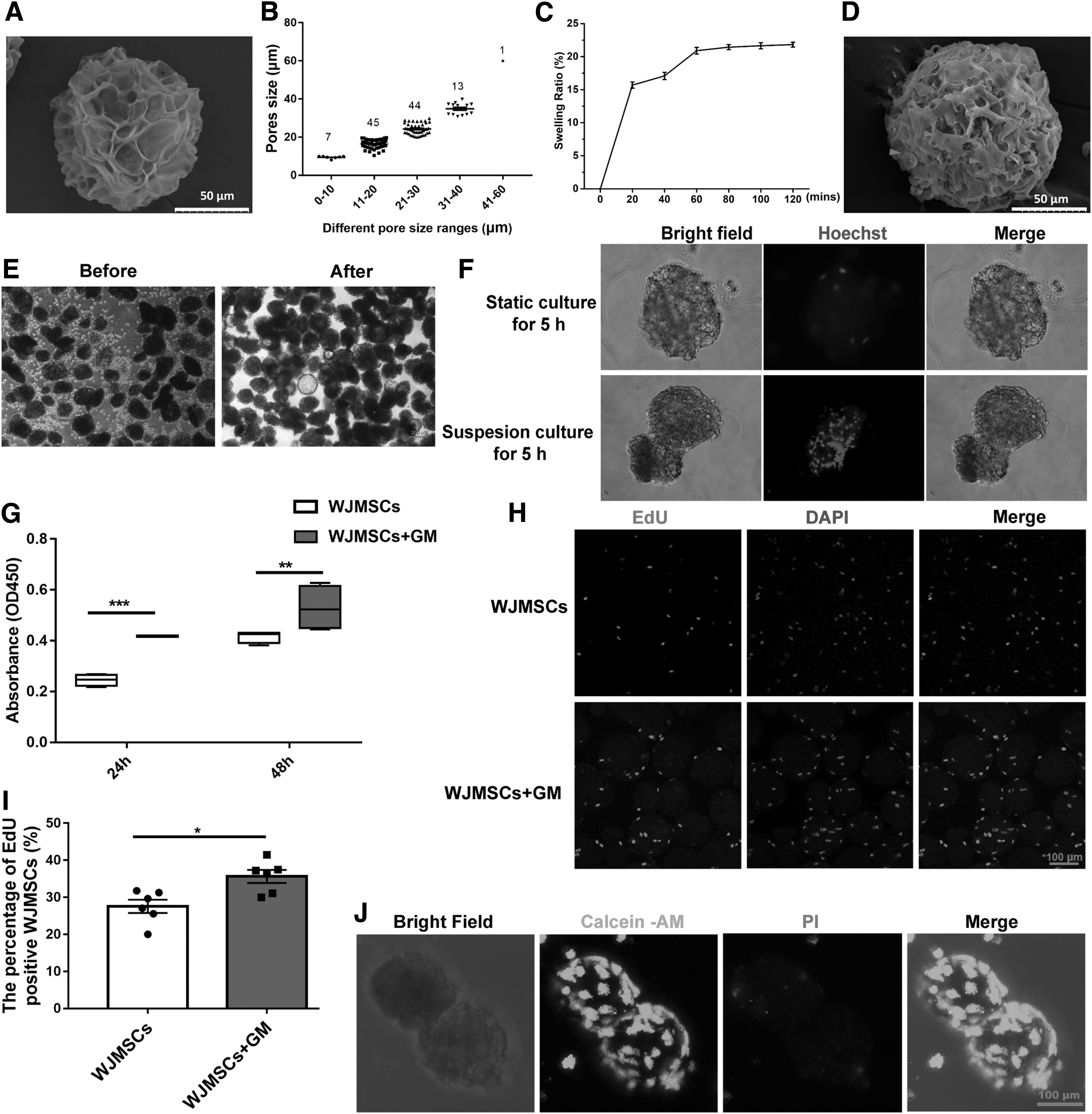
WJMSCs embedded in GM promote the viability and proliferation of WJMSCs.
To compare the effects of culture condition on GM adsorption efficiency, WJMSCs and GM were co-cultured for 5 h under static and suspension conditions, respectively. The immunofluorescence results showed that only a small amount of WJMSCs adhered to the GM surface under static culture, whereas more WJMSCs could be embedded into GM under suspension culture (Fig. 1F). Taken together, suspension culture was beneficial for WJMSCs adhering or embedding into GMs.
To identify the biocompatibility of GM, the GM and WJMSCs were co-cultured in suspension condition for 5 h, and then the viability and proliferation of WJMSCs were detected. The results showed that the cell viability in WJMSCs + GM group was higher than that in the WJMSC group at both 24 h (p < 0.001) and 48 h (p < 0.01) (Fig. 1G). Furthermore, we detected cell proliferation by EdU-labeling assays. The results showed more EdU-positive cells in WJMSCs + GM group (35.63% ± 1.77%) than those in the WJMSCs group (27.56% ± 1.80%) (Fig. 1H, I and Supplementary Fig. S2).
Besides, live-dead assay results showed that WJMSCs could effectively absorb and survive on the surface and interior of GM (Fig. 1J). Meanwhile, the percentage of dead WJMSCs embedded in GM was low (2.92% ± 0.21%) (Supplementary Fig. S3). Above all, GM was a kind of 3D encapsulation microcarrier with good biocompatibility and low cytotoxicity, which significantly increased the viability and proliferation of WJMSCs that was embedded in GM.
We further observed the adipogenesis, osteogenesis, and chondrogenesis ability of WJMSCs embedded in GM. The Oil red staining, Alizarin red staining, and Alcian blue staining results demonstrated that WJMSCs embedded in GM were able to differentiate into adipocytes, osteocytes, chondrocytes, respectively, compared with WJMSCs control group (Supplementary Fig. S4). Taken together, GM had no effect on the multilineage differentiation of WJMSCs.
GM loading further improves the viability and survival of WJMSCs in PF-127/SAP
GM is a kind of effective microcarriers for cell transplantation, but for exposed skin wounds, GM-embedded WJMSCs still could not retain at the wound site for a long time to exert therapeutic effect. Recently, our study found that transplantation of WJMSCs+PF-127+SAP can accelerate both chronic diabetic wound healing 27 and acute full-thickness skin wound healing. 28 Hydrogel PF-127 is a kind of thermosensitive hydrogel, which may serve as a scaffold to prolong the retention time of GM-embedded MSCs at wound sites. To further identify biocompatibility of combination GM with PF-127/SAP, we investigated the adhesion of WJMSCs on different materials by SEM. The results showed that only a few WJMSCs embedded in PF-127. GM loaded with WJMSCs also were fully wrapped from outside by PF-127 (Fig. 2A). The cell viability in WJMSCs + GM group was highest in all groups at 24 h or 48 h, whereas addition of PF-127 decreased the cell viability (p < 0.001). Furthermore, supplementary of 400 μM SAP in WJMSCs+GM+PF-127 could partially rescue the cell viability (p < 0.01) (Fig. 2B, C).
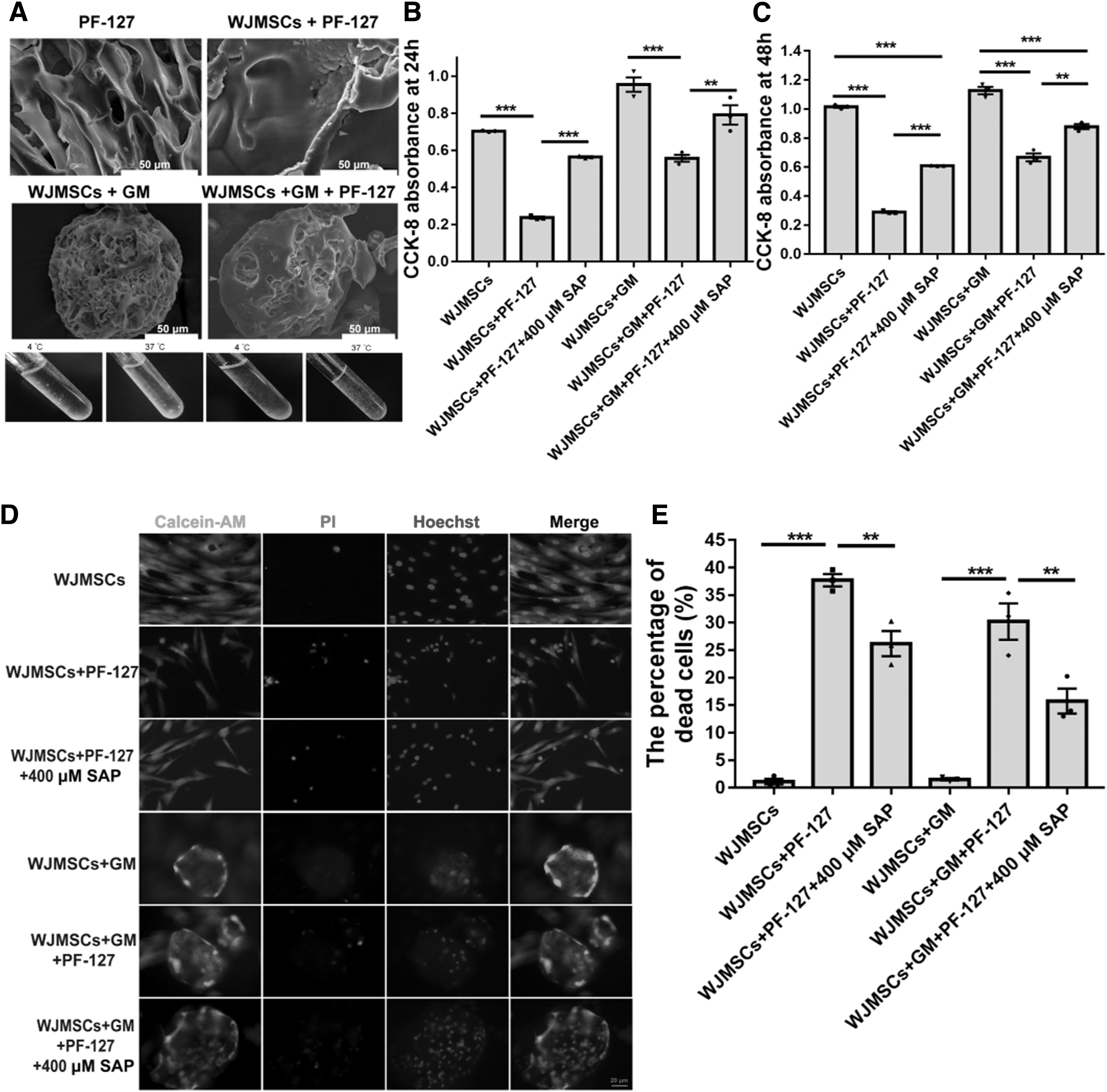
GM loaded with WJMSCs alleviated the cytotoxicity of PF-127.
In addition, Live-Dead assay results demonstrated that the percentage of dead cells in WJMSCs+GM+PF-127 was slightly decreased and supplementation of SAP further alleviated the cell dead in WJMSCs+GM+PF-127 + 400 μM SAP group (Fig. 2D, E). Above results demonstrated that GM was beneficial to WJMSC survival in vitro.
Transplantation of WJMSCs+GM+PF-127+SAP accelerates acute wound healing
Furthermore, we transplanted WJMSCs encapsulated with different combination onto wound site and observed wound healing at days 0, 3, 5, 7, and 10 after transplantation, respectively (Fig. 3A). We found that WJMSCs+GM+PF-127+SAP group (38.17% ± 2.54%) had the smallest residual wound area at day 5 post-transplantation compared with the control groups (sham, 67.61% ± 1.76%; PBS, 62.98% ± 1.64%; GM, 61.14% ± 0.92%; WJMSCs, 62.36% ± 1.53%; WJMSCs+GM, 53.00% ± 2.92%; WJMSCs+PF-127+SAP, 45.88% ± 0.92%) (Fig. 3B, C).
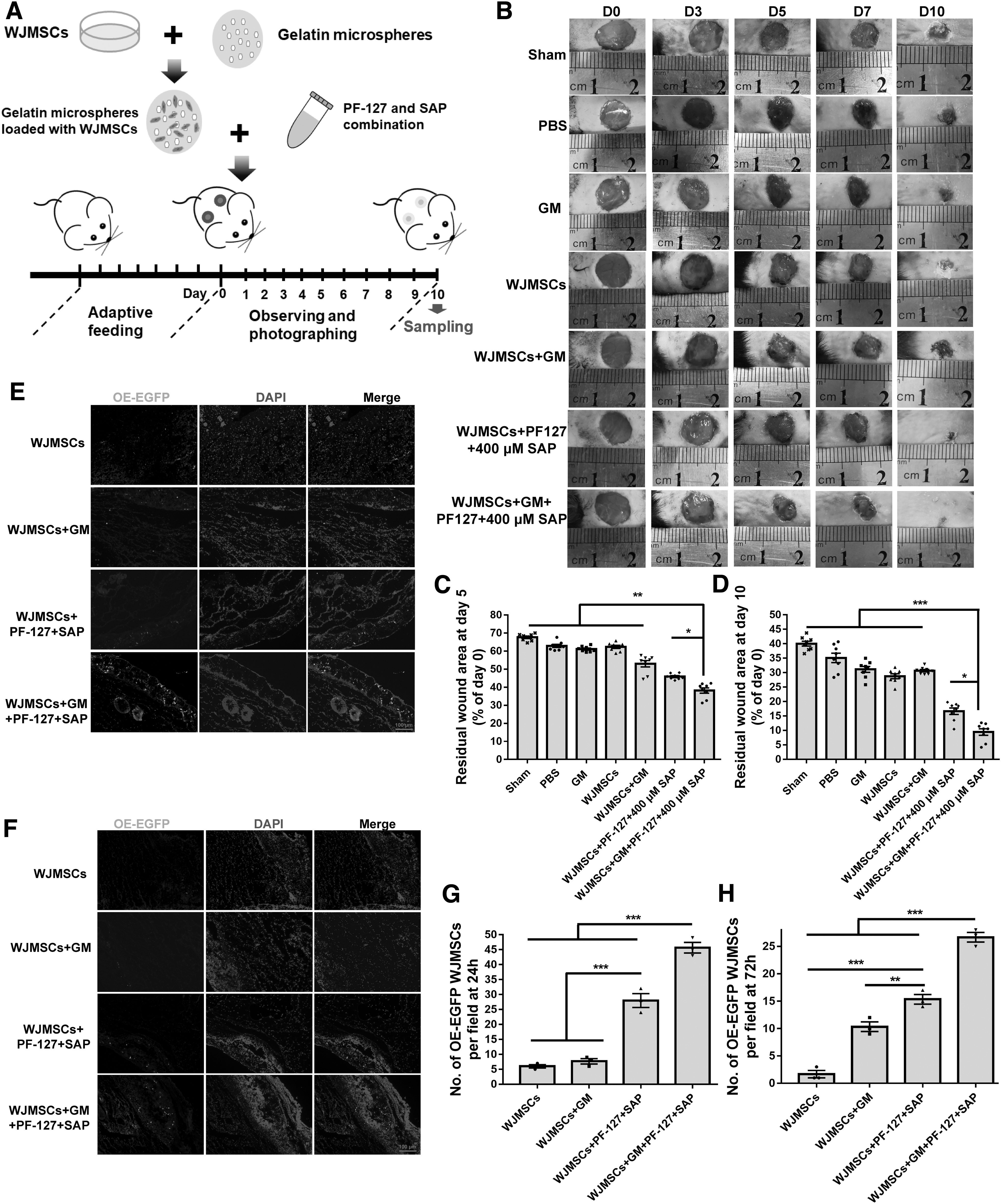
WJMSCs embedded in GM and encapsulated with PF-127 plus SAP combination facilitate wound healing and prolong the retention time of WJMSCs.
Similarly, the skin wound in the WJMSCs+GM+PF-127+SAP group was smallest and almost completely healed at day 10 post-transplantation (9.45% ± 1.99%) compared with other control groups (sham, 40.03% ± 0.84%; PBS, 35.11% ± 2.75%; GM, 31.13% ± 1.92%; WJMSCs, 28.76% ± 1.65%; WJMSCs+GM, 30.68% ± 0.83%; WJMSCs+PF-127+SAP, 16.65% ± 2.13%) (Fig. 3B, D). Above results demonstrated that WJMSCs+GM+PF-127+SAP transplantation accelerates the closure of acute skin wound.
GM plus PF-127/SAP enhance the retention and engraftment of WJMSCs
Short retention time, low survival rate, and less engraftment are the main reasons that limit the efficacy of cell transplantation therapy. 29,30 We tracked the OE-EGFP WJMSCs through in situ immunofluorescence staining to investigate the retention and engraftment of WJMSCs that was carried by GM+PF-127+SAP combination in acute skin wound (Fig. 3E, F). The number of EGFP-positive cells in the OE-EGFP WJMSCs+GM+PF-127+SAP group (45.67 ± 1.76 per field) was higher than those in the control groups (OE-EGFP WJMSCs, 6.00 ± 0.58; OE-EGFP WJMSCs+GM, 7.67 ± 0.88; OE-EGFP WJMSCs+PF-127+SAP, 28.00 ± 2.31 per field) at 24 h post-transplantation (Fig. 3E, G).
At 72 h after transplantation, the number of EGFP-positive WJMSCs in the OE-EGFP WJMSCs+GM+PF-127+SAP group (26.67 ± 0.88 per field) was also higher than those in the control groups (OE-EGFP WJMSCs, 1.66 ± 0.67; OE-EGFP WJMSCs+GM, 10.33 ± 0.87; OE-EGFP WJMSCs+PF-127+SAP, 15.33 ± 0.88 per field) (Fig. 3F, H). These results demonstrated that GM plus PF-127/SAP enhances the retention and engraftment of WJMSCs at acute skin wound site.
WJMSCs+GM+PF-127+SAP transplantation promotes dermis regeneration
We further observed the dermis regeneration in skin wound by H&E staining. The results demonstrated that the dermis thickness in WJMSCs+GM+PF-127+SAP group (1543.76 ± 120.90 μm) was thicker than those in other groups (PBS, 567.19 ± 55.93; GM, 668.97 ± 145.83; WJMSCs, 856.87 ± 62.13; WJMSCs+GM, 910.80 ± 40.11; WJMSCs+PF-127+SAP, 1122.67 ± 55.11 μm) (Fig. 4A, B and Supplementary Fig. S5A) (p < 0.01). There were more newly formed hair follicles in the WJMSCs+GM+PF-127+SAP group (48.67 ± 2.40 per field) than those in other groups (PBS, 7.00 ± 0.58; GM, 8.00 ± 0.57; WJMSCs, 13.00 ± 0.58; WJMSCs+GM, 23.67 ± 0.88; WJMSCs+PF-127+SAP, 30.00 ± 1.15 per field) (Fig. 4A, C) (p < 0.01). The width of unhealed wound in WJMSCs+GM+PF-127+SAP group (360.82 ± 32.53 μm) was narrower than other groups (PBS, 2621.37 ± 121.10; GM, 1858.05 ± 50.94; WJMSCs, 1144.31 ± 50.97; WJMSCs+GM, 961.58 ± 20.72; WJMSCs+PF-127+SAP, 549.40 ± 23.19 μm) (Fig. 4A, D) (p < 0.05).

Histology analysis the dermis regeneration of skin wound.
Sebaceous gland is a critical skin appendage that can secrete sebum onto hair follicles to lubricate the hair and maintain skin homeostasis. 31 LipidTOX staining has been widely used to investigate the regeneration of appendages in skin wound. 32 The immunofluorescence results showed that there were more LipidTOX+ sebaceous glands in WJMSCs+GM+PF-127+SAP group than other control groups (Supplementary Fig. S6). Taken together, WJMSC+GM+PF127+SAP transplantation facilitates the regeneration of dermis and appendages in acute skin wounds.
WJMSCs+GM+PF-127+SAP transplantation improves collagen deposition and type III collagen fiber formation
Collagen belongs to the large family of triple helical proteins and is the main component of the dermal extracellular matrix (ECM) in the skin tissues. 33 During the wound healing process, collagens can provide structural support to resident cells and regulate the function of both resident and inflammatory cell. 34 To investigate the collagen deposition and ECM remodeling after cell transplantation, we used Masson's trichrome staining and Picrosirius Red staining to detect the collagen deposition and subtypes of collagen fibers, respectively. The Masson's trichrome staining results showed that more collagen fibers deposited in the WJMSCs+GM+PF-127+SAP group than other groups. Besides, compared with other control groups, the collagen fibers were more compactly arranged and uniformly distributed in WJMSCs+GM+PF-127+SAP group (Fig. 5A and Supplementary Fig. S5B). Fibrotic scars contain excess type I collagen fibers synthesized by myofibroblasts, which determine the ultimate quality of wound healing outcome. 35 Picrosirius red staining has been used to investigate collagen subtypes in the skin wound. Different subtype of collagens showed various birefringence with different colors when exposed to polarized light. 36 Collagen I appears reddish or orange, and collagen III and fibronectin appear green. 37
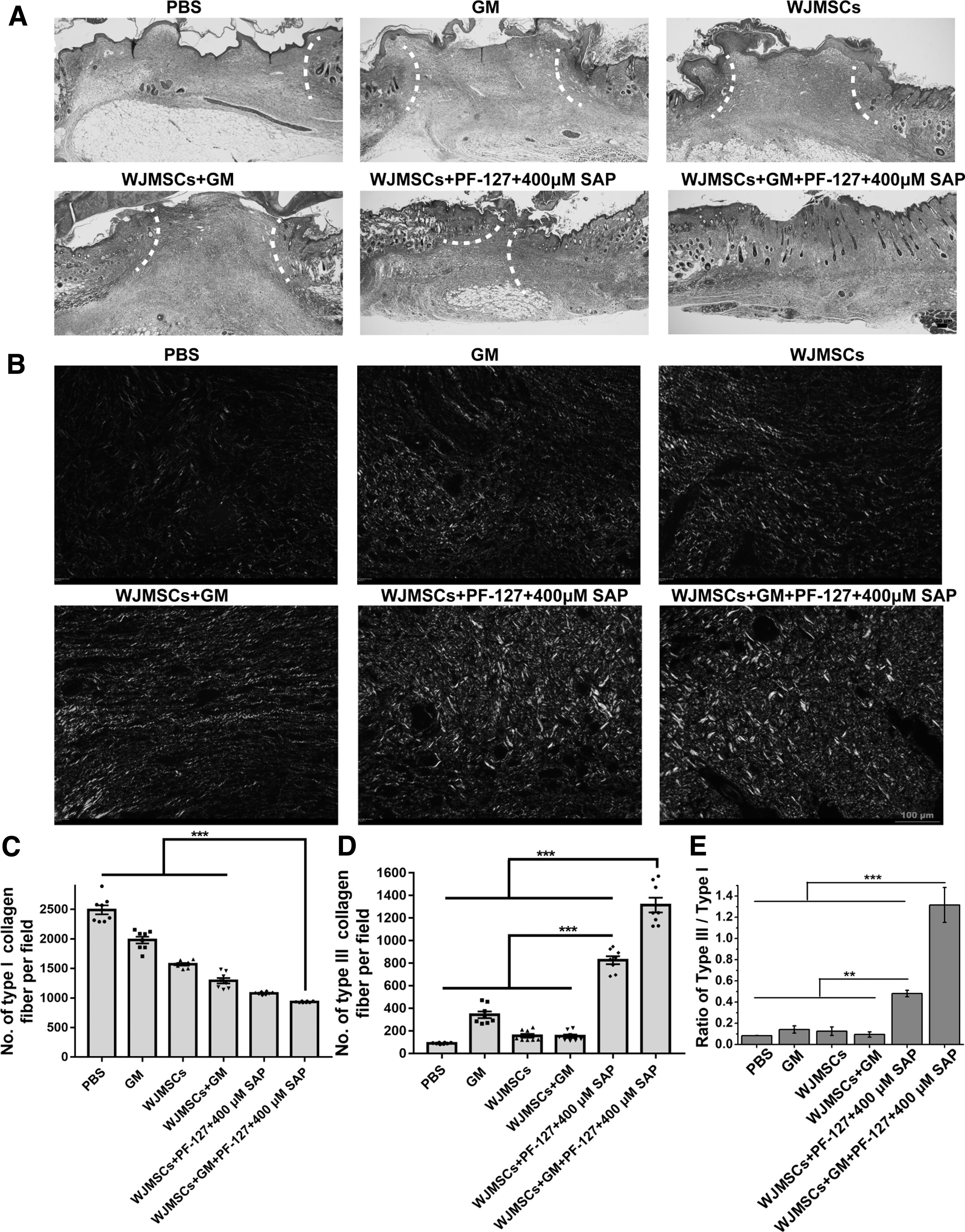
Histology analysis the collagen deposition of skin wound.
We further examined collagen composition by picrosirius red staining on day 10 to investigate fibrotic scarring of the regenerated wound. We found that there were less red and yellow collagen type I with strong birefringence (PBS, 2490.67 ± 200.67; GM, 1979.65 ± 139.43; WJMSCs, 1570.00 ± 49.96; WJMSCs+GM, 1291.66 ± 138.81; WJMSCs+PF-127+SAP, 1080.00 ± 35.93; WJMSCs+GM+PF-127+SAP, 932 ± 13.87) and more green collagen type III with weak birefringence (PBS, 90.33 ± 4.41; GM, 343.64 ± 65.17; WJMSCs, 157.66 ± 36.68; WJMSCs+GM, 153.33 ± 36.41; WJMSCs+PF-127+SAP, 826.67 ± 76.74; WJMSCs+GM+PF-127+SAP, 1315.33 ± 132.22) in WJMSCs+GM+PF-127+SAP group than that in other control groups. (Fig. 5B, D).
Furthermore, the ratio of collagen type III/collagen type I in WJMSCs+GM+PF-127+SAP group (1.32 ± 0.16) was also higher than other control groups (PBS, 0.083 ± 0.001; GM, 0.14 ± 0.03; WJMSCs, 0.13 ± 0.04; WJMSCs+GM, 0.09 ± 0.02; WJMSCs+PF-127+SAP, 0.48 ± 0.03) (Fig. 5E). Above results demonstrated that transplantation of WJMSCs+GM+PF-127+SAP indeed increases collagen deposition and formation of type III collagen fibers. Based on a higher ratio of type III/type I collagen fiber, WJMSCs+GM+PF-127+SAP transplantation might be having antifibrotic effect during wound healing.
WJMSCs+GM+PF-127+SAP transplantation promotes macrophage polarization and reepithelialization
During the inflammatory response process of wound healing, the transition of macrophages from proinflammatory M1 type into anti-inflammatory M2 type is essential for wound healing. 38,39 In this study, we evaluated the CD86-positive M1 macrophages and CD163-positive M2 macrophages in the skin wound via immunohistochemical staining. The results showed that there were less M1 macrophages (PBS, 78.67 ± 5.17; GM, 87.33 ± 1.76; WJMSCs, 71.33 ± 4.70; WJMSCs+GM, 49.67 ± 4.91; WJMSCs+PF-127+SAP, 22.00 ± 2.31, WJMSCs+GM+PF-127+SAP, 12.33 ± 1.86 per field) (Fig. 6A, B) (p < 0.001) and more M2 macrophages (PBS, 13.33 ± 1.86; GM, 24.00 ± 3.79; WJMSCs, 28.67 ± 2.85; WJMSCs+GM, 41.00 ± 4.93; WJMSCs+PF-127+SAP, 109.67 ± 2.96; WJMSCs+GM+PF-127+SAP, 178.33 ± 1.86 per field) (Fig. 6A, C) (p < 0.001) in WJMSCs+GM+PF-127+SAP group than those in other groups. These results indicated that WJMSCs+GM+PF-127+SAP transplantation accelerates the transition of the microenvironment of skin wounds from pro-inflammatory to anti-inflammatory.

Immunohistochemistry identifies the macrophages transformation, cell proliferation, and neovascularization.
During the process of the entire wound healing, newly formed vasculature play a critical role in providing oxygen and nutrients to the growing tissue. 40 In this study, anti-CD31 immunohistochemical staining results showed that there were more CD31-positive cells in the WJMSCs+GM+PF-127+SAP group (26.33 ± 2.19 per field) than those in other groups (PBS, 5.67 ± 1.45; GM, 9.33 ± 0.89; WJMSCs, 11.33 ± 0.88; WJMSCs+GM, 12.33 ± 0.88; WJMSCs+PF-127+SAP, 17.67 ± 0.89 per field) (Fig. 6A, D) (p < 0.01). Therefore, WJMSCs+GM+PF-127+SAP transplantation may facilitate angiogenesis at skin wound site to promote wound healing.
The reepithelialization process is coordinated by the migration and proliferation of various types of cells. 41,42 In this study, anti-Ki67 immunohistochemical staining results showed that there were more Ki-67-positive cells in the WJMSCs+GM+PF-127+ SAP group (44.17% ± 0.67%) than those in other groups (PBS, 5.01% ± 0.67%; GM, 5.28% ± 0.34%; WJMSCs, 9.40% ± 0.14%; WJMSCs+GM, 11.88% ± 0.58%; WJMSCs+PF-127+SAP, 34.73% ± 0.49%) (Fig. 6A, E) (p < 0.001), which imply that WJMSCs+GM+PF-127+SAP transplantation increased cell proliferation at the wound site. Overall, WJMSC+GM+PF-127+SAP transplantation promoted the macrophages transformation, cell proliferation, and angiogenesis in wound site to accelerate acute full-thickness skin wound healing.
Taken together, our results suggested that WJMSCs embedding in GM further relieved the cytotoxicity of PF-127. In vivo, WJMSCs+GM+PF127+SAP transplantation facilitated the acute full-thickness skin wound healing through enhancing cell retention and engraftment, modulating inflammatory responses, promoting reepithelialization and ECM remodeling at the wound site (Fig. 7).
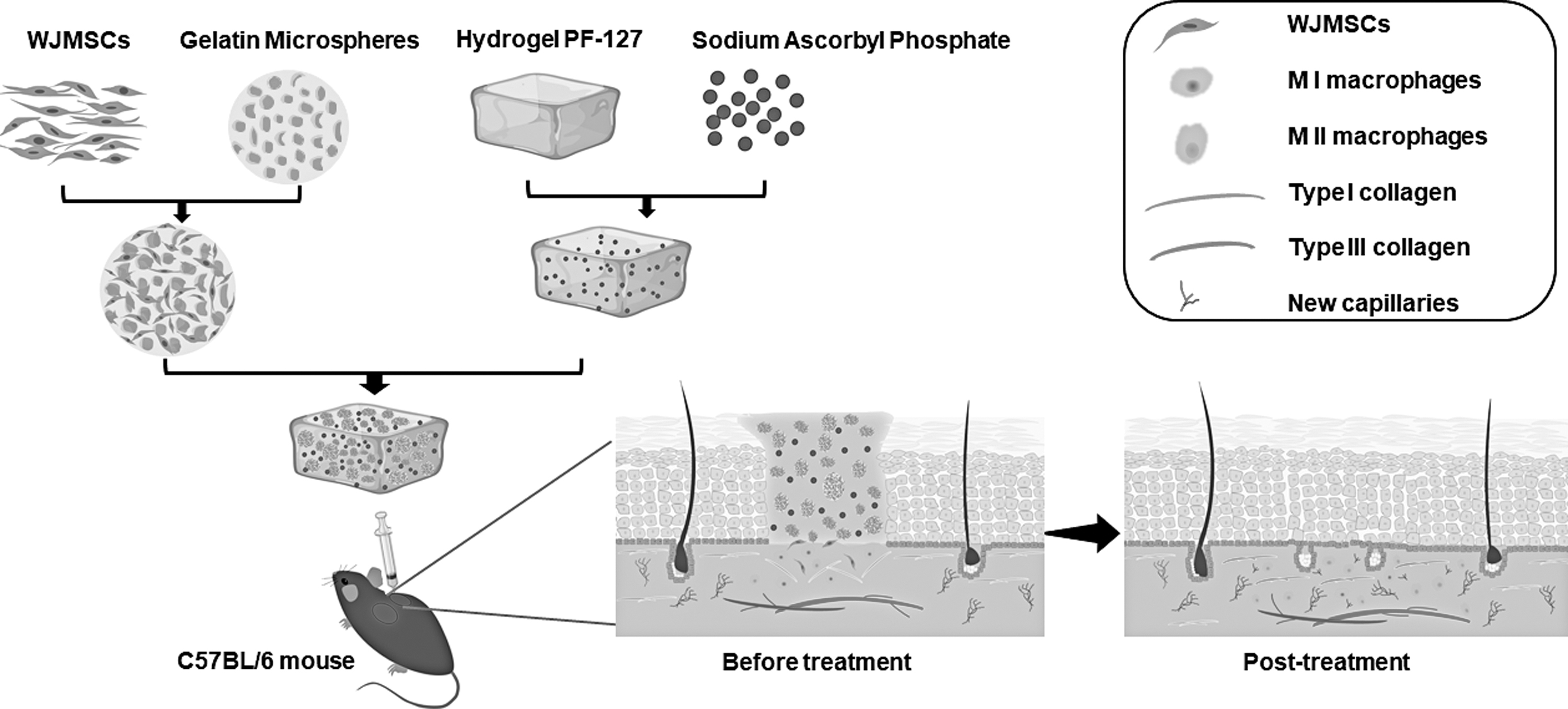
Working model of WJMSCs+GM+PF-127+SAP transplantation in acute full-thickness skin wounds. Transplantation of WJMSCs+GM+PF-127+SAP facilitates the acute full-thickness skin wound healing through promoting macrophage polarization, cell proliferation, neovascularization, collagen type III deposition, cell retention and engraftment.
DISCUSSION
Skin injuries can be divided into acute skin wounds and chronic skin wounds according to the time it takes for healing. When adult skin is injured, an early inflammatory response is activated, leading to infiltration of immune cells and macrophages. 43 Release of cytokines promotes the proliferation and migration of fibroblasts and keratinocytes, and improves reepithelialization and skin remodeling. 44 Acute skin wounds are mainly caused by injuries such as burns, surgical incisions, lacerations, and abrasions and generally will heal within 8–12 weeks. 45,46 In this study, we focused on the acute skin traumatic wounds, such as surgical incision, lacerations, and abrasions. Our results demonstrated that transplantation of WJMSCs+GM+PF-127+SAP accelerates the closure of acute skin wound, which may provide the new stem cells and biomedical material-based treatment strategy for acute skin traumatic wounds.
Recently, MSCs have been the main candidate cell type for tissue engineering and regenerative medicine applications for wound healing. To overcome the bottleneck problem during cell transplantation, many 3D encapsulation techniques for MSCs have been developed as a powerful tool for tissue injury regeneration. 47,48 Compared with traditional 2D monolayer cultures, 3D culture provides more opportunities of cell–cell interaction and closely mimics the natural microenvironment within a tissue. Previous study found that gelatin microbeads can increase the proliferation and multipotent differential ability of hWJ-MSC.
Besides, direct injection of hWJ-MSC embedded in gelatin microbeads also enhances skin wound healing in vivo. 22 Hydrogel PF-127 embedded with extracellular vesicles from adipose tissue–derived stromal cells promoted the esophageal fistula healing in procine. 49,50 Combination of PF-127 and hWJMSC exosomes also facilitated the diabetic wound healing and regeneration. 51 Adipose-derived MSCs were delivered by thermosensitive PF-127 hydrogel containing folic acid. MgO:ZnO/chitosan hybrid particles promotes the proliferation and migration of human foreskin fibroblast cells and wound healing. 52 In our study, we seeded WJMSCs into GM and then encapsulated WJMSCs+GM with PF-127/SAP. This strategy increased the viability of WJMSCs protecting by 3D microspheres and promoted the macrophage polarization, angiogenesis, cell proliferation, and collagen deposition in vivo.
Previous studies have reported that the proliferation index of 3D cultured MSCs was significantly higher than 2D cultured MSCs. 22 In our study, we found that GM loading further improved the viability and survival of WJMSCs in PF-127/SAP in vitro, but also WJMSCs+GM+PF-127+SAP transplantation increased the number of proliferating cells in skin wound, compared to WJMSCs+PF-127+SAP. Besides, OE-EGFP WJMSCs tracking results demonstrated that GM plus PF-127/SAP enhance the retention and engraftment of WJMSCs at wound site. Above results implied that GM plus PF-127/SAP combination can effectively deliver WJMSCs and solve the main problems of cell activity and residence time during MSCs-based therapy.
Compared with 2D culturing of MSCs, 3D culturing increased the expression levels of immune regulatory genes, promoted the production of ECM, and enhanced the anti-apoptotic ability. 53 Macrophages typically migrated to the wound site within 24–48 h of skin injury. 54 During the early stage of the inflammatory response, macrophages exhibit as pro-inflammatory M1 phenotype and are primarily responsible for combating infections. During the late stage of the inflammatory response, macrophages will polarize toward anti-inflammatory M2 phenotype which are associated with tissue remodeling and formation of new blood vessels. 55 –57 In our study, comparing with other control groups, there were less M1 macrophage while more M2 macrophage in the skin wound, which implied that GM+PF-127+SAP will simulate the 3D environment for WJMSCs to ameliorate the inflammatory response.
Remodeling is the last stage of wound healing that mainly involves collagen deposition and ECM remodeling. The proportions and arrangements of the various subtypes of collagen change dynamically at different phases of wound healing. In the early stage of skin wound healing, the arrangement of collagen is scattered and disorganized, whereas in the later stage of granulation and remodeling, the collagen fibers become arranged in reticular manner. 34 In our study, WJMSCs+GM+PF-127+SAP transplantation increased the collagen type III deposition and the ratio of collagen III/I compared with WJMSCs+PF-127+SAP. Our study may provide a new effective cell therapy strategy for acute skin traumatic wounds to prevent the formation of fibrotic scarring. On the contrary, it also showed that GM-simulated 3D culture environment could not only promote wound healing at the early stage, but also improve collagen reconstruction at the late stage.
INNOVATION
Our study explored an effective 3D encapsulation technology for WJMSCs, which solved the main problems of cell activity and residence time during MSC transplantation. WJMSCs+GM+PF-127+SAP transplantation facilitated the acute full-thickness skin wound healing and regeneration in mice through promoting the macrophage polarization, cell proliferation, neovascularization, collagen type III deposition, cell retention, and engraftment. Our findings may potentially provide a novel and effective therapeutic strategy for patients with acute skin traumatic wounds.
KEY FINDINGS
GM has a strong biocompatibility and promotes the viability and proliferation of WJMSCs.
WJMSCs embedded in GM and encapsulated with PF-127 plus SAP combination accelerate dermis regeneration and collagen deposition at skin wound site.
Combing 3D microcarrier with thermosensitive hydrogel PF-127/SAP complex can not only promote the survival and retention of transplanted WJMSCs, but also accelerate the acute wound healing and regeneration by improving macrophage polarization, cell proliferation, and angiogenesis.
Footnotes
ACKNOWLEDGMENTS AND FUNDING SOURCES
This study was funded by the National Key R&D Program of China (2017YFA0102801 and 2017YFC1001901), the National Natural Science Foundation of China (31971365, 32170802), the Guangdong Basic and Applied Basic Research Foundation (2020B1515120090), the Guangdong Special Support Program (2019BT02Y276), the Guangzhou Science and Technology Project (201803010032), and the Fundamental Research Funds for the Central Universities (19lgpy190), and the Foshan Medicine Dengfeng Project of China (2019–2022).
AUTHORs' CONTRIBUTION
Y.J. designed and performed the experiments, and wrote the article. Y.J., Y.N., and X.C. designed the methods, interpreted the results, and provided intellectual insights. Y.J., X.C., Y.N., M.L., and S.H. performed the experiments and collected the data. X.C., T.C., and M.L. contributed to the discussion and reviewed the article. G.S., A.W., and J.H. planned and coordinated the study and revised the article. All the authors edited and approved the final version of the article.
ABOUT THE AUTHORS
AUTHOR DISCLOSURE AND GHOSTWRITING
The authors declare that they have no conflict of interest. The contents of this article were expressly written by the authors listed. No ghostwriters were used to write this article.
SUPPLEMENTARY MATERIAL
Supplementary Figure S1
Supplementary Figure S2
Supplementary Figure S3
Supplementary Figure S4
Supplementary Figure S5
Supplementary Figure S6
Supplementary Table S1
Abbreviations and Acronyms
References
Supplementary Material
Please find the following supplemental material available below.
For Open Access articles published under a Creative Commons License, all supplemental material carries the same license as the article it is associated with.
For non-Open Access articles published, all supplemental material carries a non-exclusive license, and permission requests for re-use of supplemental material or any part of supplemental material shall be sent directly to the copyright owner as specified in the copyright notice associated with the article.
