




Abstract
Objective:
To conduct a systematic literature review to study the effects of fish-based biomaterials on wound healing in both in vivo and in vitro animal models.
Approach:
This review covers the study reported in different articles between 2016 and August 2022 concentrating mainly on the cytotoxicity evaluation of different fish-based biomaterials on inflammation, reepithelialization and wound healing.
Significance:
This review shows considerable amount of research work carried out with fish-based biomaterials and collagen for treating burn wounds. Surprisingly there are only a few commercial products developed so far in this particular regard for surgical purpose and therefore, there is a way out and need for developing medical support product from fish-based biomaterials to treat and cure wounds.
Recent Advances:
Three-dimensional skin bioprinting technique is a large-scale solution for severe burn wounds that requires collagen as a raw material for printing, wherein fish collagen can be used in place of bovine and porcine, as it is biocompatible, promotes cell proliferation, adhesion, and migration, and degrades enzymatically. In the recent times, there are a few fish-based surgical products that have been formulated by Kerecis in United States.
Critical Issues:
The different fish-based biomaterial products are all mere supplements taken in orally as food or supplements till date and there is no proper proven medications that has been formulated so far in the field of wound healing and inflammation based on fish biomaterials except the surgical products that can be finger counted.
Future Directions:
Fish-based biomaterials are known for the medicinal properties that are used throughout the world and further investigations should be carried out to understand the actual physiochemical properties of its derivatives for the discovery of novel products and drugs.

SCOPE AND SIGNIFICANCE
Manufacturing of a natural skin wound healing medical product has been a major task till date that so far only porcine and bovine-based collagen products are available in the market in the form of ointments, sponges, and surgical products for treating various types of wounds. This review shows considerable amount of work carried out on fish-based biomaterials alongside collagen for treating burn wounds. Only few products are available in the market for the above-mentioned purpose and therefore, there is an urgent need for developing a natural drug as medication from fish-based biomaterials to treat different types of wounds.
TRANSITIONAL RELEVANCE
Traditionally, consumption of fish is considered to be one of the very few major ways to heal wounds faster followed by grafting of fish skin on to the human skin. Collagen from fish skin, chitin, aqueous extracts, and oils are commonly used for skin replenishment, out of which collagen was used the most with respect to skin health and beauty. Despite the fact that fish-based biomaterials have enormous health benefits, there is a lacuna of research that has to be carried out to fabricate the different composition of fish-based biomaterials.
CLINICAL RELEVANCE
Fish-based biomaterials naturally have high wound healing potency and antimicrobial activity that has led to conduct further studies, as the till-date research is limited only to in vivo and in vitro analyses with respect to medication. All studies that have been carried out so far are limited only at the laboratory level and there is a need for taking this laboratory knowledge into the pharmaceutical industry to develop a novel drug. Given this reason, this systematic review was carried out to address both the merits and demerits to explore the extent of progress with regard to fish-based products and biomaterials.
INTRODUCTION
The aquatic environment encompasses a unique source of diverse natural products from both invertebrates (Tunicates, bryozoans, arthropods, mollusk) and vertebrates (fishes). Fishes are protein-rich diet sources containing some of the important fat contents, minerals, and vitamins. 1 Collagens, gelatin substances, fatty acids, and certain bioactive peptides derived from fishes have proven to be lifesaving metabolites since time immemorial. 1 Sponges, sea turtles, snails, ascidians, mollusks, marine fungi, bacteria, aquatic worms, mesophytes, soft corals, and fishes are among the top sources from which bioactive compounds as well as drugs—for example trabectedin, ET0743®, is a first marine anticancer drug—are derived. 2 The proteins and peptides derived from fishes possess biological activities like antioxidant, antihypertensive, anticoagulant, anti-inflammatory, antibacterial, and wound healing properties. 3
According to the recent update on Fish Base (
In recent days, fishes have been extensively used for developing drugs for treating diseases like diabetes, cardiovascular diseases, atherosclerosis, low blood pressure, and autoimmune disorders. 7
Some of the crucial growth and development nutrients found in fishes are Vit-A, Vit-D, Vit-B12, iron, protein, docosahexaenoic acid (DHA), eicosapentaenoic acid—EPA (Omega-3), calcium, iodine, and zinc. The extracts like collagen, albumin, epidermal secretions, oils, chitosan, peptidoglycans, and cartilages are some of the major sources from fish and fish extracts used for drug development. Of interest, shark cartilage and shellfish are used as medicine for the treatment of cancer in recent days. Omega-3 fatty acid is considered to be crucial for optimal functioning of the body and brain. In addition, macular impairment that leads to blindness can be cured by the intake of omega-3 fatty acids. 8
Calcium, phosphate, and hydroxyapatite (HAP) are some of the inorganic compounds, and ∼60–70% of the fish bone constituents of inorganic origin compounds of HAP is commercially used for maxillofacial surgery, alveolar bridge augmentation, and dental implants. 1 Extensive research work has been carried out and it is proven that collagen increases wound healing rate, reepithelialization through in vitro, in vivo, and human trail studies. 9,10
One of the important therapeutic applications of fish derivatives is anti-inflammatory and wound healing activities. Inflammation is a complex response of body tissues to external stimuli like physical injuries, ionizing radiation, infection by pathogens, toxins, chemical irritants, and so on. 11 Inflammation invites cells like macrophages to produce mediators important for inflammation, further leading to the increased levels of leukocyte chemoattractants. 12 Recent studies carried out using mice with certain genetically deficient specific immune cells suggest that inflammatory cells are not required for tissue repair as long as it is free of bacterial infection. 13 Systemic chronic inflammation is a risk factor for a number of illness, including cancer, wound healing, chronic kidney disease, nonalcoholic fatty liver disease, diabetes mellitus, autoimmune, and neurodegenerative disorders, which may collectively account for the major causes of disability and mortality worldwide. 14
It is estimated that 1–2% of population experience chronic wounds. 15 These wounds are mostly from venous disease, arterial insufficiency of lower extremity, diabetes, and local pressure. Given the situation of increase in type 2 diabetes, comorbid issues like diabetic foot ulcer is increasing steadily of which, 2–30 per 1,000 patients go for amputations. 16 According to the World Health Organization (WHO) data, 3.16 million people die owing to the above-mentioned injuries. 17
Physiological wound healing is characterized by four major steps like homeostasis, inflammation, proliferation, and maturation.
Homeostasis
The onset of this phase is characterized by extravasation of blood and blood components. Loss of structural integrity of blood vessels initiates coagulation cascade mechanism. 18 The platelet adheres to the sub-endothelial cells after the rupture of blood vessels. Formation of fibrin mesh begins, and simultaneously the blood is transformed from liquid to gel by the action of procoagulant and prothrombin. The different clotting cascades are initiated by the injured skin (extrinsic factors), where thrombocytes gets activated for clotting after being exposed to collagen (intrinsic factor). 19
Inflammation
Inflammation begins right after the injury leading to localized swelling and infiltration of neutrophil granulocyte to the wound cite causing removal of damaged cells, pathogens, and bacteria, followed by which, monocytes and macrophages act as key regulators for repair because they perform primary phagocytic activity and produce growth factors responsible for proliferation. 20
Proliferation
After a wound, the proliferative phase begins around day 3 and lasts for 2–4 weeks. It is characterized by fibroblast migration, extracellular matrix (ECM) deposition, and the development of granulation in tissues. The temporary fibrin/fibronectin matrix is replaced by the freshly produced granulation tissue as the proliferative phase progresses. The proliferative phase's last stage is epithelialization of the wound. 21
Maturation
The wound completely heals, and type III collagen is transformed into type I collagen. The cells that were involved in repair and no more required are removed by apoptosis. 18 Collagen is remodeled into a three-modeled structure, enhancing the tensile strength of the healing tissues. Matrix metalloproteinase are released by fibroblasts that actually converts type III collagen to type I collagen. 22 Inflammation is characterized by three cardinal signs such as redness, heat, and swelling. The inflammatory cells are triggered by infectious and noninfectious agents and cell damage.
The factors that affect wound healing are classified into comorbidities—diabetes, obesity, and protein energy malnutrition; medications—nonsteroidal anti-inflammatory drugs (NSAIDs), steroids, oncology interventions—radiation and chemotherapy. Life style habits such as smoking, alcohol intake, and accident injuries also account for the same. 23 The inflammatory cells like macrophages, adipocytes, and cytokines such as tumor necrosis factor-α (TNF-α), interleukin (IL)-1β, and IL-6 along with inflammatory proteins such as C-reactive proteins, serum amyloid-A, fibrinogen, haptoglobin, and 1-acid glycoprotein help to maintain homeostasis during injury. 11 These factors mediate inflammation through interaction with IL-1, IL-6 receptor, and TNF receptor. Receptor activation triggers intracellular signaling pathways like mitogen-activated protein kinase, nuclear factor kappa-B and Janus kinase-signal transducer and activator of transcription pathway. 11
The hypoxic condition is that the precursor of fibroblast cells, upon rupture of vasculature around the wound area, there is rapid influx of inflammatory cells to the wound. 24 These inflammatory cells accumulate in the wound area playing a vital role in granulation and reepithelialization. 25 It is estimated that the production rate of transforming growth factor-β (TGF-β) secreted by fibroblast cells increased nine times. 24 Oxygen is required in the later stages in the formation of ECM for conversion of protocollagen into stable triple helix collagen. 26 In the later stages of maturation only adequate amount of oxygen is required, which can be directly supplied to cells through blood since vascularization is complete.
Signaling from inflammatory cells at the wound site stimulates fibroblast migration inward from the wound margins at the beginning of acute wound healing. Paracrine factors, such as basic fibroblast growth factor (bFGF/FGF-2), keratinocyte growth factor (KGF/FGF-7), vascular endothelial growth factor A (VEGF-A), and insulin like growth factor-1 (IGF-1) is secreted by the fibroblast cells that trigger the adjacent keratinocytes. 27 The keratinocytes and inflammatory cells produces paracrine signaling that stimulates fibroblast to synthesize collagen and promote crosslinking of collagen to form ECM and further get differentiated into myofibroblast. 28 Angiogenic growth factors like platelet-derived growth factors (PDGFs) and VEGFs is also secreted by keratinocytes, which proliferates endothelial cell migration and angiogenesis in the bed leading to the formation of granulation tissue and restoration of epidermal barrier. 28,29
The factors for wounds in patients with other etiologies are ulcers or diabetes, which results in cellular dysfunction that affects all the stages of wound healing. 23 During homeostasis the expression of PDGF on epithelial and endothelial cells is decreased leading to delayed homeostasis. 21 Increased activity of macrophages increases the expression of inflammatory cytokines, 30 leading to prolonged inflammatory phase. Granulation tissue formation of the proliferative phase is decreased by impaired fibroblast signaling leading to delayed reepithelialization. ECM instability is caused owing to elevated expression of metallomatrix proteinases and reactive oxygen species. 23,31
Microorganisms and platelet-derived factors, such as TGF-β or ECM fragment molecules, stimulate the continuous influx of immune cells as a result of repeated tissue injury that leads to the amplification of proinflammatory cytokine cascade and persists for a prolonged period of time, resulting in elevated levels of proteases. Proteases are closely regulated in severe wounds. 32 Increase in protease levels results in destruction of ECM. In addition to preventing the lesion from progressing into the proliferative phase, the proteolytic degradation of ECM also draws more inflammatory cells, accelerating the inflammation cycle. 33 Polymicrobial consortia, which live cooperatively in highly organized biofilms, play a significant role in the inability of a wound to heal. The patient's immune system and antimicrobial medication cannot reach the harmful bacteria because the biofilm leading to chronic wounds is associated to biofilm infections. 34
Steroids and nonsteroids (NSAIDs) are the two different kinds of primary drugs used in treatment of inflammatory diseases. However, NSAIDs are proven to have side effects like blood pressure and congestive heart failure
35
; on the contrary, steroidal anti-inflammatory drugs have immunosuppressing effects.
36
Using natural biopolymer would be one the best alternative source for treating chronic wound injury. WHO (
Despite of the fact that extensive research has been carried out on natural therapeutics and drugs for wound healing, there is limited understanding about its effects and uses. The purpose of this literature review was to explore the different possible biomaterial samples of fishes used in treating inflammation and wound healing and to critically analyze the in vitro and in vivo studies supporting the anti-inflammatory (cell viability and cytotoxicity), wound healing (histology), and immunohistochemistry properties of fish biomaterials.
METHODOLOGY
Review protocol
From July to August 2022, a systematic review was carried out using PubMed and Google Scholar databases. The SYRCLE (Systematic Review Centre for Laboratory Animal Experimentation) guidelines were followed for conducting the systematic review. 39 The search was conducted in accordance with the Preferred Reporting Items for Systematic Reviews and Meta-Analysis (PRISMA). The medical subject headings (MeSH) terms used are as follows: “Anti-inflammatory” OR “Wound healing” AND “Fish.” In addition, a number of synonyms and advanced search methods such as filters according to years and exclusion of review articles, case study articles, and reports, after which the abstracts were analyzed manually based on inclusion and exclusion criteria. Furthermore, the articles were analyzed individually and added to the review. Electronic laboratory notebook was not used for the review.
Inclusion criteria
In vitro and in vivo studies specifying species name.
Manuscript to be published in the last 5 years.
Fish-based biomaterial samples in combination with other fish-based biomaterials.
Studies involving wounds, burns, and anti-inflammatory activity treated with fish-based biomaterial.
Exclusion criteria
Wound dressing, case study, and human trials were excluded.
Lack of detailing about the technique, description of wound and burns.
Animals with concomitant systemic disorders, such as osteoporosis or diabetes.
Data extraction
Wound healing and anti-inflammation were the variables to be analyzed based on cell viability, cytotoxicity in case of in vitro studies, and histology for in vivo studies. Along with this, variables like fish species, biomaterial, animal strains/in vivo model, assay (in vitro studies), dosage, implantation period (in vivo studies), and outcome of each of the study were also analyzed.
RESULTS
The PRISMA flow diagram (Fig. 1) describes the search strategy involved in this systematic review. A total of 109 articles were screened from PubMed and Google Scholar databases. After removing the duplicates, 105 articles were screened for eligibility. Furthermore, of 105 articles, 80 articles were full-text accessed and 30 articles were finally included in the systematic review considering the inclusion and exclusion criteria. Of these 30 articles, 5 articles used both in vivo and in vitro methods. Hence 16 in vivo and 19 in vitro studies were represented.
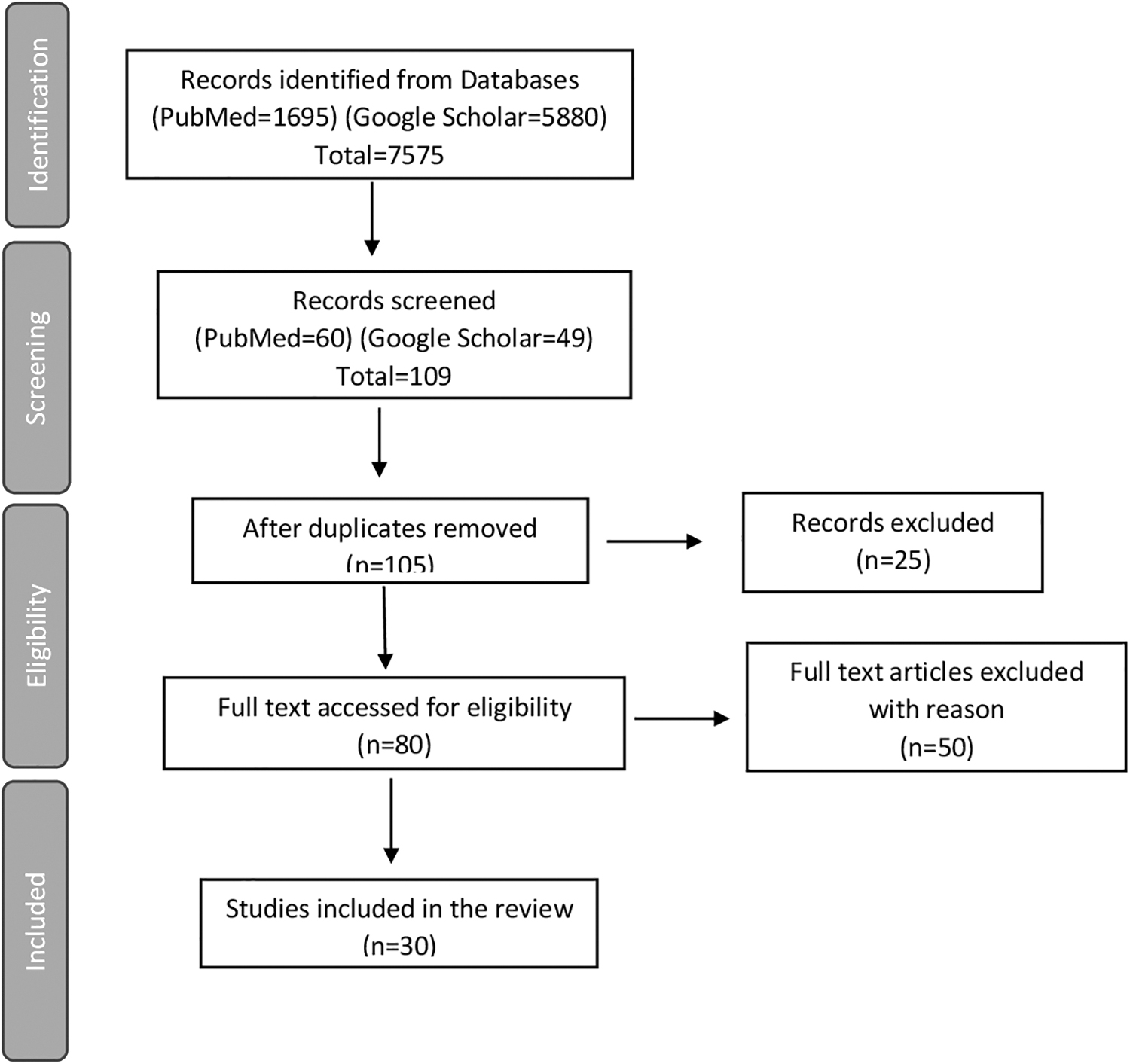
PRISMA flow diagram for systematic review. PRISMA, Preferred Reporting Items for Systematic Reviews and Meta-Analysis.
Tables 1 and 2 demonstrate the characteristics involved in the in vivo and in vitro studies showing anti-inflammatory or wound healing potencies. Among the different articles using fish species in the study, five articles were involved with Oreochromis sp., 44,45,51,55,62 four were involved with Channa striata, 41,43,57,59 three were involved with Salmon sp., 51,66,69 two were involved with extraction of biomaterial from Anguilla bicolor bicolor, 41,46 two studies were involved with Lates calcarifer, 48,58 two studies involved Hippocampus sp. 63,67 On the other hand, Lophius litulon, 42 Anabas testudineus, 70 Smooth hound triggerfish, 47 Grey triggerfish, 47 Merluccius merluccius, 49 Gnathonemus petersii, 50 Lateolabrax maculatus, 52 Acipenser baerii, 53 Lophiosilurus alexandri, 54 Cypselurus melanurus, 56 Catla catla, 56 Indian mackerel, 56 Clarias batrachus, 56 Pangasius pangasius, 56 Sparus aurata, 60 Engraulis encrasicolus, 61 Pomadasys maculatus, 64 Chanos chanos, 65 Hypophthalmichthys molitrix, 68 were used in one of the studies each. There were totally 25 different fish species explored for the study related to wound healing potencies on the whole.
In vivo analysis of anti-inflammatory and wound healing potencies of fish-based biomaterials
ASB, aqueous extract of sea bass; CG, collagen extract; CSMP, chitosan-marine peptides hydrogels; DHA, docosahexaenoic acid; GAG, glycosaminoglycan; GTSG, gray tiger fish; HHO, hake head oil; ICR, Institute of Cancer Research; LCEP, Lates calcarifer extracted peptide; MCP, marine collagen peptide; PSC, pepsin-solubilized collagen; SHSG, smooth hound fish; Ss-SCP, Salmo salar skin 18 collagen peptides; TCG, tilapia collagen; Tn-SCP, Tilapia nilotica skin collagen peptide.
In vitro analysis of wound healing potencies of fish-based biomaterials
APH, Protein hydrolysates obtained from anchovy Engraulis encrasicolus; Asbs-HC, Asian sea bass skin hydrolyzed collagen; ddH2O, double-distilled water; FDWE, freeze-dried water extracts; HaCaT, human keratinocyte–derived cells; HCSS, hydrolyzed collagen of salmon skin; HEO, 3β-hydroxycholest-5-en-7-one; HFE, Haruan fish extract; IL, interleukin; MTT, 3-[4,5-dimethylthiazol-2-yl]-2,5 diphenyl tetrazolium bromide; MSCP, collagen peptides isolated from milkfish scales; SBPHs, Salmon bone protein hydrolysate; SDWE, spray-dried water extracts.
The most common in vivo animal model used was Wistar Albino rats—five studies, 41,43,45,54,70 three studies used New Zealand rabbits, 50,55 three studies involved C57BL/6 J mice, 48,52,53 and two studies were with Swiss albino mice, 47,49 and one each with Institute of Cancer Research mice, 42 Sprague-Dawley rats, 51 and in one study there was no specified animal used. 46 Among the in vitro model studies, seven studies were found using Activated RAW 264.7 macrophages, 52,56,61,63,64,66,67 seven studies involved human keratinocyte-derived cells, 48,53,55,62,65,68,69 and four studies used L929 fibroblast cells. 44,52,60,64 On the contrary, odontoblast MDPC-23 cells, 57 MRC-5 fibroblast cells, 58 and EA.hy926 endothelial cell lines 59 were used only once.
Tables 1 and 2 also contain the biomaterial samples that were used in the studies. Collagen was used in 10 studies, 42,45,51,53,55,56,58,60,65,69 mucus was used in four studies, 43,46,54,70 and peptide extracts were used in four studies. 47,48,61,63 Three studies used oil extracts. 41,49,50 Three studies used aqueous extracts 52,57,59 and Chitosan was used in only one of the studies. 44 Fish scale was used in only one study. 62 Otoliths was used in one study. 64 Fish bone was used in two studies. 66,68 On the contrary, one of the studies used a compound 3β-hydroxycholest-5-en-7-one (HEO). 67
In the in vivo models, the manually created wound size ranged from 8 mm52 to 4 cm55 in 14 studies and was not specified in 2 studies. 48,50 In the in vitro analysis, 14 studies involved 3-[4,5-dimethylthiazol-2-yl]-2,5 diphenyl tetrazolium bromide (MTT) assay, five studies involved monolayer starch assay, one study involved crystal violet assay, and assay was not specified in one of the studies.
The progression of wound healing was analyzed using three different evaluation methods in the articles reviewed. Macroscopic analysis involved observation of healing stages directly without any aid. Immunohistochemistry analysis was used to check the specific binding pattern of antigen and antibody that helped to analyze the expression of TGF and VEGF gene. Histological analysis involved the study of cell proliferation, that is, the formation of cells for healing at different stages.
Table 3 demonstrates the overall results of in vivo studies included in the review. Of 16 in vivo studies included in this review, all of them involved macroscopic analysis for evaluation of wound healing rate, whereas 12 of them showed histological analysis 42 –47,49,50,53 –55 stating that all the samples promoted ∼90% wound healing rate with increased epithelialization, vascularization, and collagen formation. Six studies involved immunohistochemistry 45,48 –50,52,53 showing prominent expression of TGF-β, IL-6, and decreased expression of TNF-α. 52
In vivo analysis and overall result
TGF-β, transforming growth factor-β; TNF-α, tumor necrosis factor-α; VEGF, vascular endothelial growth factor.
Table 4 provides the overall analysis of in vitro studies. Of the 19 studies carried out, macroscopic evaluation was performed in 2 studies showing maximum healing rates. 55,58 Immunohistochemistry analysis of the studies showed an average of 100% cell viability and greater cell proliferation and suppression of inflammatory cytokines. One study revealed the histological analysis 62 showing progressive cell cycle.
In vitro analysis and overall results
DISCUSSION AND CONCLUSION
This systematic review evaluates the studies on the effects of fish-based biomaterial used in wound healing and anti-inflammatory activities using animal models and cell line cultures. Nile tilapia is a most common fish from which different biomaterials like chitosan, collagen, and gelatin were extracted. Studies conducted in vivo and in vitro showed that collagen accelerated tissue regeneration, increased angiogenesis, and decreased inflammation. C. striata was the next set of fish that was explored the most and biomaterials like oil, mucus, and fish extract were obtained out of which mucus showed 100% wound healing on day 8. 43 Mucus extracts of C. striata showed highest wound healing rate with minimal duration. 43
The use of fish collagen is a turning point as a substitute for porcine and bovine collagen that are used commercially, and is particularly used for the manufacture of membranes in tissue engineering. 71 Fish skin is a waste product in the fish market and innovative ideas would help in sustainable use. The extraction protocols for collagen was mostly using acetic acid and citric acid. 72
Many of the previous studies on collagen showed a lot of variation in the process of extraction as collagen has low melting point and low melting temperature. 73 This limitation can only be equated using modified extraction process and creating a hybrid composite that is thermally stable. Fish collagen has certain advantages as well like the rate of absorption of fish collagen is 1.5 times faster than other mammalian-based collagen like bovine and porcine, 73 which is because of the lower molecular weight of fish collagen. The concentration of solutions used and incubation time also differed in the protocols leading to different yields. However, the literature till date has not given a standard protocol to be carried out to optimize the parameters for collagen extraction.
It is necessary to clean other substances like calcium and lipids followed by collagen extraction using an acid. Collagen fiber has a triple helix structure that makes it insoluble in water. Therefore, acids are used to solubilize and extract collagen. Physical and morphological characteristics of the obtained collagen were analyzed by Fourier transform infrared (FTIR) spectroscopy and energy dispersive spectroscopy was performed to identify the presence of amino acid glycine and proline. Fluorescein isothiocyanate labeling was performed to analyze the molecular weight distribution of the peptide by tricine sodium dodecyl sulfate–polyacrylamide gel electrophoresis. 56 Regarding both in vivo and in vitro results, all the works demonstrated positive results. Fish collagen-based dressing, hydrogels, nanofibers, and collagen scaffolds serve as protection against infection and contamination on the skin.
With regard to the design of wound dressings, such as hydrogel-based dressings, molecular weight modulation is a crucial factor. The creation of hydrogel networks and overall mesh sizes can be significantly influenced by molecular weight, which is a typical technique for regulating the diffusional delivery rate of bioactive compounds. 74 Increased molecular weight in turn increases mesh sizes and swelling capabilities of hydrogel and decreases the mechanical property and rate of degradation. 75 Furthermore, decreasing molecular weight decreases mesh size and increased mechanical property of hydrogel, but it also sometimes shows rapid degradation of hydrogel owing to increased activity of reactive cross-linking sites. Thus, depending on the wound type, respective molecular weight can be selected. 25
Collagen I and IV have been identified as a chemoattractant for neutrophils and enhancing phagocytosis and immune response modulating gene expression. 76 Collagen-I stimulates angiogenesis in vitro and in vivo through specific integrin receptors. Studies have also proved the antioxidant and COX-2 gene expression capacity from fish mucus. 77 Fish mucus is known to be acidic as it contains antimicrobial activity 78 and this acidic nature can help to neutralize the alkaline environment of the wound.
With reference to both in vivo and in vitro results, all the works demonstrated positive results. Fish collagen–based dressing, hydrogels, nanofibers, and collagen scaffolds serve as protection against infection and contamination on the skin. 79 It also accelerated cell proliferation and stimulated fibroblast DNA synthesis and supports cell growth. 80 The absorbed amino acid could provide the elements for protein synthesis and increase the healing process. Wound healing is a complex process where decellularized organ has to be recellularized, hence, the currently available collagen-based biomaterials like sponges and films are the mere primitive biomaterials and therefore a complex structure has to be established and methods also have to be used to obtain a commercialized product that can be used in the field of medicine for a longer period of time.
Table 5 demonstrates the bovine and porcine-based products that are available in the form of collagen mostly not fish based. This gives a clear understanding that the fish-based collagen is still not actively used by the industries to develop commercial products. Table 6 provides the fish-based products that are mostly used as supplements that can be consumed orally. The fish-based biomaterials have slightly picked attention in certain countries like the United States. Kerecis has introduced a produced named Kerecis® Omega-3 SurgiBind™, which can be implanted in place of weak soft tissues in patients with plastic and reconstructive surgery. Table 7 represents some of the commercially manufactured products from United states for surgical purpose.
Commercial products of porcine and bovine-based collagen
Products of fish-based biomaterial
Represents products manufactured by Kerecis
Three-dimensional (3D) skin bioprinting technique can be used as a large-scale solution for severe burn wounds. 81 The capacity to immediately produce customized tissue for a patient with injury using 3D bioprinting is very impressive. To mimic skin, it handles a large wound surface using biomaterial. Using this method, a skin structure with improved esthetic qualities is produced. Custom software with the necessary architecture is used to manage skin cell deliveries. Using cells to create skin in three dimensions, 3D bioprinting is a powerful additive manufacturing technique. Essential chemical and physical cues are provided by bioink to promote cell interactions. These characteristics are great for cell growth and aid in carrying out the necessary tasks for a 3D bioprinted skin. Bioink should nourish skin cells, be biocompatible, and allow skin cells to function. It requires naturally occurring substances like fibrin, collagen, and alginate, which are used to make hydrogels. 82
The essential biomaterial for skin printing is the bioink that can be obtained from materials like collagen, gelatin, chitin, fibrin, and hyaluronic acid. 82 Collagen is biocompatible that promotes cell proliferation, adhesion, migration, and degrades enzymatically. 83 The above-mentioned limitation of collagen having low melting temperature is an advantage of 3D printing technique. 84 The limitation of collagen in 3D bioprinting is its mechanical strength and low gelation speed as well. 85 Agarose is also a natural biopolymer that has high gelation speed and can be combined with collagen. Collagen is an abundant biopolymer that is present in the scales of fishes and these scales are discarded as waste.
Moreover, different species of fish mucus has been tested to have significant antimicrobial activity against various strains of Gram-positive and Gram-negative bacteria. 86 A preliminary stage for wound healing is to avoid growth of bacteria or fungus. 87 Fish mucus is an excellent agent with antimicrobial activity, which is tested both in vivo and in vitro. 43 Biomaterials like mucus, 88 which has high healing potency and antimicrobial activity, can also be used and requires attention for further studies, as research is limited only to in vivo and in vitro analyses. All the studies that have been carried out until date concentrate mostly on collagen alone. However, fish mucus is known for its medicinal property with proven records and therefore there is a typical need for further investigation to be carried out to understand the physiochemical properties of mucus for novel discoveries in the future.
SUMMARY
Fishes are one of the major sources of natural protein that is consumed worldwide. Omega-3 fatty acid, collagen, and chitin are some of the commercially marketed products for its health benefit. Of interest, collagen supplement has a huge demand in cosmetic industry as it is a major building block of the skin. Despite having a proven effect of fish biomaterial on wound healing property and replenishment of skin, there is no clear understanding about why all these biomaterials have been used to develop products only for surgical purpose and not as medications. Based on the above scientific findings of the studies the purpose of this systematic literature review was to study the effects of fish-based biomaterial on wound healing in both in vivo and in vitro models.
This systematic review was carried out according to the protocol of PRISMA. Databases like PubMed and Google scholar were used for collection of data and MeSH terms used were “Anti-inflammatory” OR “Wound healing” AND “fishes.” The review covers the study reported in different articles between 2016 and August 2022. The results mainly concentrate on cytotoxicity evaluation of different fish biomaterials for inflammation, reepithelialization, and wound healing. It was also seen no medication is available as a product that is based on fish biomaterial and to treat wound care. The fish-based collagen is consumed as a secondary supplement or surgical purpose and not as medications. This paves way for further investigations and also to the light of pharma industry.
TAKE-HOME MESSAGES
There is no commercial fish-based biomaterial product for wound healing at present.
Collagen has certain limitation for its direct use, and these limitations are advantageous in case of 3D skin bioprinting.
In general, other than collagen, mucus extracts of C. striata has a maximum wound healing capacity/property with shorter duration.
Other than collagen, biomaterial like chitin, fish oil, chitosan, bone powder, otoliths have also shown wound healing potencies.
Footnotes
AUTHORs' CONTRIBUTIONS
M.G.: Conceptualization (lead), data curation (lead), formal analysis (lead), methodology (equal), supervision (equal), writing—original draft (lead), writing—review and editing (supporting). V.K.: Conceptualization (supporting), supervision (equal), writing—review and editing (supporting). A.J.P.: Conceptualization (equal), formal analysis (equal), supervision (lead), writing—review and editing (lead). A.J.P. takes full responsibility for the work, the study design, had access to data, and made the decision to submit and publish the article.
ACKNOWLEDGMENTS AND FUNDING SOURCES
No funding was obtained from any organization to carry out this review.
AUTHOR DISCLOSURE AND GHOST WRITING
There is no conflict of interest between the authors. The content of this article was expressly written by the authors listed. No ghostwriters were used to write this article.
AUTHORs' CONTRIBUTIONS
R.H.: Methodology, investigation, formal Analysis, writing—original draft preparation. P.T.: Methodology, preparation, investigation, formal analysis, writing—review and editing. B.M.: Conceptualization, methodology, writing—review and editing, supervision. O.G.: Conceptualization, writing—review and editing, supervision. A.H.: Writing—review and editing, supervision.
ABOUT THE AUTHORS