




Abstract
Significance:
Colorectal cancer is currently ranked third in terms of the global cancer incidence. Enterostomy, a common surgical procedure for colorectal cancer treatment, creates a temporary or permanent stoma in the abdominal wall for waste excretion. Cancer itself and the associated treatments, such as chemotherapy and radiation therapy, increase the likelihood of various types of peristomal skin damage.
Recent Advances:
Recent research has focused on developing more targeted treatment approaches for peristomal moisture-associated skin damage (P-MASD). In addition, studies are investigating the potential of novel wound care products and therapies to enhance healing and reduce the risk of complications. There is also growing interest in understanding the different types except P-MASD during chemoradiotherapy. Different types match the varied treatments. Thus, we aimed to comprehensively review the most prevalent types of peristomal skin damage during chemoradiotherapy and their associated risk factors.
Critical Issues:
The five prevalent types of peristomal skin damage that occur during chemoradiotherapy are peristomal radiodermatitis, P-MASD, peristomal acneiform rash, peristomal pyoderma gangrenosum, and peristomal abscess/infection/fistula. The risk factors vary depending on the type; however, they include the radiation dose, ileostomy surgery, chemoradiotherapy-associated diarrhea, use of epidermal growth factor receptor inhibitors, inflammatory bowel disease, and unclear factors.
Future Directions:
This review guides the clinical identification of peristomal skin damage during chemoradiotherapy, laying a solid foundation for developing effective strategies to prevent this condition.
SCOPE AND SIGNIFICANCE
The aim of this review was to establish and categorize the types of peristomal skin damage occurring during chemoradiotherapy to aid in the quick identification, prevention, and management of this condition. To do so, we examined the prevalent types of peristomal skin damage and their risk factors during chemoradiotherapy. Overall, we hope that this review offers insights into the development of preventative stoma care and standardized care protocols for such patients.
LaiJuan Li

TRANSLATIONAL RELEVANCE
Most cases of peristomal skin damage are broadly classified as P-MASD. Unfortunately, this classification may lead to misdiagnosis and nonoptimal treatment or management effectiveness. Therefore, we aimed to identify the most prevalent types of peristomal skin damage during chemoradiotherapy and their associated risk factors to aid in the development of early prevention strategies. This has significant translational relevance as it can help to improve the care and management of patients with ostomy during chemoradiotherapy.
CLINICAL RELEVANCE
Our findings lay a solid foundation for developing effective strategies to prevent peristomal skin damage during chemoradiotherapy treatment. Overall, these findings should aid in the ongoing wound care management following ostomy surgery and help to improve patients’ quality of life.
BACKGROUND
Colorectal cancer, including colon and rectal cancers, currently ranks third in terms of the global cancer incidence. With the societal lifestyle changes that have occurred in recent years, the incidence rate is steadily rising; there are an estimated 1.2 million new cases annually worldwide and the mortality rate is approximately 50%. 1 Enterostomy, a common surgical procedure for colorectal cancer treatment, involves creating a temporary or permanent opening (stoma) in the abdominal wall for waste excretion to preserve the patient’s life. The prevailing treatment paradigm comprises a multidisciplinary integrative approach, where surgery is the primary treatment, supplemented by radiation therapy, chemotherapy, targeted therapy, and immunotherapy.2,3 The fragility of the peristomal skin may change due to direct causes (e.g., repeatedly sticking on or removing the ostomy bag) or indirect causes (e.g., radiation therapy, the pharmaceutical agents used in chemotherapy or targeted therapy). Consequently, the likelihood of various types of peristomal skin damage, such as P-MASD, adhesive dermatitis, fungal infections, and peristomal acneiform rash, increases. This presents significant challenges for patients, their families, and stoma care nurses. For stoma care nurses, the early prevention or identification of peristomal skin damage during chemoradiotherapy is crucial for subsequent treatment and care. However, the literature summarizing the types of high-risk peristomal skin damage during chemoradiotherapy is scarce. Therefore, in this review, we examined the prevalent types of peristomal skin damage and their risk factors during chemoradiotherapy to offer insights into developing preventative stoma care and standardized care protocols for such patients. In this article, the term “prevalent types” refers to those that (1) are likely to occur during chemoradiotherapy, (2) carry a high risk of adverse outcomes, or (3) occur infrequently but can lead to severe consequences.
To identify relevant articles, we searched the PubMed, Medline, Web of Science, Cochrane Library, SinoMed, WanFang Data, and CNKI databases for articles published from database inception until October 14, 2024. The search terms were as follows: “chemoradiotherapy,” “chemoradiotherapy reaction,” “chemoradiotherapy, adjuvant[MeSH Terms],” “radiotherapy reaction,” “chemotherapy reaction,” “anti-tumor chemotherapy[MeSH Terms],” “ileostomy[MeSH Terms],” “enterostomy[MeSH Terms],” “surgical stomas[MeSH Terms],” “ileostomy, loop[MeSH Terms],” “colostomy[MeSH Terms],” “cecostomy[MeSH Terms],” “complication,” “stoma complication,” “radiodermatitis,” “moisture-associated skin damage,” “moisture-associated skin dermatitis,” “pyoderma gangrenosum,” “peristomal abscess,” “peristomal rythema,” “peristomal infection,” “peristomal Acneiform rash,” “peristomal fistula,” “peristomal skin complications,” The inclusion criteria included patients who had undergone enterostomy and patients who had received chemoradiotherapy (including chemotherapy reaction, radiotherapy reaction, chemoradiotherapy adjuvant treatment, and antitumor chemotherapy) before or after enterostomy. Studies that clearly involved complications related to enterostomy and chemoradiotherapy, such as peristomal infection, abscess, peristomal radiodermatitis, moisture-associated skin damage, erythema, pyoderma gangrenosum (PG), fistula, and acneiform rash, among others, were included. Randomized controlled trials, cohort studies, case–control studies, case series studies, and systematic reviews were eligible for inclusion. However, animal experiments were excluded. In total, we searched 3783 articles, and we screened 43 most relevant articles.
DISCUSSION
Types of peristomal skin damage
Peristomal radiodermatitis
Definition
Peristomal radiodermatitis refers to radiation-induced harm to the peristomal skin in patients undergoing radiation therapy. 4
Epidemiological features
Studies have shown 4 that the incidence of peristomal radiodermatitis is as high as 53% and usually occurs 2–3 weeks after radiation therapy commences.
Clinical features
Peristomal radiodermatitis manifests as redness, swelling, erosion, and ulceration of the skin around the stoma (Fig. 1), and patients may experience varying degrees of pain, discomfort, irritation, itching, and burning sensations.

Peristomal radiodermatitis, male, 58 years old, ileostomy status, received radiation therapy 11 times.
Diagnostic criteria
Currently, the extent of radiation damage to the patient’s stoma mucosa and peristomal skin can be assessed based on the acute reaction evaluation criteria of the Radiation Therapy Oncology Group. 5
Risk factors
The risk factors for peristomal radiodermatitis are radiation therapy related. The literature 6 shows that 87% of patients undergoing radiotherapy experience erythema and more severe radiation-induced skin reactions. As the radiation dose increases, basal cell damage can lead to dry desquamation, wet desquamation, and ulcerative necrosis. Patients with a stoma who undergo radiotherapy require careful care of their peristomal skin because the target organs are primarily located in the pelvis, which increases the risk of peristomal radiodermatitis. Radiation-induced peristomal skin reactions can cause changes in the fragility of the peristomal skin, which increases the likelihood of adhesive dermatitis and fungal infections.
Peristomal moisture-associated skin damage
Definition and clinical manifestations
P-MASD is a form of contact dermatitis that occurs in the peristomal skin (Figs. 2–4) and is often toxic in nature. It manifests in 30–67% of all patients with a stoma. 7 P-MASD can be induced by various factors, including contact with various fluids emanating from the stoma, such as feces, urine, or mucus, which may cause toxic contact dermatitis.

Peristomal moisture-associated skin damage, female, 35 years old, ileostomy status, received chemotherapy for a month.
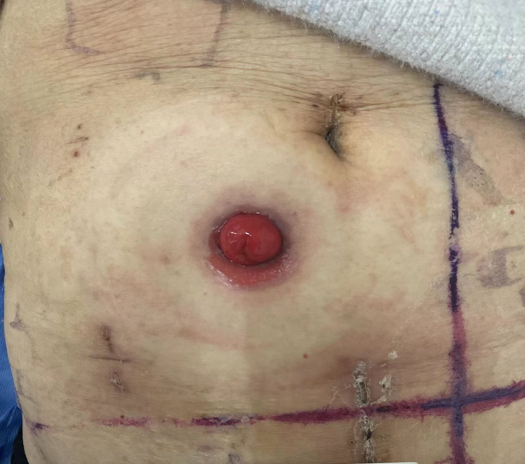
Peristomal moisture-associated skin damage, female, 71 years old, ileostomy status, received radiotherapy three times.

Peristomal moisture-associated skin damage, female, 80 years old, ileostomy status, received chemotherapy for 1.5 months.
Incidence
To date, the incidence of P-MASD in patients undergoing chemoradiotherapy has not been reported. However, multiple studies have reported an increased risk of P-MASD in such patients. Zhu et al. 8 demonstrated that undergoing radiation therapy or chemotherapy (odds ratio [OR] = 7.281; 95% confidence interval [CI] = 3.309–16.024) was an independent risk factor for the onset of P-MASD.
Risk factors
Patients with ileostomy who undergo radiation therapy are at a higher risk of developing P-MASD. Nagano et al. 9 showed that 90.4% of temporary stomas (n = 52) were ileostomies (n = 47), and multiple linear regression analysis showed that patients who underwent ileostomy surgery were three times more likely to develop P-MASD than those with a colostomy (OR = 3.782; 95% CI = 1.34–10.64; p = 0.012). The analysis also revealed that patients who received postoperative chemotherapy were more than 2.5 times more likely to develop P-MASD than those who did not (OR = 2.702; 95% CI = 1.02–7.18; p = 0.046).
Moreover, chemoradiotherapy-associated diarrhea significantly contributes to P-MASD. This condition is a common complication in patients with stomas who undergo cytotoxic drug treatment and pelvic abdominal radiation therapy, which then cause painless diarrhea or mild abdominal pain with watery stools. Chemoradiotherapy may result in increased excretion in patients with stomas, leading to fecal leakage around the stoma baseplate and subsequent P-MASD. Chemoradiotherapy-associated diarrhea may also result in weakness, electrolyte imbalances, kidney failure, hypovolemia, shock, and life-threatening conditions, such as neutropenia and infections. When the diarrhea occurs in conjunction with neutropenia and infections, it may delay treatment cycles, increase the length of hospital stay, increase hospital costs, exacerbate the psychological burden on patients, reduce treatment compliance, or even cause treatment discontinuation, thereby delaying the entire treatment plan. 10 Diarrhea caused by intravenous chemotherapy drugs, especially those that are highly diarrheagenic, such as 5-fluorouracil, irinotecan, methotrexate, and oxaliplatin, is of high clinical concern. According to previous randomized clinical trials, the incidence of grade 3–4 diarrhea ranges from 5% to 47% during different chemotherapy protocols.11,12
Peristomal acneiform rash
Molecular targeted therapy represents a breakthrough and revolutionary development in cancer treatment by prolonging patients’ lives and improving their quality of life. Epidermal growth factor receptor (EGFR) inhibitors are currently widely used. 13 For example, cetuximab (trade name: Erbitux)—the first approved molecular targeted therapy drug—has shown favorable outcomes in the treatment of metastatic colorectal cancer. 14 Combination treatment with cetuximab and irinotecan is approved in the United States and Europe for the treatment of patients with metastatic colorectal cancer who express EGFRs after failing irinotecan-based cytotoxic therapy. 15 However, EGFR inhibitors can cause toxic reactions in the skin, and there are reports of peristomal acneiform rashes induced by EGFR inhibitors in patients undergoing chemotherapy. Papulopustular rash is the most common cutaneous adverse event in patients undergoing EGFR therapy and occurs in 50–100% of patients. 16 In relation to a single drug, 24–62%, 49–67%, and 75–91% of patients experience this rash after receiving gefitinib, erlotinib, and cetuximab, respectively. 17
Definition and clinical manifestations
Peristomal acneiform rash refers to acne-like lesions around the stoma. 18 Clinically, it manifests as follicular papules and pustules, often initially appearing in areas rich in sebaceous glands. The associated symptoms include itching, stinging, and pain.
Incidence
According to a study conducted outside of China, 19 up to 90% of patients with cancer receiving EGFR inhibitor treatment experience adverse skin reactions. However, specific reports on the incidence of peristomal acneiform rash are lacking. In China, Xu et al. 18 reported six cases of peristomal acneiform rash occurring after the use of cetuximab (an EGFR inhibitor). In all six cases, the appearance of an acneiform rash around the stoma made it difficult to attach the stoma baseplate, shortened its usage time, and affected the patient’s quality of life. Overall, only scattered case studies reporting peristomal acneiform rashes are available.
Risk factors
The most common skin toxicity caused by EGFR inhibitors is acneiform rash, which presents as a follicular papular rash. 19 During the first few weeks of treatment, it typically occurs on the head, back, and upper chest. When it occurs around the peristomal skin, it is referred to as a peristomal acneiform rash. 18 The lesions disappear after discontinuing the EGFR inhibitor and do not cause any sequelae. After 1–2 months of treatment, patients commonly experience sebum accumulation, epidermal atrophy, pruritic eczema, dry skin, paronychia, and hair changes (such as changes to the scalp hair and eyelashes). 20 In addition, skin toxicity caused by EGFR inhibitors is often accompanied by severe pain and intense itching, leading to physical discomfort and psychological stress. 21
Peristomal PG
Definition
The presence of necrotic skin lesions around the stoma is referred to as peristomal PG (PPG). 22
Diagnosis
PPG is diagnosed based on the clinical features, wound characteristics, and collective clinical experience. To make a diagnosis, at least two major and two minor criteria need to be met. The major criteria include the following: (1) the presence of rapidly progressing painful wounds, necrotic skin ulcers, and ulcerated surfaces forming proud flesh with irregular violaceous borders and subcutaneous extension; and (2) the exclusion of other causes of skin ulcers. The minor criteria are as follows: (1) the presence of cribriform scars on clinical examination; (2) the presence of systemic diseases associated with PG; (3) sterile dermal neutrophilia on histological examination; and (4) rapid relief of symptoms with glucocorticoid treatment. 23
Clinical manifestations and features
PPG predominantly occurs at ileostomy sites but may occasionally appear in cases of colostomy and urostomy. It exhibits a disease duration ranging from 2 months to 25 years, showing significant individual variations. 24 PPG can develop at any time after ostomy surgery, 23 and the course of PPG is long, often exceeding 1 month. Cases typically occur in patients between 20 and 50 years of age, although reports of PPG have been documented in patients younger than 18 and older than 50 years.25,26 PPG can occur at any site covered by an ostomy baseplate and most ulcers have a maximum diameter of less than 3 cm, although there are reports of ulcers with a maximum diameter of up to 30 cm.25–27 PPG can manifest as deep nodules, hemorrhagic pustules, or small wounds that gradually expand into the subcutaneous tissue, forming a subcutaneous and suppurative ulcer (Fig. 5). There can be one or many ulcers, and skin bridges may appear above the wound base. 28 The ulcers are often very painful 26 and can rapidly expand by 1–2 cm per day; this highlights the progressive nature of PPG. 29 After healing, the ulcers commonly leave reticular atrophic scars that are prone to recurrence when under stress or in the event of new trauma. 23
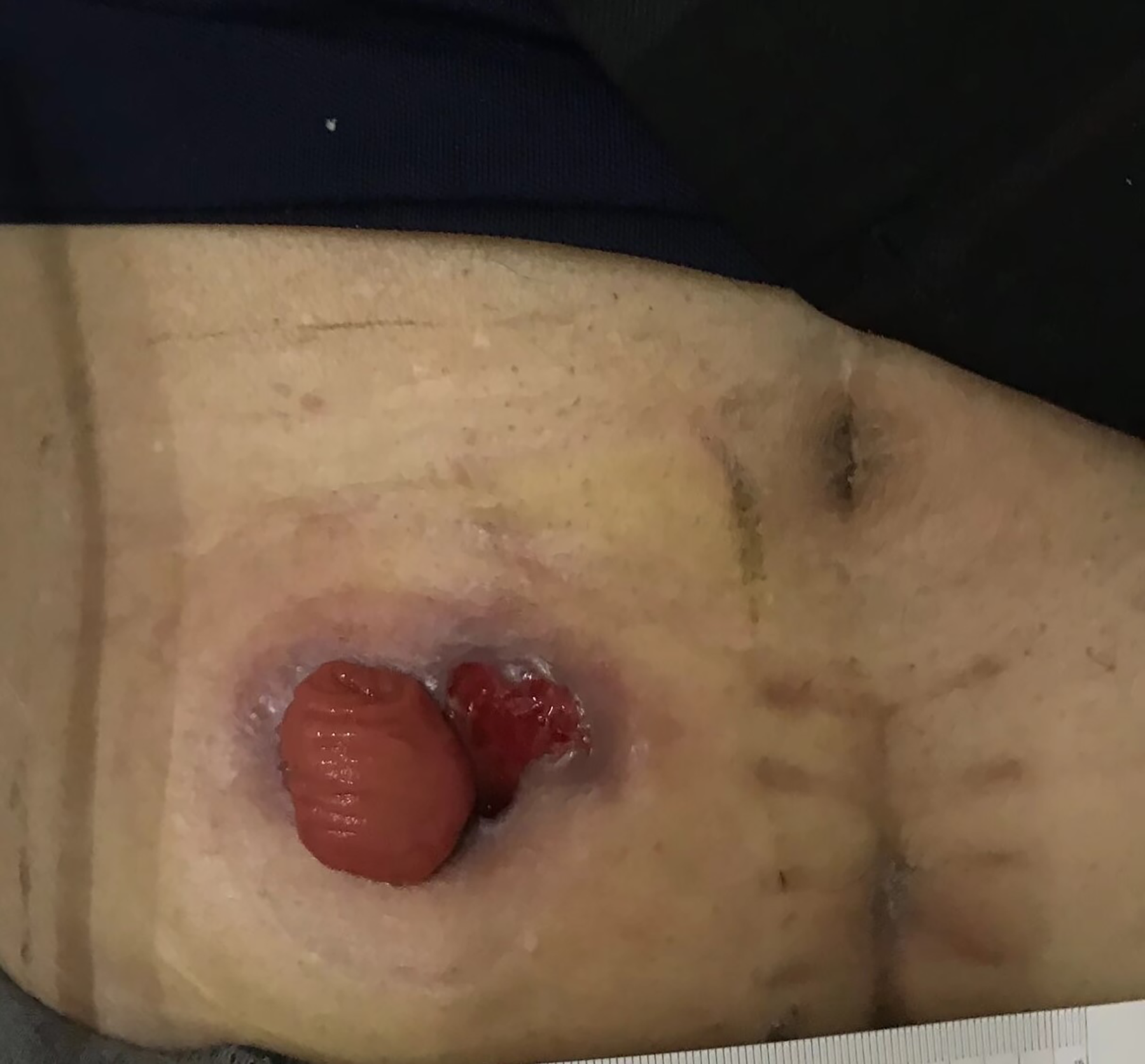
Peristomal pyoderma gangrenosum, male, 58 years old, ileostomy status, received target drugs for 6 weeks.
Uchino et al. 30 described two types of PPG. The R type is characterized by the sudden onset and rapid spread of ulcers. It usually occurs within the first 6 days after surgery and is strongly associated with ulcerative colitis. In contrast, the G type on average occurs 52 days after ostomy surgery, spreads more slowly, and is closely associated with Crohn’s disease.
Incidence
PPG accounts for 15% of all cases of PG. 31 PPG was first identified in three patients with Crohn’s disease in 1984; however, the exact cause of the disease remains unclear. 32 Studies have shown 33 that PPG is relatively rare among peristomal skin issues (0.3%), with approximately 80% of PPG cases occurring in patients with inflammatory bowel disease and a smaller percentage occurring in patients with diverticular disease, abdominal malignancies, and neurological disorders. 34 The incidence of PPG in patients with Crohn’s disease who have undergone ostomy surgery ranges from 2.0% to 4.3%. 23 It is relatively rare in patients who have undergone ostomy surgery after rectal cancer surgery; sporadic case reports have been published although its incidence remains unclear. Carlos et al. 35 reported the case of a female patient who received prophylactic ileostomy after undergoing neoadjuvant chemoradiotherapy and low anterior resection of the rectum. Moreover, a review article 32 mentioned four suspected cases of PPG that were not diagnosed owing to confusion with other peristomal skin issues or because they were deemed unsuitable for publication. Although PPG is rare in patients with stomas undergoing chemoradiotherapy, it is a significant source of distress in these patients when it does occur. Therefore, PPG was included in this review as a type of peristomal skin damage in patients undergoing chemoradiotherapy.
Risk factors
The etiology and risk factors for PPG remain unclear. Histological biopsy typically reveals a high density of polymorphonuclear cells in the superficial epidermis. Elevated levels of proinflammatory mediators, such as interleukin-2, tumor necrosis factor-α, interleukin-6, and interleukin-8, are believed to recruit polymorphonuclear cells to the area, leading to papules, pustules, and eventually peristomal skin damage. 33 During tumor chemoradiotherapy, certain executioner proteins in the apoptotic pathways are activated because of tumor cell death, leading to the release of many inflammatory mediators from dying tumor cells. 34 Therefore, patients with an ostomy who undergo chemoradiotherapy may have an increased susceptibility to PPG. However, further research is needed to gain deeper insight into the underlying biological mechanisms.
Regardless of the underlying cause, inadequate or delayed treatment is the main reason for seeking medical care among patients experiencing PPG. Fecal stimulation is a potential contributing factor to the occurrence and reoccurrence of PPG. 35 Therefore, it is important that patients undergo comprehensive assessments and prioritize stoma care to prevent stoma bag leakage.
Peristomal infections, abscesses, and fistulas
Definition
Peristomal infections, abscesses, and fistulas are partially related; however, there are some differences. Peristomal infection is often the result of local folliculitis or recurrent inflammatory bowel disease in the clinical setting. A peristomal abscess is a collection of purulent material beneath the skin. 36 After an abscess has been drained, the subsequent development of a fistula is not uncommon. Peristomal fistula becomes evident upon the issuance of enteric contents from the exposed abscess cavity with subsequent skin excoriation.
There is no standard way of categorizing peristomal infection/abscess/fistula severity.
Diagnosis
A skin scraping and bacteria culture can be performed for diagnostic confirmation of infection. Peristomal fungal infection is most commonly due to Candida albicans and presents as itchy, bright erythema with satellite lesions in the setting of an advancing border. Bacterial infections originate from bacterial folliculitis, which is most commonly due to Staphylococcus aureus and characterized by erythematous, dome-shaped papules and pustules surrounding hair follicles. 37
Clinical manifestations and features
Peristomal infection/abscess/fistula is a less prevalent but challenging complication. 38 Peristomal infection/abscess/fistula in the immediate postoperative period is most commonly seen in the setting of stoma revision or reconstruction of a stoma at the same site, mainly owing to preoperative colonization of the peristomal skin and perioperative seeding of the surgical site. They may also be seen because of an infected hematoma or an infected suture granuloma.39,40 (Figs. 6–8)

Peristomal abscess/infection/fistula, male, 68 years old, colostomy status, received chemotherapy.

Peristomal abscess/infection/fistula, male, 68 years old, ileostomy status, received chemotherapy.

Peristomal abscess/infection/fistula, female, 64 years old, colostomy status, received chemotherapy.
Incidence
In the early postoperative period, peristomal infections and abscesses are relatively uncommon; the incidence may differ between countries, with a reported incidence of 0% to 14.8%.38,39 At present, there are few studies on the incidence of peristomal infection/abscess/fistula in the Chinese population during chemoradiotherapy. However, although peristomal infections are rare, they can become extremely problematic when they do occur.
Risk factors
In a cross-sectional study 41 of 232 Italian patients with a stoma, class 2 obesity and atopic dermatitis were associated with an increased risk of peristomal infections. Moreover, frequent physical activity and nonautonomy were found to be common risk factors for infections. In contrast, the only protective factor identified was the use of a one-piece device. However, the use of a one-piece device was found to be a risk factor for dermatitis. Once ileostomy effluent causes peristomal skin breakdown, skin ulceration and infection can occur, resulting in peristomal infection/abscess/fistula. 42
Peristomal infection/abscess/fistula is a common early complication of stoma creation. However, epidemiological studies on peristomal infection/abscess/fistula during or after chemoradiotherapy are rare. According to the current literature review, during the early postoperative period, peristomal infection/abscess/fistula is associated with preoperative localization, mainly because of preoperative colonization of the peristomal skin and perioperative seeding of the surgical site. 41 In addition, peristomal fistulas are attributed to many causes, including inflammatory bowel disease, PG, infection, and trauma. Early signs of fistula formation include pain and difficulty with appliance placement.
Limitations
This review has several limitations. First, we included a limited number of articles on the topic. Second, we did not apply a systematic review or meta-analysis approach, and additional studies are needed to confirm the prevalent types of peristomal skin damage during chemoradiotherapy and their risk factors. Third, unpublished articles were not included. Fourth, we were unable to obtain detailed photographs due to time constraints. There are no published examples that we could use in this article with permission. In the future, we will collect photographs of the different types of peristomal skin damage and describe the specific features in more detail. Fifth, we were unable to discuss the pathophysiology of peristomal skin damage in this article due to our professional limitations. However, we will consult the relevant experts or the literature to gain more knowledge on this aspect and improve our future work. Sixth, for some types of peristomal skin damage, the number of studies was still relatively small, and it was difficult to obtain more data on the risk factors. This scarcity of research means that our understanding of certain aspects of peristomal skin damage remains incomplete. We will continue to follow up and explore this area to gather more information over time. Lastly, we believe that it is valuable to discuss the appropriate wound care products, dressing techniques, preventive measures for complications, and the importance of patient education. However, this review only focuses on the prevalent types of peristomal skin damage and the associated risk factors. In the future, we will review the latest research findings and clinical guidelines to make appropriate recommendations for wound management.
FUTURE DIRECTIONS
Conducting cross-sectional studies to gain deeper insights into peristomal skin damage during chemoradiotherapy to enrich the research data
To date, only a limited number of cross-sectional studies have examined the incidence of peristomal skin damage during chemoradiotherapy In the future, cross-sectional research studies with large sample sizes should be conducted to identify the prevalent types of peristomal skin damage and their influencing factors (e.g., types of chemotherapy drugs, the interval time between radiation treatments, the total treatment time of chemotherapy or radiotherapy, and the self-care/family-care ability).
Early identification of the types of peristomal skin damage during chemoradiotherapy and the implementation of preventive care measures and standardized care pathways
In the future, studies should aim to develop preventive and management protocols for the specific types of peristomal skin damage that occur during chemoradiotherapy. Moreover, preventive stoma care measures and standardized care pathways should be developed.
SUMMARY
Overall, the five prevalent types of peristomal skin damage that occur during chemoradiotherapy are peristomal radiodermatitis, P-MASD, peristomal acneiform rash, PPG, and peristomal infection/abscess/fistula (Table 1). The risk factors vary depending on the type but may include the radiation dose, ileostomy surgery, chemoradiotherapy-associated diarrhea, EGFR inhibitor use, and inflammatory bowel disease. This review aids in the clinical identification of peristomal skin damage during chemoradiotherapy, thereby laying a solid foundation for developing effective strategies to prevent these types of skin damage.
Summary of the prevalent types of peristomal skin damage and their risk factors

TAKE-HOME MESSAGES
It is important to distinguish the types of peristomal skin damage that occur during chemoradiotherapy to avoid misdiagnosis and improve treatment or management. In this article, we categorized the different types of skin damage to aid in quick identification, prevention, and management. The five types of skin damage include peristomal radiodermatitis, P-MASD, peristomal acneiform rash, PPG, and peristomal infection/abscess/fistula. Future studies are needed to develop appropriate prevention, treatment, and management guidelines for the different types of skin damage.
AUTHORS’ CONTRIBUTIONS
Y.L. contributed to the conception of the study. L.Z. contributed to data collection and article preparation. Y.C. and L.J.L. performed the data analysis and wrote the article. All the authors have approved the article.
Footnotes

FUNDING INFORMATION
This study was supported by the Soft Research Project of the Lianyungang Science and Technology Association (grant number:
AUTHOR DISCLOSURE AND GHOSTWRITING
All authors of this article directly participated in the planning, execution, or analysis. The contents of this article have not been copyrighted or published previously. They declare no conflict of interest.