




Abstract
Significance:
Alginate, sourced from seaweed, holds significant importance in industrial and biomedical domains due to its versatile properties. Its chemical composition, primarily comprising β-D-mannuronic acid and α-L-guluronic acid, governs its physical and biological attributes. This polysaccharide, extracted from brown algae and bacteria, offers diverse compositions impacting key factors such as molecular weight, flexibility, solubility, and stability.
Recent Advances:
Commercial extraction methods yield soluble sodium alginate essential for various biomedical applications. Extraction processes involve chemical treatments converting insoluble alginic acid salts into soluble forms. While biosynthesis pathways in bacteria and algae share similarities, differences in enzyme utilization and product characteristics are noted.
Critical Issues:
Despite its widespread applicability, challenges persist regarding alginate’s stability, biodegradability, and bioactivity. Further understanding of its interactions in complex biological environments and the optimization of extraction and synthesis processes are imperative. Additionally, concerns regarding immune responses to alginate-based implants necessitate thorough investigation.
Future Directions:
Future research endeavors aim to enhance alginate’s stability and bioactivity, facilitating its broader utilization in regenerative medicine and therapeutic interventions. Novel approaches focusing on tailored hydrogel formations, advanced drug delivery systems, and optimized cellular encapsulation techniques hold promise. Continued exploration of alginate’s potential in tissue engineering and wound healing, alongside efforts to address critical issues, will drive advancements in biomedical applications.

INTRODUCTION
Alginate was first described in the 19th mid-century and was first used for industrial applications for stabilizing, thickening, and gelling food products. 1 More recently, alginate has been widely applied for pharmaceutical and biotechnological applications such as wound healing, drug delivery, and cell-based treatments. 2 As a linear polysaccharide containing β-D-mannuronic acid (M) and C-5 epimer α-L-guluronic acid (G) residues, alginate is water soluble with a negative charge polymer that allows for its polymerization. 3 Considered to be a natural substance, alginate is also commonly extracted from either oceanic seaweed or particular types of bacterial species. It is natural biocompatibility, and as such, has contributed to a wide variety of biomedical and clinical applications. 4
ALGINATE CHEMICAL COMPOSITION AND PHYSICAL PROPERTIES
Composition of alginate
Alginate is a linear anionic carbohydrate polymer that can be isolated from various species of brown algae as well as bacteria. 5 The alginate polymer is composed of units of uronic acids, specifically B-D-mannuronic acid (M) and a-L-guluronic acid (G). These are organized into homopolymeric blocks of either M or G units (MM blocks and GG blocks, respectively), or heteropolymeric blocks of M and G units (MG and GM blocks). The M unit exists in a 4C1 chair conformation and the G unit exists in a 1C4 chair conformation. These organic conformations influence the polymer structures they create when bound to other units within the alginate composite. MM blocks contain linkages in diequatorial positions, which result in flat sheet-like structures. On the other hand, GG blocks contain linkages in diaxial positions, resulting in folded structures. Lastly, MG blocks contain equatorial/axial linkages and GM blocks contain axial/equatorial linkages, which produce unique helical structures (Fig. 1). 6

Schematic of alginate showing the molecular structure of the uronic units and configuration of the uronic blocks, b-D-mannuronic and a-L-guluronic acid, that makeup alginate polymers. The physical and chemical properties are indicated for each block configuration (GG, MM, MG, and GM blocks).
Alginate polymer composition varies depending on the source organism. Varied composition patterns confer differing physical and chemical properties. Prior reports have thoroughly characterized alginate M:G ratios as well as their composition of monomers (M and G), diads (MM, GG, and MG), and triads (MMM, GGG, MMG, MGG, GMM, GGM, GMG, and MGM). 5,7 As demonstrated with proton and carbon nuclear magnetic resonance analyses of brown algae, there is a wide variation in the fraction of G units ranging from 0.2 to 0.75. Alginates with higher G unit fractions are derived from bacterial such as Laminaria hyperborea, while alginates with lower G unit fractions are observed in Ascophyllum nodosum, Laminaria digitata, and Macrocystis pyrifera.
Chemical and physical properties of alginate
The molecular weight of alginate varies depending on its source organism as well as the method of its extraction. Typically, commercially available alginates have an average molecular weight of ∼200,000, but the molecular weight can range from several hundred thousand to several million Daltons. 5,7 The average molecular weight of alginate can be determined using techniques such as gel permeation chromatography or size-exclusion chromatography. Since alginate is a highly hydrated polymer, typically its molecular weight value is obtained as a dry polymer. 8
Due to variations in molecular conformation, as well as additional steric and electrostatic interactions, the composition of the polymer results in variations in compound flexibility and rigidity, viscosity, and solubility. 9 Heteropolymer blocks of MG or GM blocks confer the most flexibility and least rigid, while GG blocks are the least flexible and most rigid (Fig. 1). 7 Alginate in its natural alginic acid form, and therefore is insoluble in water and organic solvents. On the other hand, commercially available alginate is commonly distributed in the form of sodium alginate, which is water soluble. 10
In addition, alginate polymer stability can be impacted by various factors. For example, in low pH environments, alginate can lead to a reduced rate of polymerization and reduced polymer stability. On the other hand, higher pH environments can increase the rate of alginate polymerization. 11 Studies have demonstrated that alginate typically shows stability in close to acidic environments. Excess acidification of sodium alginate solutions can lead to precipitation of alginic acid, which occurs below the pKa for the G and M units. The pKa value determines how strong or weak an acid is, and molecules with a pKa <1 are considered to have strong acidic properties, which are not preferred for alginate gelation and stability. M units have a pKa of 3.38 and G units have a pKa of 3.65, lending varying solubility in low pH solutions depending on the alginate M:G ratio (Fig. 1). 5 Therefore, environments with a pH >8 can compromise the stability and gelation of alginate. While the relationship between gelation and pH can present unique challenges when alginate is used in biological environments, for the most part, alginate is considered to otherwise be highly biocompatible and suitable for use within normal physiological conditions. In fact, in some situations, pH modulation may enhance the biological properties of alginate and its capacity to facilitate tissue regeneration. For example, a few studies have demonstrated that pH modulation of methacrylated alginate hydrogels that encapsulate fibroblasts can impact deposition and alignment of collagen fibers. A process that mimics the natural tissue environment and is proposed as a method to impact tissue healing and capacity to regenerate. 12
Others have investigated zwitterionically modified alginates as a way to improve biocompatibility when it is implanted in tissue. 12,13 By modifying the alginate polymer chains with zwitterionic groups, alginate implants are found to repel cellular infiltration into the polymer and prevent cellular overgrowth, while still maintaining cell viability and functionality. These findings suggest that zwitterion-modified alginates may offer delivery of biologically active growth factors that promote tissue regeneration and healing, while avoiding tissue overgrowth and potential scarring. 12,14
ALGINATE SOURCING AND SYNTHESIS
Sources of alginate
In brown algae (Phaeophyceae), alginate exists as sodium, calcium, and potassium alginic acid salts and functions as a structural polysaccharide in the cell wall and extracellular matrix of these algae. Alginic acid is also synthesized and utilized in the polysaccharide capsule of some bacteria, including Azobacter vinelandii and certain species of Pseudomonas. 15 During the extraction process this alginic acid, an insoluble salt, is converted to soluble alginate, which is then used in various downstream applications. 16 Not all brown algae species produce sufficient amounts of alginate, and commercially it is most commonly extracted from species such as L. digitata, L. hyperborean, Saccharina japonica (formerly Laminaria japonica), M. pyrifera, A. nodosum, Ecklonia maxima, Lessonia nigrescens, Durvillea antarctica, and species of Sargassum. 15 Although each of these species produces abundant alginate, the alginate composition with respect to the M:G ratio and the MG configuration varies, with each species producing various amounts of homopolymer blocks (MM and GG) and heteropolymer blocks (MG and GM). 17 Alginate composition can also vary within each species based on its geographical location and season of harvest. 15
Bacterial production of alginate was first described in 1966, the first source being Pseudomonas aeruginosa. 18 Additional bacterial sources of alginate have been widely reported, including additional Pseudomonas species (P. mendocina, P. putida, and P. fluorescens) 19 and A. vinelandii. 20 A. vinelandii has been the preferred bacterial source of alginate, due to concerns about the pathogenicity of Pseudomonas as well as its alginate composition properties. The composition of the alginate produced by A. vinelandii is often preferred for its gelling properties, lending it to be more useful in both food and biochemical applications. 21 Although these bacteria are a potential source of alginate, the majority of commercially available alginates continue to be sourced from brown algae. 22
Biosynthesis of alginate
The overall steps in the biosynthesis of alginate by both bacteria and brown algae include: the synthesis of a GDP-mannuronic acid precursor through the conversion and metabolization of carbohydrates, production of the glycan chain, and the modification of the glycan chain. 23 In bacteria, such as P. aeruginosa, a series of biochemical reactions then lead to the production of mannose 6-phosphate by phosphomannose isomerase, which is then polymerized into polymannurnate. 2 The biosynthesis of alginate has been well characterized within the bacterial producers of alginate, such as P. aeruginosa and A. vinelandii. 24,25 On the other hand, this biosynthesis within brown algae is not as well defined, but is thought to contain similar steps and using different enzymes than those observed in bacteria. 26
Investigation of brown algae Fucus gardneri and its production of alginate (or alginic acid) led to exploration of the following synthesis pathway: D-mannose → D-mannose-6-phosphate → D-mannose-1-phosphate → GDP-D-mannose → GDP-D-mannuronate → alginic acid. 26 Michel et al. identified the potential enzymes within the brown algae Ectocarpus siliculosus that support this synthesis pathway. 27 The alginate biosynthesis pathway facilitates the production of the alginate precursor GDP-mannose through a multistep synthesis pathway (Fig. 2). The precursor molecule, GDP-mannose, is converted into GDP-mannuronic acid, which then undergoes polymerization to form a mannuron chain and then finally alginates. Bacterial biosynthesis of alginate occurs in the cytoplasm of species such as P. aeruginosa 28 and A. vinelandii, and utilizes the same multistep biosynthesis pathway. 24,25 The alginate synthesized in bacteria differs from alginate produced by brown algae as it contains O-acetyl groups. 29 While the impact of the acetylation of bacterial alginate has not been fully explored, it has been shown to produce a higher molecular weight product and may impact immunogenicity.
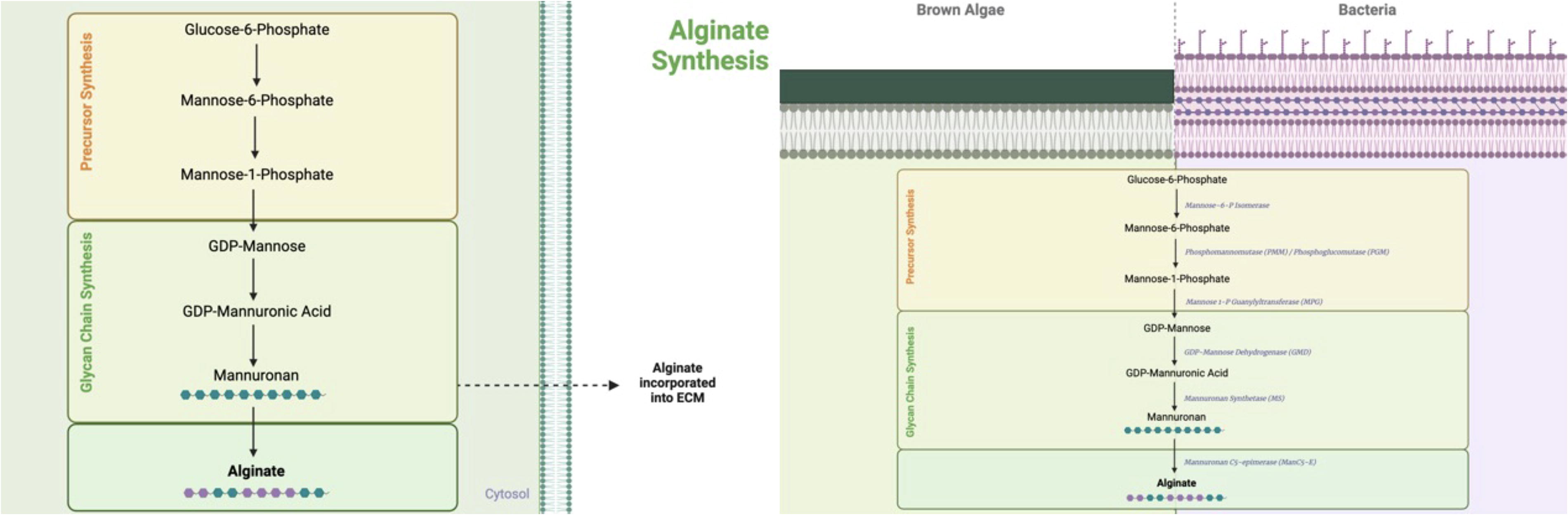
Schematic illustration of the biosynthesis steps of alginate within algae cell cytoplasm and within bacteria. Known bacterial enzymes involved in alginate biosynthesis are indicated.
EXTRACTION AND ISOLATION OF ALGINATE
The commercial extraction of alginate from algae is in the form of sodium alginate through a protocol initially described by Calumpong et al. 31 The protocol converts the insoluble alginic acid salts within the algae into soluble sodium alginate through a series of biochemical steps. These include collecting, processing, and cleaning of the algae, a formaldehyde pretreatment, and an acid pretreatment with hydrochloric acid. This is then followed by an alkali extraction, a neutralization step, alginate precipitation, filtration, and finally drying and milling of the dried alginate components (Fig. 3). 30

Schematic illustration of the extraction, isolation, and purification process of obtaining alginate from brown algae.
In recent decades, L. nigrescens, sourced from Chile and Peru, has been the highest yield of industrial source of alginate. 31 In these industrial settings, harvested algae are cleaned from general impurities, and chemically pretreated with formaldehyde and hydrochloric acid. 32 Resultant insoluble alginic acid salts are then separated through filtration and/or centrifugation, from contaminating components such as other algal polysaccharides. 15 The insoluble residue produced by the pretreatment steps is then treated with an alkaline solution (sodium carbonate, sodium hydroxide, or aluminum hydroxide), which leads to the production of soluble sodium alginate. This product is then neutralized, filtered (through filtration or centrifugation), and precipitated with an alcoholic solution (calcium chloride or HCl precipitation is also used). 32 The product is then dried, milled, and stored as a powder for various applications. 15
PROPERTIES OF ALGINATE
Alginate compound cross-linking
Alginate hydrogels can be cross-linked in diverse ways, resulting in different properties and applications. Three common methods for cross-linking alginate hydrogels are ionic cross-linking, covalent cross-linking, and thermal cross-linking. Ionic cross-linking is the most commonly used method for cross-linking alginate hydrogels. For this method, divalent cations, such as Ca2+, are added to the alginate solution to form ionic bonds between the carboxylic acids and the macromere chain. This creates a physical network of cross-linked alginate chains that give the hydrogel its mechanical properties. 33 Covalent cross-linking involves the formation of covalent bonds between the alginate chains, resulting in a more stable and permanent hydrogel. This can be achieved by introducing reactive groups, such as amine or thiol groups, onto the alginate chains, which can then react with other reactive groups in the presence of a cross-linking agent. 33 Thermal cross-linking, also known as phase transition, involves the use of temperature-sensitive polymers that undergo a reversible phase transition in response to changes in temperature. Thermo-sensitive polymers such as poly (N-isopropylacrylamide) are grafted onto the alginate macromer chains in this method. 34
Gelation of alginate compound
Alginate compounds are amenable to a gelation process. This process involves mixing a solution of alginate with a divalent cation such as calcium or strontium ions. The cations crosslink with the negatively charged carboxyl groups on the alginate molecules, which ultimately polymerizes in the form of a soft gel. The gel can then be formed into different shapes and particle sizes. For example, Saether et al. demonstrated that alginate can be formed into different shapes and sizes using different extrusion method. 35 Others have also demonstrated that the size and shape of alginate particles can be controlled by adjusting factors such as the concentration of the alginate material, the concentration of the supplemented cations, the viscosity of the alginate solution, and the rate of extrusion of the gelling alginate. 36 Examples of structures such as alginate hydrogel pouches, 37 strings, 38 discs, 39 and beads. The different sizes of alginate particles have been used in a range of biomedical applications, including drug delivery, tissue engineering, as well as food ingredients. 40 Additional applications for alginate particles and beads have recently emerged to facilitate the delivery of bioactive pharmaceuticals and cells, in both preclinical and clinical settings. 41 The versatility of alginate to be excluded and formed into different forms allows it to be useful for a variety of purposes.
Cellular encapsulation properties
Prior studies have demonstrated that alginate can facilitate cellular encapsulated implants for the purpose of tissue engineering. Alginate particles can be used to deliver cells for therapy through implantation to various intended sites throughout the body. The particles can be used in the forms of pouches, beads, and/or discs (Fig. 4). Recent use of alginate composites containing stem cells have demonstrated enhanced cellular survival and successful differentiation into various tissue types including bone, cartilage, cardiomyocytes, hepatocytes, and neuronal tissue. 42,43 In addition, bone marrow-derived stem cells, adipose-derived stem cells, myoblasts, and osteoblasts have shown to be differentially regulated. In one study, osteoblast-alginate scaffolds supported osteoblast proliferation and growth. 57 Similarly, alginate gels can be used as a vehicle for growth factor-controlled release and delivery. Growth factors such as vascular endothelial growth factor (VEGF), bone morphogenetic protein-2 (BMP-2), and fibroblast growth factor-2 (FGF-2) have been used in vivo to stimulate angiogenesis, wound healing, and osteogenesis. 44 –46 A recent study demonstrated that hydrogel-mediated delivery of VEGF and BMP-2 was sufficient in inducing human umbilical vascular endothelial cell (HUVEC) proliferation compared to standard basal medium (p < 0.05). 46
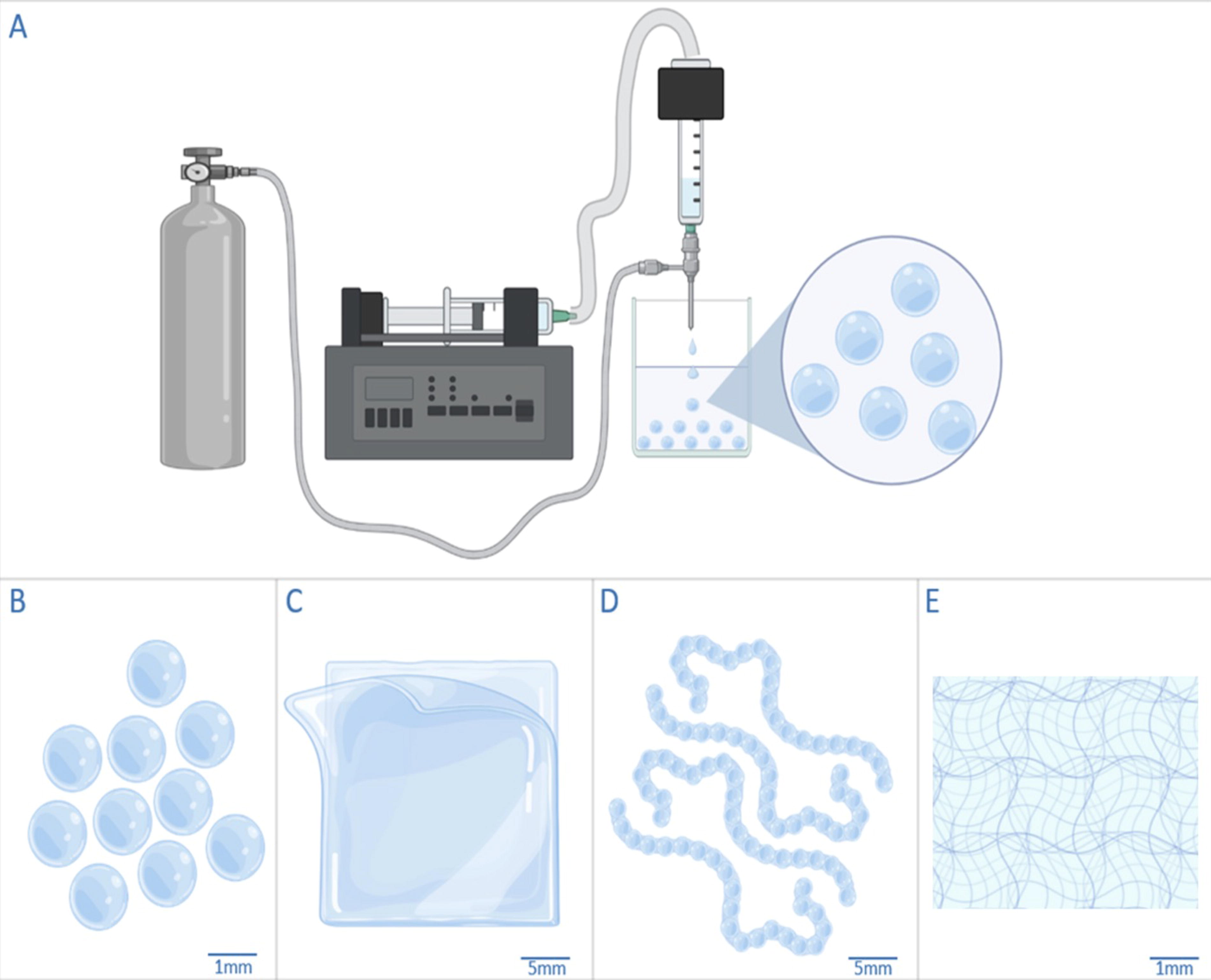
Apparatus for alginate bead formation.
These emerging body of investigation demonstrates that alginate composites can potentially mimic the traditional extracellular matrix and support cell adhesion, proliferation, as well as differentiation. 42 It is anticipated that wider applications of alginate matrix in vivo can help further differentiate its use in different microenvironments and may lead to novel future regenerative therapies.
IN VIVO APPLICATIONS OF ALGINATE
Alginate wound dressing application
A number of studies have investigated the role of alginate as a substrate that can facilitate wound healing. An overview of these studies demonstrates promise across a diverse set of wound conditions, showcasing various types of alginates with distinct applications (Table 1). 47 –52 A common theme is the biocompatible nature of alginate matrixes, which allow it to become suitable for wound applications and help reduce the risk of adverse reactions. One primary studied application is the use of alginate dressings, which are highly absorbent and capable of effectively managing exudate levels in open expressing wounds. 53 When applied to the wound bed, alginate dressings form a gel-like consistency upon hydration that creates a moist environment for fibroblast proliferation to occur within 2–3 days of wound injury. Furthermore, recruited fibroblasts appear to differentiate into myofibroblasts to seal the wound site and to aid in the formation of a new epithelial skin layer. 54 The moisture-retentive environment has also been demonstrated to promote rapid wound re-epithelialization, formation of granulation tissue, and autolytic debridement. During this process the wound bed’s own endogenous enzymes proteolytically digest necrotic debris within the wound and allow it to be removed with dressing changes. This overall process is thought to reduce bacterial overload and accelerate the healing process. 55
Summary of alginate uses for wound care compositions and applications
Alginate dressings are also reported to have inherent hemostatic properties, making them particularly beneficial for managing a bleeding wound bed. The formation of a gel-like composite matrix upon contact with the wound surface helps to promote clotting factors and reduce bleeding. 56 Figure 5 demonstrates the different alginate forms: hydrogel, foam, and film for lymphatic, venous, and arterial wounds, respectively. Small platelet plugs can also form onto the alginate matrix that can facilitate blood coagulation by exchanging the Ca2+ that are present with sodium ions in the wound site. These Ca2+ play a vital role in activating the coagulation cascade thereby accelerating hemostasis. 57 Given the flexible and gel-like nature of alginate, it is also well suited to conform well to the contours of the wound site, providing a comfortable and secure covering that promotes ease of application and comfort with mobility—particularly if the wound site is near an area that experiences dynamic forces with extension and flexion, like a foot ulcer or post operative wound. 58 This conformability can further minimize the risk of maceration, post operative adhesion and trauma to the surrounding skin, and further supports the healing process.
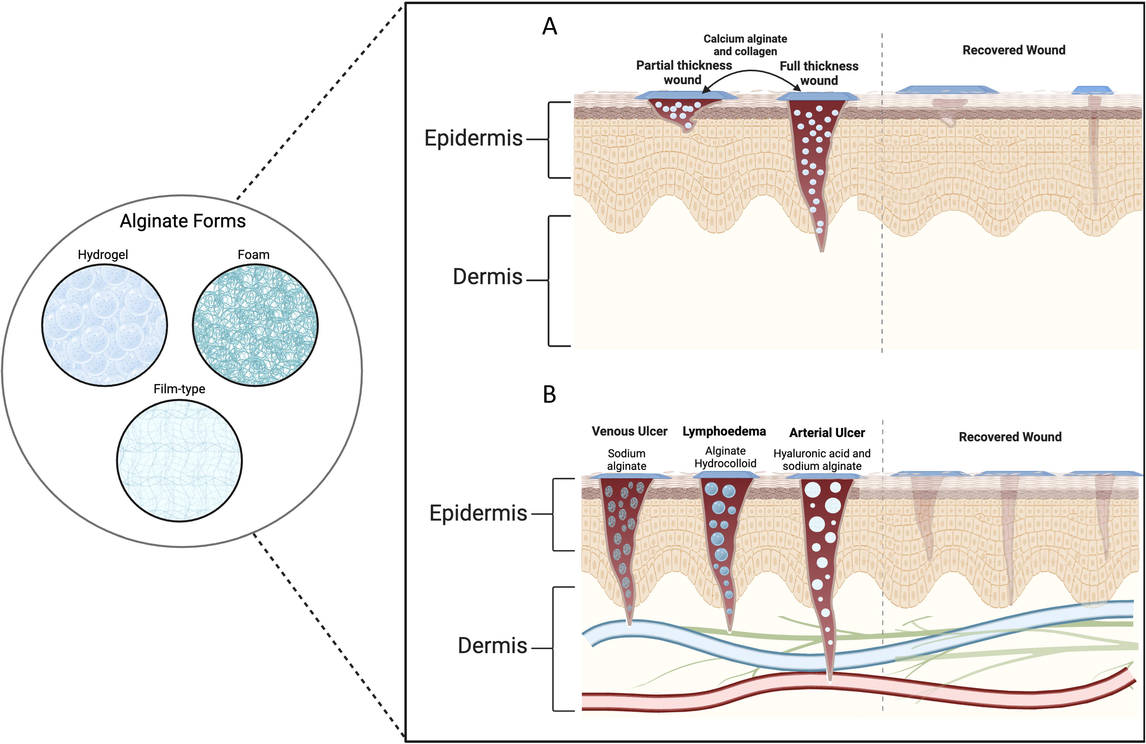
Illustration of a range of alginate formulations designed for application across different wound types: including lymphatic, arterial, and venous ulcers.
The breakdown of alginate in the wound bed into harmless byproducts also reduces the need for frequent wound dressing changes and reduces the risk of wound bed disruption. Biocompatibility also makes alginate dressings suitable for potential use on a wide range of wound types and in various clinical settings, including chronic wounds, surgical wounds, pressure ulcers, and traumatic injuries. 59 Additionally, alginate dressings have been recently reported to possess antimicrobial properties, attributed to the release of Ca2+ during the gelation process on to the wound. These ions create an environment that is hostile to microbial growth and helps prevent bacterial overgrowth and wound super-infection. 59
It is proposed that alginate biocompatibility is in part due to promoting macrophage polarization, which refers to the transition of macrophages from a pro-inflammatory (M1) state to a pro-healing (M2) state. 60 This macrophage transition is particularly important for the foreign body response because M2 macrophages promote tissue repair and remodeling, while M1 macrophages secrete substances that propagate inflammation and lead to further tissue damage. 60 By creating a local wound microenvironment that is conducive for macrophage polarization, alginate appears to not only serve as an anti-inflammatory, but also a promoter of tissue regeneration and stabilization. 60,61
Drug delivery & cellular implantation
Multiple reports show that alginate polymers can be used for local or systemic delivery of pharmacological agents in vivo. Alginate implant formulations have been shown to successfully deliver antibiotics, chemotherapeutics, and immunosuppressants (Table 2). 34,62 –68 Similarly, a few reports have evaluated the potential role of alginate implants to deliver tissue regenerative growth factors and even cell clusters that can promote wound healing. 49
Summary of selected drugs for different animal species, their indications, and duration of treatments
It is commonly understood that growth factors play a pivotal role in the various stages of wound healing, including regulation of tissue inflammation, cellular proliferation, and tissue remodeling. Accordingly, a number of studies have explored whether alginate can be used as a vehicle for controlled release administration of growth factors into a wound bed with sufficient bioavailability. 62 One study demonstrated that an alginate microcapsule matrix can facilitate extended and sustained release of growth factors within a local wound site. 69
Cellular implantation using alginate for wound healing also offers a potentially promising avenue in regenerative wound healing. Since alginate encapsulation provides a semipermeable barrier, it can theoretically protect an implanted cell from a host immune response, while still allowing the exchange of essential nutrients, growth factors, and signaling molecules. 40 It has been demonstrated that alginate can be used to successfully encapsulate various cell types including mesenchymal stem cells (MSCs), fibroblasts, keratinocytes, and endothelial progenitor cells. 59 In one study, the viability of encapsulated MSCs was maintained up to 93% (p < 0.05). While there was a transient nonsignificant decline in metabolic activity after 24 h following encapsulation, the cells appeared to make a full recovery after 7 days. 43 A number of studies have demonstrated that alginate-encapsulated cell clusters can facilitate the delivery of trophic growth factor, stimulate angiogenesis, and tissue regeneration. 46 –48 In one study that evaluated alginate microencapsulation of human (MSCs), it was observed that implants were able to significantly enhance wound bed angiogenesis compared to controls that were not treated with human MSCs.14,70 Despite these encouraging results, additional studies are needed to evaluate the allogenic and host immune response profiles associated with alginate cellular implants particularly in vulnerable tissue sites such as open recalcitrant wounds.
It is proposed that the impact of cellular implants is multifactorial. In some instances, embedded cells appear to modulate the host local immune response in the wound bed. 44 However, in other circumstances the implants appear to impact extracellular matrix deposition, angiogenesis, and efficiency of tissue remodeling. 45 Recent findings have suggested that orchestrated functions between pluripotent stem cells, capillary endothelial cells, and growth factors (such as platelet-derived growth factor) play vital roles in the various stages of wound healing and dermal regeneration.71 Additional studies focusing on determining what cellular implant types are needed to optimally facilitate wound healing at different stages. Rather than having a one size fits all, a menu of different regenerative cells that can be deployed at different stages of wound healing can provide significant promise to future clinical translation and application.
CONCLUSION
Alginate is a highly unique biopolymer that is derived from seaweed and specific types of bacteria. It has proven to be versatile in the field of biomedicine due to its ability to form various structures, including hydrogels, microspheres, nanofibers, and scaffolds to support a variety of biological applications. Alginate matrix structures can be tailored to have specific physical and chemical properties to support targeted drug delivery and cellular implants that have the potential to improve and accelerate wound healing. Although, alginate-based technologies have a variety of important opportunities for clinical application, additional studies are needed to further explore methods of enhanced alginate stability and biodegradability, while also improving its bioactivity and clinical efficacy.
Footnotes
ACKNOWLEDGMENTS AND FUNDING SOURCES
This work was facilitated by a generous grant from the
AUTHOR DISCLOSURE AND GHOSTWRITING
The authors declare no conflicts of interest or financial disclosures. The authors declare there is no ghostwriting to disclose.
AUTHORS’ CONTRIBUTIONS
Conceptualization: L.A., D.A., S.M., S.R., M.A.Z. Funding acquisition: T.A., M.R., Y.L., M.A.Z. Project administration: M.A.Z. Resources: T.A., M.A.Z. Supervision: M.R., M.A.Z. Writing original draft: L.A., D.A., S.M., S.R., M.Z., W.A., B.A. Writing review/editing: L.A., D.A., S.M., S.R., M.Z., W.A., B.A., T.A., M.R., Y.L., M.A.Z.
ABOUT THE AUTHORS