




Abstract
Significance:
The study of extracellular vesicles (EVs), especially exosomes, has unlocked new avenues in understanding cellular communication and potential therapeutic applications.
Recent Advances:
Advancements in EV research have shown significant contributions from the International Society for Extracellular Vesicles (ISEV), in establishing methodological standards. The evolution of the Minimal Information for Studies of Extracellular Vesicles (MISEV) guidelines from 2014 to 2023 reflects enhanced research rigor and reproducibility. The launch of EV-TRACK platform promotes uniformity and reproducibility by providing a centralized repository for data sharing and standardization practices. Furthermore, databases like EVpedia and ExoCarta have facilitated data sharing and collaboration within the scientific community. Concurrently, exosome-based therapies have emerged as a forefront area within regenerative medicine and targeted drug delivery, showcasing the potential of exosomes in promoting tissue regeneration.
Critical Issues:
Despite advancements, the field grapples with challenges such as vesicular heterogeneity, EV isolation complexity, and standardization. These issues impact research reproducibility and clinical applications. The inconsistency in exosomal preparations in clinical trials poses significant challenges to therapeutic efficacy and safety.
Future Directions:
The review outlines critical areas for future research, including the need for technological innovation in EV isolation and characterization, the establishment of standardized protocols, and a deeper understanding of exosome biology. The review also highlights the need to reassess guidelines, develop new EV isolation and characterization technologies, and establish standardized protocols to overcome current limitations. Emphasis is placed on interdisciplinary research and collaboration to address the complexities of EV biology, improve clinical trial design, and ultimately realize exosome’s therapeutic and diagnostic potential. Continued evaluation and rigorous scientific validation are essential for successful exosome integration.

SCOPE AND SIGNIFICANCE
The scope of the review involves research on extracellular vesicles (EVs), highlighting their evolution from initial observations of their existence in blood coagulation processes to their present-day recognition as distinct biological units with functional capabilities. The significance lies in understanding EV biology, particularly its importance in cellular communication, physiological regulation, and potential therapeutic applications. The article emphasizes the importance of methodological precision, scientific inquiry, and standardization in EV research to enhance transparency, address current and emerging challenges, and improve reproducibility. The evolving nature of EV research, particularly the need for ongoing reassessment of guidelines and standardization practices within the field, is underlined as crucial for advancing scientific understanding and applications of EVs.
TRANSLATIONAL RELEVANCE
The translational relevance of this article is deeply anchored in the therapeutic applications of EVs, particularly exosomes. Exosome therapy, identified as a pivotal developmental nexus for clinical implementation, is substantiated by extensive preclinical investigations demonstrating efficacy across various disease outcomes. However, the translation from bench to bedside requires meticulous scientific substantiation to ensure safety, efficacy, and regulatory compliance. The review discusses the burgeoning potential of exosomes in fields such as regenerative medicine, oncology, and neurology and the challenges and questions arising from exosome-based clinical trials because of the complexity of vesicular heterogeneity, which can lead to varied therapeutic outcomes.
CLINICAL RELEVANCE
The increasing number of clinical trials investigating EV emphasizes their significance in biomarker identification and their potential as cell-free therapeutic agents in drug delivery. This article reviews the current state of clinical research, primarily in early phases, focusing on safety, optimal dosing, and preliminary efficacy. It addresses challenges such as the purity and consistency of exosomal preparations, vesicular heterogeneity, and the complexities in resolution, recovery efficiency, scalability, and purity of EV subtype differentiation. In addition, it underscores the need for stringent evaluation processes, standardized methodologies, and detailed reporting protocols to facilitate multicenter studies, enhance scientific knowledge, and ensure safe and effective clinical integration of EV.
Research on EVs as distinct biological units with functional capabilities began to take shape during the 1980s and 1990s, 1 building on earlier studies that pointed out their existence and roles, particularly in blood coagulation processes. 2 The foundational work of Chargaff in the 1940s, who observed accelerated coagulation owing to high-speed sediment, laid the groundwork for this field. 2 Subsequent milestones include Peter Wolf’s identification of “platelet dust” in 1967 3 and Neville Crawford’s characterization of microparticles containing lipids and proteins in 1971. 4 These early explorations and electron microscopy studies revealing vesicle-like structures in various biological contexts have significantly contributed to understanding EV biology, highlighting their importance in cellular communication, physiological regulation, and the potential for therapeutic applications. 5 –8
BACKGROUND EV RESEARCH GUIDELINES
As the field of EV research continues to evolve rapidly, methodological precision, scientific inquiry, and standardization remain a critical challenge. 9 The International Society for Extracellular Vesicles (ISEV), established in 2011, 10 advocates adopting a provisional nomenclature without the precise verification of the origin of the vesicles. The International Society for Extracellular Vesicles is strategically positioned to develop and disseminate consensus on optimal methodologies and pertinent scientific questions within this dynamic field. 11 This dynamic realm of EV research necessitates the ongoing reassessment of ISEV guidelines. To standardize practices within the field, enhance transparency, address current and emerging challenges, and improve reproducibility, the EV-TRACK platform recommends the examination of diverse markers in collective EV samples, as outlined in the Minimal Information for Studies of Extracellular Vesicles (MISEV) guidelines. 12
Evolution of MISEV guidelines from 2014 to the present day
The evolving nature of EV and exosome research underscores the necessity for ongoing reassessment of ISEV guidelines. 13 These guidelines also propose modifications in the vocabulary of EV reporting, initially promulgated in 2014 (MISEV2014) 14 detailing categorizations based on vesicle size, density, biochemical signature (e.g., presence of specific surface proteins), and cellular origin. MISEV2014 was aimed to bolster the rigor of EV research. 14 The International Society for Extracellular Vesicles endorses the EV-TRACK knowledge base as an invaluable, dynamic platform for the comprehensive documentation of EV experimental research. EV-TRACK, designed to facilitate the submission of detailed information on EV isolation and characterization processes, uses a structured online template to collect data. 15 –17 Each submission, associated with a specific research project or publication, is evaluated to produce an “EV-METRIC.” 13 This metric reflects the comprehensiveness of the information provided rather than serving as a score to be maximized. The emphasis is on ensuring that the level of detail supports the transparency and reproducibility of the methods used rather than meeting a predefined standard. 10 This approach recognizes the variability inherent in the requirements of basic versus clinical research. Researchers are encouraged to deposit data on EV profiling into public repositories, including EVpedia, Vesiclepedia (previously known as ExoCarta), and the exRNA Atlas. 18 These platforms serve as vital resources for the scientific community, offering access to a wealth of data on exosome and vesicle proteomics. EVpedia and ExoCarta were among the first databases to provide comprehensive information on EVs and exosomes, with EVpedia showcasing proteome datasets from numerous EV studies. 19,20 ExoCarta features extensive listings of exosomal constituents, such as proteins, lipids, mRNAs, and miRNAs. 20 Vesiclepedia, like EVpedia, hosts a wide array of EV-related entries, covering both protein and lipid components. 21,22 These databases significantly contribute to the field by facilitating the sharing and dissemination of crucial EV data, thereby providing a structured framework to guide research on EV biology and fostering advancements in research methodologies. 23
In 2014, several publications first documented the size distributions of EVs within human plasma and urine, revealing a broad spectrum of diameters ranging from <100 nm to over 1 μm. 10 Such variation underscores the significant impact of isolation methodologies on the observed dimensions and subtypes of EV, highlighting the absence of discrete size demarcations for categorizing “small EV” and “large EV.” 9 Furthermore, the density of EV significantly influences their isolation efficiency, especially in media such as blood plasma and serum, where the low-density contrast with the surrounding microenvironment complicates EV isolation via centrifugation techniques. 24,25 The complexity is further exacerbated in isolating plasma-derived EVs through density gradient centrifugation because of the copresence of non-EV particles such as lipoproteins. 26 Although compositional analyses have provided insights into the aggregate composition of EV populations, critical confounding factors remain unidentified. 27 Figure 1 shows the heterogeneity and biogenesis of EV subtypes and highlight the size and density overlap between exosomes and lipoproteins.

MISEV2018 offered a meticulous examination of separation methodologies, suggesting experimental strategies to surmount persisting obstacles and refine EV characterization. The MISEV2018 guidelines recommend classifying EV and their subsets according to dimensions (small EV <200 nm; medium/large EV >200 nm), density gradients (low, medium, high), biochemical properties (e.g., CD9/CD81/CD63-positive EV), and progenitor cell lineage. 14,22 The MISEV2023 guidelines, while retaining previous position statements, advocate for improved experimental design and meticulous reporting in EV-related research across different matrices such as cell cultures, bodily fluids, and tissue samples. 28 –31 This iteration not only addresses and refines issues from MISEV2018 but also introduces recommendations for new research areas including nomenclature, preprocessing considerations, separation techniques, characterizations, advanced methods, the expanding scope of the field, and concise overviews of in vivo EV studies. 32,33 It also highlights the importance of using a standardized set of markers relevant to either the isolation methodology or the source cell lineage. 14 The MISEV2023 document integrated feedback from ISEV’s expert panels and the broader research community, delineating the current landscape of EV research and assisting practitioners in adopting and refining best practices tailored to each EV source, variant, research focus, or application. 9 A meticulous analysis of the MISEV survey responses reveals the presence of divergent viewpoints among scientists regarding the physiology of EV analysis in vivo, particularly regarding release and uptake kinetics. 9 Table 1 represents the consensus of MISEV2023 survey respondents. For example, regarding EV analysis in vivo, whereas approximately two-thirds of the EV community completely agreed with the MISEV2023 guidelines, one-fifth of the people share the enthusiasm with clear reservations. Figure 2 compiles the timeline illustrating the main discoveries related to exosome-based research and PubMed record of publication in this field as of March 2024.
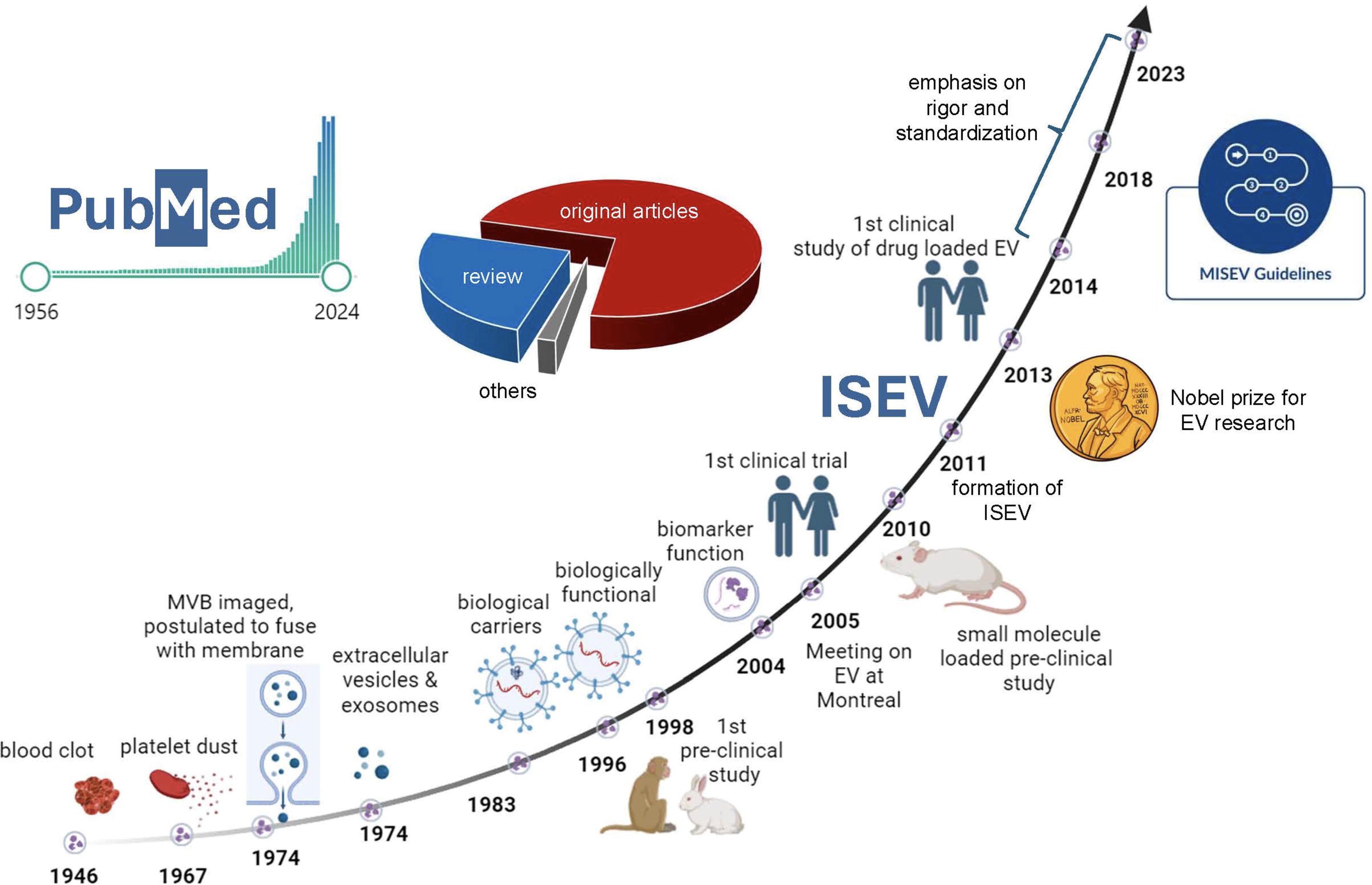
As of March 2024, timeline illustrating main discoveries related to exosome-based research. PubMed shows the number of publications published as original articles, reviews, and others. This figure was created with BioRender.com
The consensus of MISEV2023 survey respondents
The table is generated from the recent publication in Journal of Extracellular Vesicles (JEV). 9
EV, extracellular vesicles; MISEV, Minimal Information for Studies of Extracellular Vesicles.
Building bridges: Unifying guidelines across consortia
The area of extracellular RNA (exRNA) biology was established by the discovery that EV may carry RNAs between cells, indicating an as-yet-unacknowledged function in intercellular communication. 34 Launched in 2013, the Extracellular RNA Communication Consortium (ERCC), an initiative financed by the National Institutes of Health (NIH) Common Fund, aimed to accelerate advancements in this emerging field. 35 The consortium, launched to explore the roles of exRNAs and their carriers, particularly EVs, has focused on understanding the mechanisms of exRNA biogenesis, cargo selection, secretion, and uptake. Key achievements include the identification of exRNA biomarkers, development of exRNA/EV-based therapeutics, and the creation of robust isolation and analysis methods. A collection of ERCC papers, an exRNA-focused blog, and descriptions of ERCC initiatives may all be found on the exRNA Portal and the exRNA Atlas for data sharing and analysis. 36 Recognizing that exRNAs carried by EVs can mediate intercellular communication, the consortium has made strides in elucidating the complex mechanisms underlying exRNA biogenesis and function. 37 Through collaborative efforts, ERCC has laid a solid foundation for future research, addressing fundamental gaps and paving the way for new diagnostic and therapeutic applications, including the development of novel drug delivery systems leveraging the natural properties of EV. 38 Despite these advancements, the ERCC also faced challenges such as the heterogeneity of exRNA carriers and the need for standardized methods remain. Addressing these issues is essential for the clinical translation of exRNA research. 36
Combining the guidelines from the MISEV and the ERCC can significantly enhance the rigor, reproducibility, and utility of EV research. A comprehensive strategy to integrate these guidelines can be categorized into five key areas:
Standardization of experimental procedures
Adopt MISEV-recommended isolation and characterization methods as standard practices, using ERCC-validated protocols where applicable.
Use ERCC-developed computational tools to process and analyze data, ensuring consistency and reproducibility across studies.
Comprehensive EV characterization
Use a combination of physical and molecular characterization techniques as suggested by MISEV, using ERCC resources for more detailed analysis.
Include ERCC’s reference profiles to enhance the understanding of the RNA cargo in EVs, correlating physical characteristics with molecular content.
Data reporting and sharing
Report experimental details following MISEV standards and use ERCC platforms for data sharing.
Process and analyze EV-associated RNA data with ERCC tools, ensuring uniformity in data reporting.
Biomarker discovery and therapeutic applications
Validate potential EV biomarkers identified by ERCC studies using MISEV-recommended validation protocols.
Explore therapeutic applications of EVs, integrating ERCC findings with MISEV guidelines for functional studies and clinical translation.
Collaborative research and resource sharing
Engage in collaborative projects that align with both MISEV and ERCC goals, leveraging shared resources and expertise.
Participate in ERCC-organized events and use their platforms to share findings and resources, fostering a collaborative research environment.
This integrated approach will enhance the reliability of EV research findings, facilitate the discovery of clinically relevant biomarkers, and advance the development of EV-based therapeutics.
EVERYTHING IS NOT EXOSOME
Exosomes, a distinct subset of EV, originate from the endosomal system. 39 They are formed as intraluminal vesicles (ILVs) within multivesicular bodies (MVBs) through a series of maturation events within the endosome. 40 The MVB ultimately merges with the plasma membrane, discharging the ILVs into the extracellular matrix. 41 The secreted ILVs are reclassified as exosomes, highlighting their unique biogenetic pathway, unlike membrane-originated ectosomes. 42 –44 In addition, specialized nomenclatures have been adopted to describe EVs associated with cellular processes, including “migrasomes,” related to cell migration, 45 and “apoptotic bodies,” linked to programmed cell death. 46 The ISEV recommends caution in using biogenesis-based nomenclature for EV subtypes because of the absence of universally accepted molecular markers. The terms “tiny EV” and “exosomes” are not interchangeable 47 ; the former encompasses a diverse population, including small ectosomes and exosomes. “EV mimetics” are described as EV-analogous particles produced through cellular disruption, synthetic generation, or liposome fusion, with nomenclature that should ideally underscore their genesis mechanism, differentiate them from native EVs, and avoid implying specificity to a particular biogenetic pathway. 48 To avoid misunderstanding, phrases like “exosome-like vesicles” that allude to exosomes or biogenetic characteristics should be avoided. Furthermore, MISEV2023 discusses the nomenclature for nonvesicular extracellular particles that are often co-isolated with EVs, highlighting the intricacy and developing knowledge in this area. 9 Examining the literature on nanovesicles revealed notable variations in the separation, characterization, and source of nanovesicles from where they are formed. 9 Most of the examined papers defined the nanovesicles used in wound healing research as exosomes. 49,50 Exosomes serve as a channel for cellular communication and offer insight into the physiological condition of a particular tissue or organ. 49 Exosomes are gaining prominence as pivotal nanosized carriers for biomolecular signals, including functional proteins, metabolites, and nucleic acids, through intricately regulated molecular processes within the in vivo systems biology, orchestrating intercellular communication across various biological scales, from tissues to entire organisms. 51 The surfaces of these nanosized vesicles are often specifically modified to facilitate targeted recognition and uptake by recipient cells, enhancing the specificity and efficiency of intercellular signal transmission. 52 This evolving paradigm posits that exosomes, derived from specific cell types, are meticulously engineered to carry distinct assortments of proteins, lipids, and RNAs, destined for precise recipient cells to induce targeted functional alterations. 53 Furthermore, the horizontal transfer of miRNAs encapsulated within exosomes instrumental in mediating posttranscriptional gene silencing exosomes has emerged as essential mediators in intercellular communications. 54,55 Such intercellular transfers via exosomes have significant diagnostic and therapeutic implications. 56,57 This is notably demonstrated in cutaneous wound healing, where specific miRNAs like miRNA-155, upregulated during the inflammatory response phase, and miRNA-21, known for its anti-inflammatory properties and increased presence in macrophages during inflammation mitigation, play pivotal roles. 57,58 Furthermore, absence of specific miRNA payload such as miR-425-5p has been reported to have detrimental effect in tissue repair, especially in diabetes. miR-425-5p was found to be associated with regulating adiponectin, a protein with insulin-sensitizing properties. 59 According to Prasai et al., 42% of research published up to this point has referenced at least one interesting cargo molecule found within the exosome. 49 This research documented miRNA cargo validates the critical function of miRNA. 49 They have also addressed the concentration of exosomes to be used in the studies for therapeutic outcomes and the mechanism involved. 49 The diagnostic superiority of exosomal miRNAs over their nonexosomal counterparts in disease detection underscores their potential as biomarkers. 60
EXOSOME THERAPY
Exosome therapy is identified as being at a pivotal developmental nexus for clinical implementation, substantiated by an extensive array of preclinical investigations demonstrating its efficacy across a diverse spectrum of disease outcomes. 61 –66 The exosome therapeutic applications segment was the leading market category in 2022, valued at USD 146.7 million. 67 The growth in this segment is fueled by its burgeoning potential in fields such as regenerative medicine, oncology, and neurology, including chronic pain, musculoskeletal disorders, and cardiovascular diseases. 68,69 In addition, the significant impact of exosomes in targeted drug delivery, especially for cancer and neurological disorders, has markedly contributed to the expansion of this market. The exosome market was valued at approximately USD 250.8 million in 2022 and is projected to expand at a compound annual growth rate (CAGR) of 29.9% from 2023 to 2032, reaching an estimated value of USD 3.2 billion. 67 This robust growth is primarily driven by industry-initiated initiatives worldwide such as VesiCURE Therapeutics, Exopharm, RION, The Cell Factory (Esperite), Codiak Biosciences, ILIAS Biologics, and Evox Therapeutics. 70 In 2022, the largest market share belonged to the kits and reagents segment, which accounted for 44.9% of the total market. 67 This dominance is due to the requirement of different isolation and analysis reagents, which are fundamental to developing exosome-based commercial product lines. 68,70
The past half decade has witnessed a substantial upsurge in the quantity of leading clinical trials scrutinizing exosome dynamics, biomarker identification, and the application of these nanovesicles as cell-free therapeutic agents in drug delivery. 71,72 Given their nascent recognition as critical mediators in physiological and pathological processes, these diminutive vesicles are increasingly favored for their therapeutic and diagnostic capabilities. 73 This surge is particularly noted in targeted disease domains such as tissue regeneration, COVID-19, cancer, neurodegeneration, inflammation, and immunology. 68,69
According to the data retrieved from ClinicalTrials.gov, a significant number of ongoing trials are currently in the initial stages of clinical research, such as Phase I or Phase II, as shown in Figure 3. These stages primarily focus on assessing safety, optimal dosing, and preliminary efficacy of treatments. Transitioning from these early-phase trials to widespread clinical application demands extensive time and meticulous scientific substantiation. As of the latest analysis, 345 trials have been registered, distributed across various phases: 11 in early Phase 1, 53 in Phase 1, 57 in Phase 2, 6 in Phase 3, and 3 in Phase 4. Furthermore, 98 trials are categorized under the Food and Drug Administration (FDA)-defined phases marked as “Not Applicable.” Given the early stage of many therapies involving exosomes and the imperative for thorough clinical trials to ascertain their safety and efficacy, it is critical to adopt a rigorous and objectively defined approach. This rigorous evaluation is indispensable to guarantee that such innovative treatments adhere to the stringent standards expected for clinical applications. 64,74
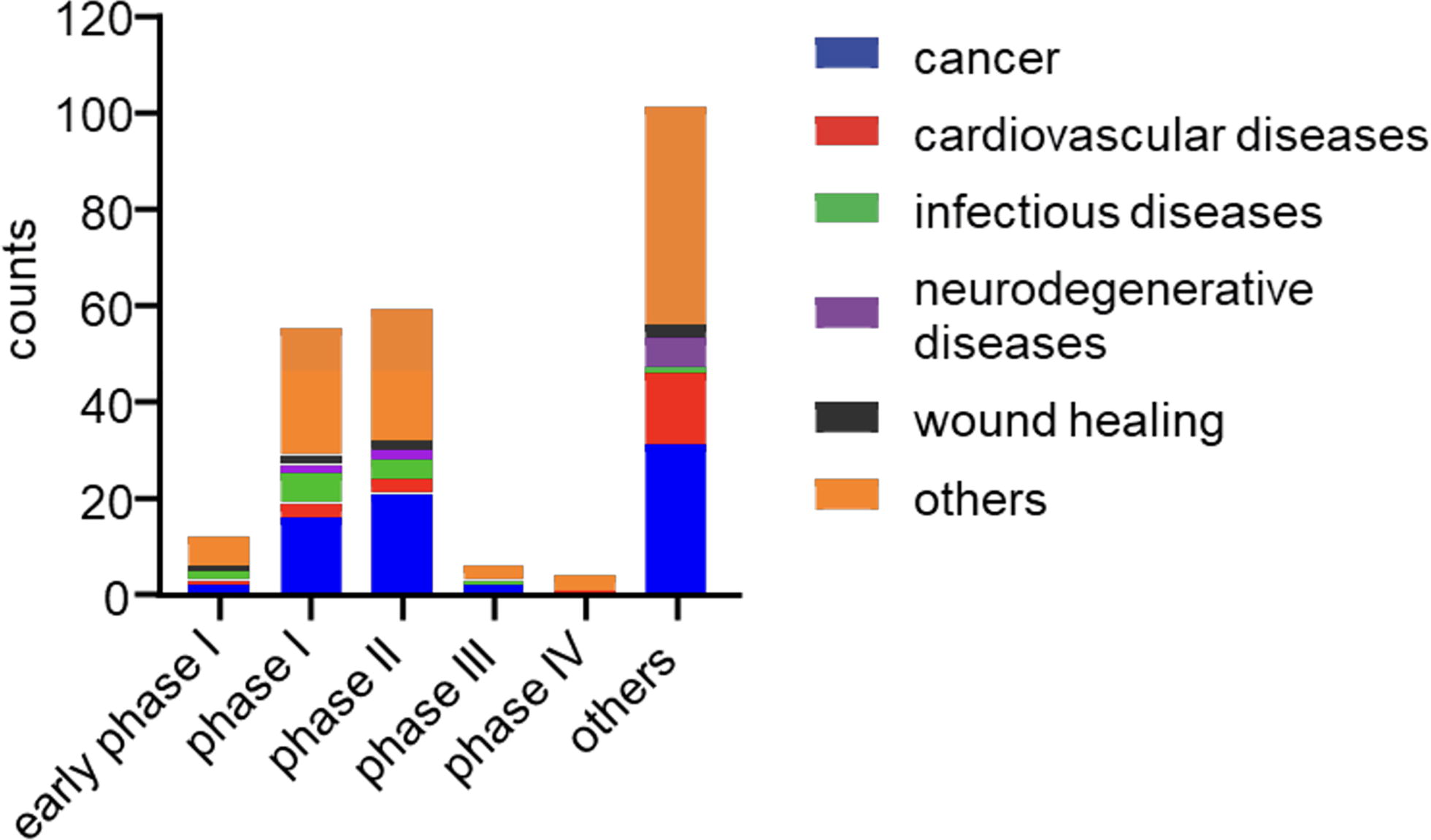
Exosome-related clinical trials based on an analysis of clinical trials listed in clinicaltrials.gov as of March 2024 using the keyword exosomes and diseases as shown in the bar graph. This graph was created with a GraphPad prism.
Clinical trials focusing on so-called exosome-based interventions grapple with multifaceted challenges and questions, mirroring the complexity of this burgeoning research domain. Standardized methods play a pivotal role in clinical applications of exosomes. There is a need for more efficient and reasonable separation technologies. High preliminary preparation and long centrifugation time limit its clinical applications. The purpose of current clinical trials is to demonstrate the feasibility and short-term safety of administering autologous exosomes; however, safety issues for treatments based on exogenous exosome-based products would likely require a more stringent approach. 65 Even though several studies have detailed procedures for producing exosomes in bulk and improvements in biocompatibility, further preclinical and clinical testing are required for verification. Exosome quality and purity standards ought to be more rigorous. To guarantee that the target is receiving an appropriate therapeutic dose of exosomes, it is advised that future research investigate the aforementioned criteria. 65,75 This field has certain hurdles and limits, and additional research is required to validate these findings and establish a gold standard for exosome-based studies. We conducted a search using keywords such as exosomes, biomarkers, and therapeutics and discovered that 17 clinical trials used exosomes as biomarkers and 101 researches used exosomes as treatments. When we explored deeper, we discovered that 25 trials had performed exosome profiling only, 51 trials had administered exosomes in human subjects, and 25 trials did not mention exosomes or any vesicles (Fig. 4). Table 2 represents the clinical trials using the intervention method of delivering exosomes. Several techniques are used to isolate exosomes, but ultracentrifugation is the most common.
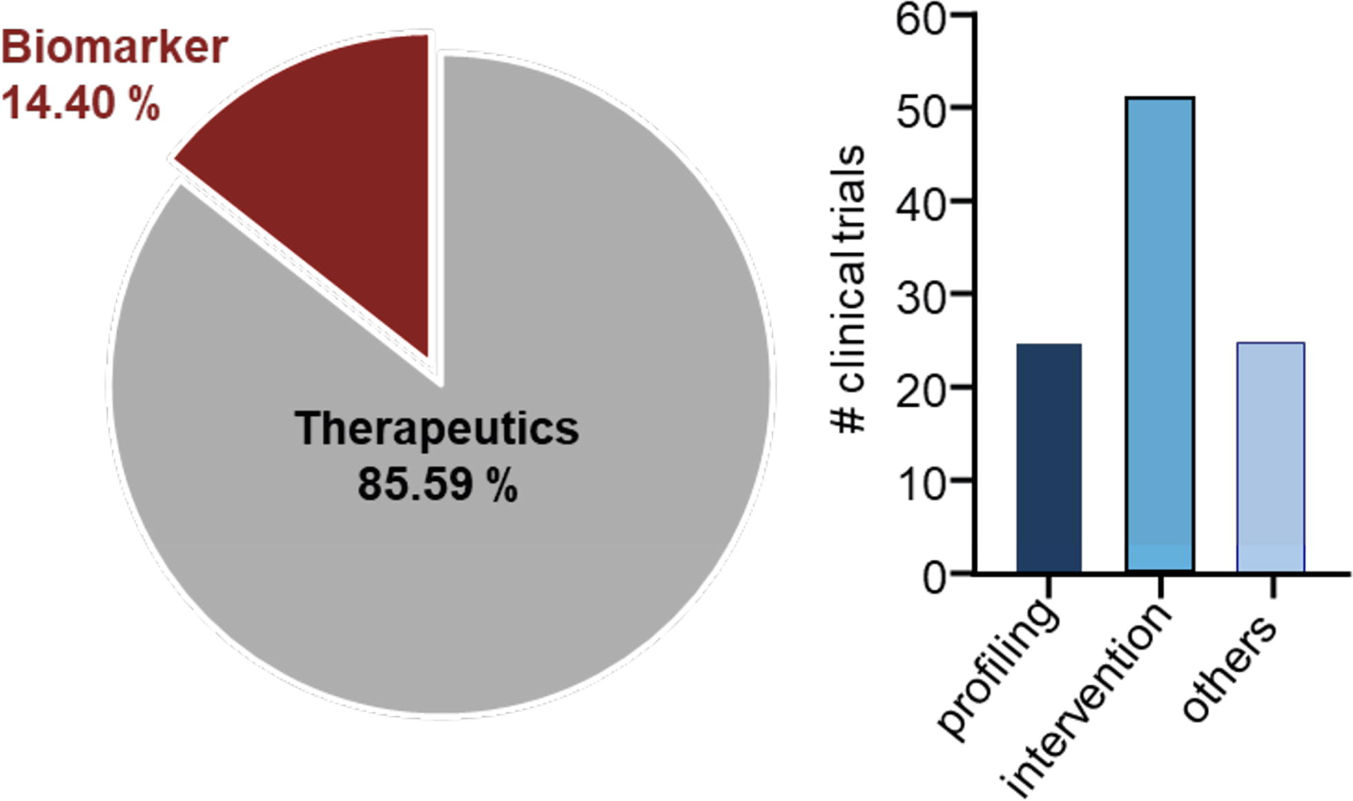
According to a search on clinicaltrials.gov, using keywords such as exosomes, [AND] biomarkers, [AND] therapeutics, as of May 23, 2024, there were 17 and 101 clinical trials on exosomes as biomarkers and exosomes used for therapeutic intervention. Out of these 101 clinical trials, 25 trials had performed exosome profiling only, and 51 trials had administered exosomes in human subjects.
List of clinical trials (from clinicaltrials.gov) of exosomes where exosomes were delivered intravenously, via inhalation, or using some other intervention method
A principal issue pertains to the purity and consistency of the exosomal preparations used in these studies. 76,77 The phenomenon of “vesicular heterogeneity” presents significant obstacles stemming from variations in the vesicles’ sizes, origins, compositions, cargo contents, and surface markers. 43 This heterogeneity can lead to disparate therapeutic outcomes across different clinical trial settings because of the diverse biological activities of the exosomes. 78 In 2022, the understanding of exosomal diversity, particularly highlighting the heterogeneity inherent in exosomes of keratinocyte origin, was further advanced from the work of Brown et al. 79 Although the exosomes were of keratinocyte origin from murine wound edge, Brown et al. could successfully identify and characterize up to 20 distinct subpopulations of keratinocyte-derived exosomes using charge detection mass spectrometry (CDMS). This technique enabled precise measurements of individual exosomal particles’ charge (z) and mass-to-charge ratio (m/z), facilitating a nuanced analysis of their physical properties. The research encompassed the collection of day 5 wound-edge (WE) exosomes of keratinocyte origin from both diabetic mice and their heterozygous nondiabetic littermate controls, revealing a comprehensive dataset of 54,974 single exosome particles across six datasets (split evenly between diabetic and nondiabetic sources). A targeted mass range of 1–200 megadaltons (MDa) was set for the direct analysis of these exosome particles. Using a two-dimensional Gaussian model for statistical modeling, the study achieved partial resolution of ∼10–20 unique exosome subpopulations, distinguished by their mass and charge. Further investigations, incorporating electron microscopy, mass spectrometry-based proteomics, immunofluorescence, and electron microscopy techniques, corroborated significant differences in these keratinocyte-derived exosome subpopulations between diabetic and nondiabetic mice. Notably, subpopulations with high charge (z > 650) and high mass (m > 44 MDa) were identified as markedly different, being more prevalent in nondiabetic specimens—a variation potentially linked to differential exosomal cargo packaging. These findings underscored a shift in the overall mass distribution, with keratinocyte-derived exosomes from nondiabetic animals exhibiting an average mass ∼10% higher than those from diabetic counterparts. They hypothesized that the observed variance in high mass and charge subpopulations among nondiabetic keratinocyte-derived exosomes might have underlying biological implications. A significant aspect of their findings was the lower expression of heterogeneous nuclear ribonucleoprotein Q in diabetic mice, suggesting compromised exosomal packaging efficiency in these exosomes. This work opens new avenues for physically characterizing exosome particles, leveraging mass and charge metrics. This approach holds promise for enhancing our understanding and treatment strategies for wound healing and other disorders by providing a more detailed picture of exosomal heterogeneity and function. 79 In the context of diabetes, Park et al. demonstrated the importance of intercellular communication between immune and stromal cells at the site of injury. By using proteome analysis to design the EV payloads, they have prepared Serpin-loaded EVs to rescue the Serpin deficiency identified by proteomics to augment wound healing in diabetic mice. 80 Moreover, the methods to delineate EV subtypes, including exosomes and ectosomes, through various separation strategies present a nuanced landscape of advantages and challenges regarding resolution, recovery efficiency, scalability, and purity. 81 Despite the inherent biological distinctions between these subtypes, accurately differentiating exosomes from membrane-originated ectosomes poses a significant challenge because of the overlap in their biophysical attributes. 81 This variability may lead to unpredictable and diverse patient responses to the therapeutic interventions, thereby complicating the treatment’s overall efficacy assessment. 82 Predominantly, current separation methodologies prioritize size as a distinguishing factor, inadvertently sidelining the critical aspect of biogenetic pathways. 51,82 Inconsistencies in the composition of exosomal cargo compounds contribute to the difficulty in identifying reliable biomarkers for disease tracking or diagnosis. 51 This oversight and a dearth of markers specific to EV subpopulations complicate the identification and isolation processes. This challenge could obstruct the advancement of dependable diagnostic assays, essential for the clinical application of exosome-based strategies. 83 Technical impediments further exacerbate the issue, notably suboptimal recovery rates and difficulties in precise quantification. These limitations are particularly critical in functional studies, where comparative analyses of diverse vesicular entities are essential. Moreover, the extant techniques for particle quantification exhibit inadequate sensitivity for detecting smaller vesicles, underscoring a gap in the current technological capabilities. 84,85 However, single vesicle flow cytometry effectively examines the EVs at the single-particle level. 86 In addition to determine the size distribution of vesicles, it can validate the presence of EV-specific markers by using fluorescent markers, 87 such as antibodies against EV surface proteins. Such approach can address heterogeneity, identify subpopulations, and differentiate between EVs and other particles such as protein aggregates or lipoproteins. 88 An additional technical limitation is the unsatisfactory recovery rate of EV from tissues and body fluids, compounded by the inadvertent co-isolation of nonvesicular entities such as protein complexes, aggregates, and lipoproteins. 84 These contaminants may inadvertently contribute to the observed functional activities attributed to EVs, thereby obscuring the true biological effects of pure vesicular fractions. 85,89
THE PERSISTENT CHALLENGES OF EV SEPARATION
The prevailing challenges inherent in existing separation techniques underscore the urgent need for technological innovation and enhancement in detection and isolation. 85 Table 3 represents various techniques for EV isolation, their advantages, and challenges. Concurrently, the establishment of standardized methodologies and the promotion of comprehensive reporting practices are paramount. 90 Such advancements are anticipated to bolster the reproducibility of experimental outcomes and facilitate multicenter studies, thereby enriching the scientific understanding and utility of EVs in biomedical research. The lack of definitive and universally accepted biomarkers for ectosomes and exosomes exacerbates the challenge of distinguishing between these entities. Consequently, characterizing and acknowledging the heterogeneity of exosomes will facilitate the elucidation of their molecular composition and functional roles, thereby enhancing the classification of EVs. 91
A comparative chart or infographic detailing various techniques for EV isolation, their advantages, challenges, and typical applications. This could include density gradient centrifugation, size exclusion chromatography, immunoaffinity capture, etc.
UC, Ultracentrifugation; sEV, small Extracellular Vesicles.
The study of EVs released near their site of action predominantly involves interactions between cells and EVs within tissues. The investigation of tissue-derived EVs is rendered complex by variabilities in tissue harvesting, preservation techniques, and cellular and extracellular matrix composition. 92 Despite these challenges, foundational methodologies for analyzing tissue-derived EVs have been established. Following tissue collection, it can either be maintained in culture or subjected to EV extraction before or after preservation. 93 The ex vivo culture of certain tissues may extend over several days, during which EVs from the original tissue, EVs generated during culture, and byproducts of cellular death, such as apoptotic bodies, can be isolated. 9,92 Alternatively, tissues are processed immediately following excision or after being stored frozen. 9 Preliminary investigations have indicated minimal differences in the EV composition between fresh and frozen tissues. 9 Expert recommendations emphasize the importance of preserving tissue in conditions as close to its physiological state as possible when employing ex vivo culture methods. Given that cells and cellular debris constitute primary contaminants in tissue-derived EV preparations, the characterization of such EVs should focus on identifying and quantifying cellular components expected to be minimally present in EVs. 9,93,94
Given the variety of bacteria, bacterial EVs, and other source materials, providing general guidelines for bacterial samples is challenging. Many bacterial species and strains impact EV heterogeneity on multiple levels. 9 In vitro microbial cultures, in vivo bodily fluids, and environmental samples can all yield bacterial EVs. Nonspecific techniques can co-isolate and agglomerate undesired non-EV components, such as complexes of proteins, lipoproteins, and flagella. 95 The availability of proven, commercially accessible affinity reagents to bacterial markers for a restricted range of species limits the detailed characterization of bacterial EV. 96 Lipoteichoic acid (LTA, gram-positive bacteria) and lipopolysaccharide (LPS, gram-negative bacteria) are universal markers for these broad types of bacterial EVs. 96 Because in vivo samples may include LPS, suitable controls must be used. Recommendations include minimizing storage before EV concentration and disclosing the bacterial growth phase during harvest. It is noteworthy that host EVs or EVs from nontarget species are probably present when acquiring bacterial EVs from in vivo and environmental sources. 9,95,96
Exosome heterogeneity also muddles the interpretation of data from clinical trials, making it challenging to formulate definitive conclusions regarding the efficacy and safety of exosomal therapies. This ambiguity in data analysis can potentially delay the development of reliable diagnostic tools and the incorporation of exosome-based treatments into standard clinical practice, underscoring the need for refined methodologies to ensure the uniformity and purity of exosomal preparations in therapeutic settings.
68
To facilitate the successful incorporation of exosome-based modalities into clinical practice, both for therapeutic and diagnostic applications, it is imperative to address the challenges posed by exosome heterogeneity meticulously. Overcoming these hurdles necessitates a multifaceted approach: Standardization of isolation and characterization techniques: Variability in the methods used for exosome isolation and characterization significantly impacts exosome preparations’ quality, purity, and therapeutic potential. The foundation of exosome research is the proper isolation technique. These days, greater attention is being paid to this area of study, and significant advancements are being made in the isolation technique that preserves exosome bioactivity and morphological integrity. The advantages of simplicity, ease, high yield, high purity, and cheap cost should be brought about by combining approaches. Several challenges along the lengthy road led to high throughput, standardization, and integration of isolation equipment. Therefore, establishing standardized, reproducible protocols ensures consistent results and facilitates comparability across studies.
97
Rigorous evaluation of source cells: Exosomes biological properties are inherently influenced by their cell of origin. Careful selection and characterization of source cells can help mitigate heterogeneity and enhance the specificity and efficacy of exosome-based interventions. Comprehensive characterization of exosome populations: A detailed analysis of exosome populations, including their size distribution, molecular composition, and functional attributes, is crucial. Such characterization aids in understanding the therapeutic potential and safety profile of exosome preparations. Efficient and targeted cargo loading: Developing techniques for effectively loading therapeutic molecules into exosomes while maintaining their structural integrity and biological functionality remains challenging. Achieving targeted delivery and release of the therapeutic cargo at the disease site is paramount for maximizing efficacy. Understanding exosome pharmacokinetics and elimination: A thorough grasp of exosomes biodistribution, metabolism, and clearance mechanisms is vital for optimizing their therapeutic application. This understanding can help design strategies to enhance targeting efficiency and reduce potential off-target effects. Safety and immunogenicity assessment: A comprehensive evaluation of the safety profile and immunogenic potential of exosomes, especially in chronic conditions or repeated administrations, is indispensable. Such assessments ensure the long-term viability and acceptability of exosome-based therapies. Determining therapeutic dosage: Identifying the optimal dosage of exosome-based treatments, considering patient-specific factors and the nature of the exosomal cargo, poses a significant challenge. Dosage determination is critical for balancing efficacy with safety. Scalable manufacturing and regulatory compliance: Establishing scalable production processes that comply with regulatory standards is crucial for transitioning from research to clinical application. This includes addressing ethical considerations related to exosome sourcing and minimizing the risk of unintended consequences.
Addressing these challenges through interdisciplinary research, collaboration, and innovation is critical for harnessing the full potential of exosomes in medicine, ensuring their effective, safe, and ethical application in treating and diagnosing diseases.
CONCLUSION
It’s prudent to recognize that applying exosomes in medical treatment cannot be universally beneficial. For example, exosomes isolated from synoviocytes in patients with osteoarthritis (OA) have been shown to increase the secretion of proinflammatory cytokines, exacerbating the condition. 98 In addition, exosomes emanating from senescent cells can disrupt cartilage synthesis in nonsenescent cells, thereby facilitating the transmission of OA phenotypes and fostering pathology in otherwise healthy cells. It is also crucial to note that, till date, the FDA has not approved exosomes to diagnose, treat, or prevent any medical conditions. 99 Consequently, using exosomes in clinical settings should be strictly limited to research conducted under FDA biologics license applications or duly approved clinical trials. The FDA issued a consumer advisory in 2020 warning of the risks and unsubstantiated therapeutic claims associated with unregulated exosome treatments. 100,101 Numerous clinics across the United States currently offer exosome-based treatments. In the absence of regulatory approvals certifying specific healthcare claims, greater responsibility rests with providers and consumers. 91 It is imperative to conduct further research to advance the field of exosome-based therapy safely and effectively. This requires a concerted effort among researchers, clinicians, regulatory bodies, and industry stakeholders to overcome existing challenges and limitations, ensuring that future exosome therapy applications are safe and effective. The diverse origins of EV populations have been acknowledged in previous studies but remain inadequately characterized by the prevailing binary classification or origin (membrane or endosomal origin), which does not encapsulate the full spectrum of EV diversity. We propose a conceptual shift from this binary framework to a more nuanced understanding, introducing the concept of selective cargo packaging in response to microenvironmental cues. We redefine the conventional EV into a dynamic messenger–recipient–effector network, shaped by repackaging and rereleasing exosomal contents. A multisystem-based interdisciplinary approach highlighting the significance of rigorous methods of exosome identification is recommended.
Footnotes
ACKNOWLEDGMENT AND FUNDING SOURCES
This work is primarily supported by NIH grant R01DK129592 to S.G. and GM143572 to Y.X. and partially by DK135447 to C.K.S.
AUTHOR DISCLOSURE AND GHOSTWRITING
There is no conflict of interest and no ghostwriters are involved in the writing of this paper.
AUTHORS’ CONTRIBUTIONS
A.Y.: Writing original draft and visualization. Y.X.: Review and funding acquisition. C.K.S.: Writing original draft, review, and funding. S.G.: Conceptualization, writing original draft, visualization review, funding acquisition, and supervision.
ABOUT THE AUTHORS