
Abstract
Abstract
Magnetic resonance imaging (MRI) and computed tomography (CT) are noninvasive medical imaging techniques used for the detailed visualization of internal organs of the human body. Because CT uses X-rays for imaging, there is a risk of radiation exposure. In contrast, MRI uses radiowaves and magnetic fields for imaging; thus, there are no reported biological hazards. However, neither MRI nor CT is suitable as a noninvasive imaging tool applicable in small laboratory animals such as zebrafish embryos or larvae. The recently established micro-CT scanner is only suitable for scanning adult fish and a staining procedure is required for imaging. In addition, CT-based scanning is generally more suitable for skeletal imaging but not for visualization of soft tissues because of its lower contrast. In this study, we evaluated whether 633 nm HeNe laser-coupled confocal microscope allows simulating MRI/CT scan and imaging soft tissues such as brain and eye in zebrafish embryos/larvae. We show that the 633 nm HeNe laser can penetrate well into intact brain and eye of zebrafish. It represents a noninvasive imaging method with high resolution while not requiring contrast agents, enabling the detection of differential signals from normal and pathological organs such as brain and eye.
Introduction
Materials and Methods
All zebrafish protocols were approved by the Institute of Animal Care.6,7 Optical imaging using Zeiss LSM 510 confocal microscope via 633 nm HeNe laser line (Carl Zeiss) was used for imaging the intact brain and eyes of zebrafish embryos/larvae. Although the most important features of a confocal laser scanning microscope (CLSM) are its capability of isolating and collecting a plane of focus from within a fluorescent sample and precise colocalization for double- and triple-labeled samples, we did not apply the feature in this study. We used an alternative way by simply placing intact unstained zebrafish embryos/larvae into a plate under CLSM, followed by using 633 nm HeNe laser and optical sectioning properties for scanning. CLSM is an optical sectioning microscope. The imaging method requires a CLSM equipped with 633 nm HeNe laser. A microscope without optical sectioning properties and 633 nm HeNe laser is unsuitable for this method. The imaging method does not require any fluorescent or nonfluorescent dye stainings, and therefore, any staining procedure for detection is not required. To immobilize living zebrafish embryos or larvae, we fixed them in 4% paraformaldehyde as previously described 6 and arrayed them in a 24-well plate (one piece/well). To minimize the risk of drying out the samples during the scanning procedure, we used a plate cover throughout the scanning procedure. To differentiate normal brain from its pathological counterpart, we also implicated the technique to the brains and eyes of our zebrafish Glut1 knockdown model in which alterations of cerebral 7 and ocular 8 structures have taken place.
Results and Discussion
Generally, the optical properties of tissues/organs are determined by light scattering and the absorption coefficients of the particular area. 9 Changes in light scattering or absorption can provide useful information about the altered compositions and the optical properties of a particular region. We evaluated age-paired controls (n = 60) and Glut1 knockdown zebrafish embryos/larvae (n = 60) at various stages of postfertilization (days post-fertilization [dpf]). In this study, we focused on brain and eye of zebrafish embryos/larvae. The differential signals from normal and diseased zebrafish embryos/larvae are shown in Figure 1.
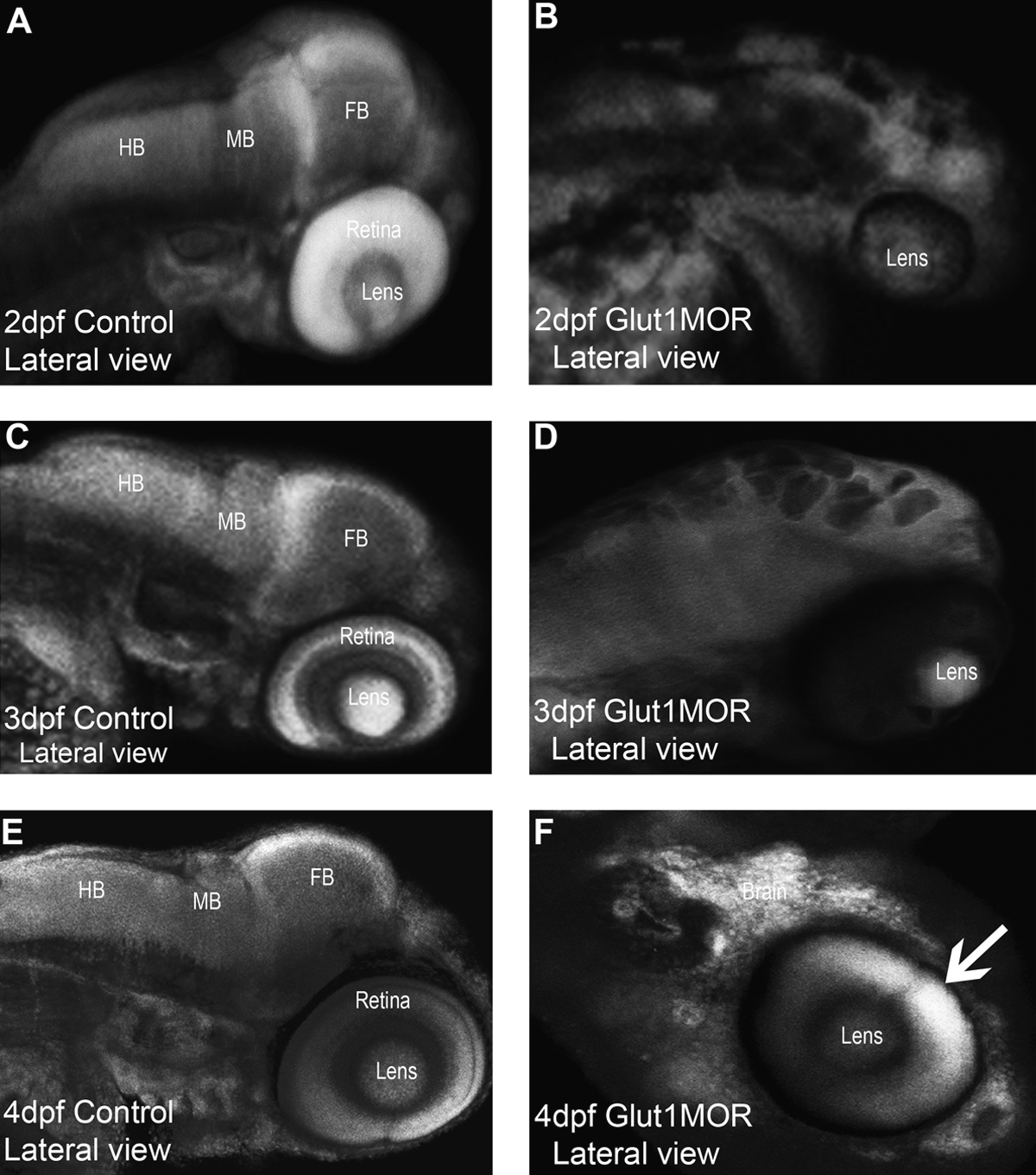
Detection of differential signals of normal and diseased brain and eye of zebrafish.
This study illustrates that the 633 nm HeNe laser penetrates deep enough to visualize an intact organ of a normal small laboratory animal. The detection of differential signals from the normal and pathological brain and eye is possible with this noninvasive technique. Using the 633 nm HeNe laser-coupled confocal microscope enables to adequately simulate imaging at least as good as the images of MRI/CT scanning of brain and eye. The optical method provided good spatial resolution, sufficient soft tissue contrast without using any contrast agents, noninvasive detection, and localization of soft tissue alterations in intact brain and eye of zebrafish embryos/larvae. We expect that the method may be further extended for the imaging of other organs/tissues of zebrafish or other small laboratory animals for research purpose.
Footnotes
Disclosure Statement
No competing financial interests exist.