
Abstract
Abstract
A novel protocol using gluten as a carrier material was developed to administer chemicals to adult zebrafish, per os (p.o.). To evaluate the capacity of gluten to retain chemicals, we prepared gluten granules containing eight types of chemicals with different Log Pow values and immersed them in water. Less than 5% of chemicals were eluted from gluten granules within 5 min, a standard feeding time for zebrafish. Although retention capability was dependent on the hydrophilicity and hydrophobicity of the chemicals, the gluten granules retained 62%–99% of the total amount of chemical, even after immersion in water for 60 min. Vital staining dyes, such as 4-Di-2-Asp and Nile red, administered p.o., were delivered into the gastrointestinal tract where they were digested and secreted. Subsequently, we conducted a pharmacokinetic study of oral administration of felbinac and confirmed that it was successfully delivered into the blood of zebrafish. This indicates that chemicals administered using gluten granules are satisfactorily absorbed from the digestive tract and delivered into the metabolic system. The absorption, distribution, and pharmacokinetics of chemicals given by oral administration were also compared with those of chemicals given by alternative administration routes such as intraperitoneal injection and exposure to chemical solution.
Introduction
Methods have been developed for chemical administration to zebrafish. Embryos are exposed to a chemical bath or are microinjected with chemical solutions directly into the yolk sac. 8 Larval zebrafish are commonly used for chemical screening, and are often placed into multi-well dishes to swim in water containing various dissolved chemical compounds.9–13 Chemical screening can also be performed in adult zebrafish using water-borne exposure or intraperitoneal injection (i.p.). For water-borne exposure, fish are immersed in chemical solutions at a density of one fish per 100–200 ml, with chemical solutions changed daily where long-term exposure is required.14–18 For intraperitoneal injection, fish are anesthetized and chemical solutions are injected into their peritoneum in a small volume (generally <10 μl).19–21 However, almost all of the methods described above involve water-borne exposure; methods for treatment with chemicals possessing little or no solubility in water need to be established.
Oral administration methods for zebrafish have also been reported. Toxicology studies have developed a purified casein-based diet formulated for oral administration of chemicals to medaka (Oryzias latipes), 22 and these have been modified for zebrafish. 8 Additionally, an oral perfusion system was developed to permit continuous electrocardiographic recording outside of the normal aqueous environment. 23 However, these methods can be difficult to manipulate and are labor-intensive.
Here we describe a simple and novel protocol for oral administration of test chemicals to adult zebrafish using gluten as a binding agent. We confirmed the capacity of gluten granules to retain chemicals of different solubility when immersed in water, and compared this method with other methods such as i.p. or exposure administration.
Materials and Methods
Animals and maintenance
Zebrafish were supplied by the Zebrafish International Resource Center (University of Oregon, Eugene, OR) and maintained in our facility according to established protocols. 24 “Adult zebrafish” used in this study were 4–6-month-old males of AB line and rose mutant line which were bred in our facility. Fish were fed twice daily with commercial flake food (Hikari Tropical Fancy Guppy, Kyorin, Hyogo, Japan) and once daily with live Artemia nauplia. During the chemical screening experimental period, fish were fed the untreated or chemically treated gluten granules in addition to the two diets described above.
Preparation of gluten granules
Water was added to gluten powder (Wako, Osaka, Japan) and this mixture was kneaded together using a medicine spoon. The dough was stored overnight at −80°C. The frozen dough was then lyophilized (DC-400, Yamato Scientific Co., Ltd., Japan). After complete drying, gluten was transferred to a mortar and carefully ground to granules (not to powder). To adjust the granule size as suitable for consumption by adult zebrafish, the grinding process was repeated until all of the granules could pass through a 700 μm mesh sieve. Granules were stored at 4°C in an air-tight container prior to use.
Preparation of chemical-containing gluten granules
The relevant physiochemical properties of the chemicals used in this elution study are given in Table 1. Pyranine was obtained from Alfa Aesar (Heysham, UK). Toluidine blue O, C.I. Food Yellow 3 free acid and Coumarin 7 were obtained from Sigma Aldrich (St. Louis, MO). Phloxine B was obtained from Acros Organics (Geel, Belgium). Hematoporphyrin was obtained from ChromaDex (Irvine, CA). Solvent Red 197 and Solvent Yellow 43 were obtained from Canon Inc. (Tokyo, Japan). Stock solutions of Pyranine, Toluidine blue O, and C.I. Food Yellow 3 free acid were prepared by dissolution in sterile distilled water; the remaining five chemicals were dissolved in dimethyl sulfoxide (DMSO; Nacalai Tesque, Kyoto, Japan) at 50 mg/ml. Working solutions were prepared from each stock solution by adding sterile distilled water to provide a final concentration of 10 mg/ml.
Estimated values obtained from EPI Suite 4.10 software, provided by the U.S. Environmental Protection Agency and available from http://www.epi.gov/opptintr/exposure/pub/episuite.htm CAS, Chemical Abstracts Service.
For fluorescent dye-containing gluten preparation, stock solutions of 4-Di-2-Asp (4-(4-diethylaminostyryl)-N-methylpyridinium iodide; Sigma Aldrich) and Nile red (9-(diethylamino)-5H-benzo[a]phenoxazin-5-one; Wako) were prepared respectively in DMSO and acetone at 10 mg/ml and diluted with water to make a 1 mg/ml working solution.
BPAA (Biphenyl-4-ylacetic acid), also known as felbinac (Xunda Pharmaceutical Co., Ltd., Hubei, China), was dissolved in methanol to produce a 15 mg/ml stock solution which was then diluted with water to make a 2.625 mg/ml working solution.
Each chemical working solution was added to gluten powder at a 1:1 (ml:g) ratio, respectively. The wet dough was kneaded, freeze-dried, and ground to granules as described above.
Confirmation of elution from gluten granules
To determine the peak absorption wavelength, the chemical working solutions were further diluted with water to 0.05 mg/ml and the absorption spectrum between 300 and 1000 nm was scanned using a Varioskan (Thermo Fisher Scientific, Waltham, MA). The optimum absorbance wavelength (λ) was selected for each chemical (Table 1), for use in determining the concentration of chemicals eluted from the gluten granules. Gluten granules containing 0.5 mg of chemical were immersed in 100 ml of water with gentle stirring, and a 1 ml water sample was collected at 5, 10, 20, 30, and 60 min after immersion. The absorbance of water samples at the pre-determined wavelength (n=5) and of standard solutions were analyzed. A calibration curve was constructed by plotting the absorbance of a series of standard solutions (concentration range 0.156–10 μg/ml) and used to estimate the concentration of each chemical.
Fluorescent dye administration
Adult male zebrafish of the rose mutant line at 6 months of age (3.5–4 cm body length; average weight: 0.38 g) were used and fasted overnight before experiments.
On the day before an oral administration test, fish were divided randomly (one fish per 0.5 L water tank). Gluten granules containing 1 μg/mg of 4-Di-2-Asp or Nile red (2 mg per fish, n=5) were added to each tank and fish consumed them freely within 5 min. Untreated gluten granules were also administered as a control. For intraperitoneal (i.p.) administration, fish were anesthetized with 0.16 mg/ml Tricaine (Wako) and injected with 5 μl of 4-Di-2-Asp or Nile red solutions (0.4 mg/ml, diluted with saline). Fish in the control group were treated with the vehicle only (4% DMSO or acetone). For exposure treatment, five fish were transferred into 500 ml of 4-Di-2-Asp (10 μg/ml) or Nile red (0.1 μg/ml) solution, and incubated in the dark for 1 h. After incubation, the fish were rinsed three times in 500 ml of fresh water for 5 min.
Fish were anesthetized either immediately or 6 h after being treated (as described above) and observed under a fluorescence microscope (MZ 16F, Leica, Wetzlar, Germany) with GFP3 (4-Di-2-Asp) or GFP2 (Nile red) filters (Leica). Part of the zebrafish abdominal skin was removed for observation of visceral organ morphogenesis.
Felbinac administration
Adult male zebrafish of AB strain at 5–6 months of age (2.9–3.5 cm body length, average weight: 0.26 g) were fasted overnight before experiments. Fish were randomly divided into three groups, each containing 30 fish. On the day before p.o. administration, fish were placed into 0.5 L water tanks (one fish per tank). Gluten granules containing 2.625 μg/mg of felbinac were fed to the ‘p.o.’ group at 2 mg per fish, approximately equivalent to 20 mg/kg body weight. In the ‘ i.p.’ group, each fish was weighed to calculate the required dose. Felbinac solution (1.05 mg/ml) was administered intraperitoneally at a dose of 20 mg/kg. Injection volumes ranged from 3.5 to 5.5 μl. In the exposure treatment group, fish were immersed in 1 μg/ml felbinac solution at a density of 1 fish per 100 ml. One hour later, fish were rinsed several times in 1 L of fresh water; this point was determined as 0 h after exposure treatment.
At 0.5, 2, 5, 12, and 24 h (n=5 at each point) after felbinac treatment, 5 μl of blood was collected through dorsal artery puncture from each fish by using a heparinized glass capillary needle. Ethanol was added to blood samples to adjust the final volume to 250 μl. This sample was then vortexed and centrifuged at 7700 g for 10 min. Blood from fish treated with either untreated gluten granules or vehicle were also collected as controls and processed in the same manner.
High performance liquid chromatography (HPLC) analysis
Felbinac was dissolved in ethanol to produce a 5 mg/ml stock solution for calibration standards preparation. The stock solution was diluted with ethanol to produce 0.2, 0.4, 0.6, 0.8, and 1.0 μg/ml of calibration standards.
A 20 μl aliquot of each sample supernatant was analyzed by an HPLC system consisting of a L-7100 pump, L-7200 autosampler, L-7300 column oven, L-7400 UV detector (Hitachi High-Technologies, Tokyo, Japan), and a Shimadzu CR-7A computing integrator (Shimadzu, Kyoto, Japan). The system was equipped with a 4.6 mm×250 mm C18 column (6.5 μm particle size, Wako) maintained at 40°C. Elution was carried out isocratically with a mobile phase consisting of acetonitrile/phosphoric acid (0.01 M) (1:1, v/v) at a flow rate of 1 ml/min. The eluate was monitored at a wavelength of 254 nm. Blank data were obtained from the blood of the control fish described above. Drug concentrations were determined by measuring the peak area and comparing it to the peak area of known standards.
Results
Properties of chemical-containing gluten granules
Wheat gluten is easily obtained as a by-product of wheat flour. Purified gluten powder is white and odorless and can absorb approximately its own weight of water. We confirmed that 1 g of gluten could absorb approximately 1 ml of water. Gluten granules were prepared as described in the Materials and Methods section. These granules are insoluble, float on the surface of water, and can retain their form and remain afloat for up to 30 min. To test whether gluten is preferred by zebrafish, the adult AB line zebrafish were fed gluten granules and their feeding behavior was observed. The average time needed for a single fish to completely consume 2 mg of gluten granules was 79 seconds (n=40). Furthermore, the fish consumed 4 mg of gluten granules within 4 min, and approximately 80% of the 5 mg feeding was consumed within 5 min. Since it is recommended that all food should be eaten within 5 minutes, 24 we determined that the optimal amount of gluten granules for administration of a test chemical to zebrafish should be below 5 mg per fish and should be set at 2–4 mg per fish.
To investigate the amount of chemicals eluted from gluten granules, we selected eight chemicals (fluorescent dyes) covering a range of log octanol-water partition coefficients (Log Pow) from −3.97 to +6.45 (Table 1). Gluten granules containing each chemical were prepared and immersed in water. Water samples containing chemicals eluted from these gluten granules were collected and their absorbance at a pre-determined wavelength was measured (n=5). The concentration of each chemical in the water samples was calculated from the regression equation of the calibration curve. The time courses for elution of each chemical from the gluten granules are shown in Figure 1. Up to 5 minutes, all chemicals were eluted less than 5% or not at all. Pyranine, toluidine blue O, and C.I. Food Yellow 3 free acid were soluble in water, and the elution curves exhibited an increased modal average over time. Pyranine showed a maximum elution rate of 38%, and the elution rate of chemicals with Log Pow values of −3.97 to +1.4 ranged from 15%–38% when gluten granules when immersed in water for up to 60 min. For chemicals with Log Pow values of 2–5 (Solvent Red 197, Coumarin 7, and Solvent Yellow 43), the elution curves showed a slow increase over time and less than 10% of the chemical was eluted from the granules within 60 min. Chemicals with Log Pow values above 5.0 (e.g., Phloxine B (5.47) and Hematoporphyrin (6.45)) were only minimally eluted from gluten granules.
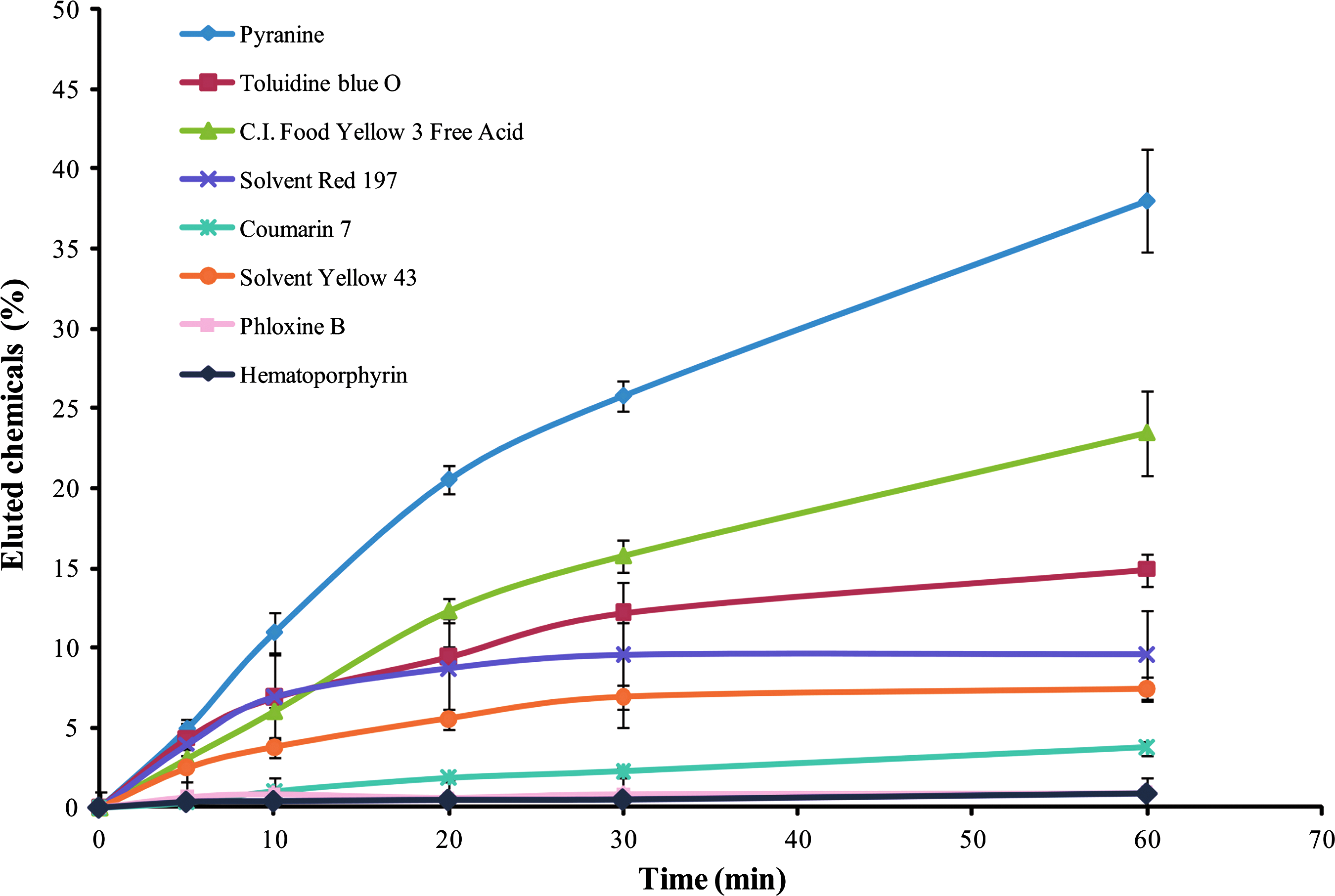
Elution of test chemicals from gluten granules. Time-dependent change in the ratio of eluted test chemicals to the original amount of test chemical contained in the gluten granules (mean±S.D., n=5). Water samples containing eluted chemicals from gluten granules were collected at 5, 10, 20, 30, and 60 min after immersion in water. The concentration of each chemical was determined by measuring the absorption at the respective peak wavelengths given in Table 1.
Absorption and distribution of test chemicals
We used two fluorescent dyes, 4-Di-2-Asp and Nile red, to observe the absorption and distribution of test chemicals contained in gluten granules. The rose mutant zebrafish, derived from the AB strain and carrying a mutation in the endothelin receptor B gene (ednrb1) that leads to a pigment deficiency phenotype, 25 were treated with both dyes via p.o., i.p., and exposure administration routes (Fig. 2). Since the adult rose mutant zebrafish have no pigmentation in the ventral skin, a sensitive fluorescence image can be derived.

The uptake and distribution of 4-Di-2-Asp and Nile red after p.o., i.p., and exposure administrations. Fluorescence images of 4-Di-2-Asp
4-Di-2-Asp is a green fluorescent vital staining dye used for labeling hair cells, 26 has a log Pow value of 0.38, and was used here as a representative hydrophilic chemical. Zebrafish treated orally with gluten granules containing 4-Di-2-Asp showed strong, localized fluorescence in the gastrointestinal tract (Fig. 2A, p.o.). After 6 h of digestion, the signal weakened and was relocated to the posterior intestine and the anus. A low-intensity label was observed in the liver, indicating that the 4-Di-2-Asp absorbed from the intestinal wall was metabolized in the liver (Fig. 2B, p.o.). The intraperitoneal injection of 4-Di-2-Asp resulted in widespread vital staining in the peritoneal cavity, including in the gastrointestinal tract, heart, liver, and testis (Fig. 2A, i.p.). After 6 h, the fluorescence signal became weak and irregularly distributed (Fig. 2B, i.p.). Water exposure of zebrafish to 4-Di-2-Asp solution caused a remarkable concentration of fluorescence at the gills and a relatively weak fluorescence in the gastrointestinal tract (Fig. 2A, exposure), each of which had mostly disappeared after 6 h (Fig. 2B, exposure).
Nile red has a strong yellow-gold emission and is commonly used as a vital staining dye for neutral lipid droplets in vivo.27,28 In this study, we used Nile red as a representative hydrophobic chemical having a Log Pow value of 4.38. The fluorescence morphologies after p.o. and i.p. administration were similar to that shown at 0 h and 6 h after 4-Di-2-Asp vital stain. Fish exposed to Nile red solution exhibited intense fluorescence across the whole body, because Nile red is a lipophilic dye and can stain the intracellular lipid droplets at the body surface. The laparotomy photograph revealed that the fluorescence was not localized in either the gastrointestinal tract or the other internal organs. In contrast, the mesenteric adipose tissue was strongly stained by Nile red (Fig. 2C, exposure). After 6 h of metabolism, only low fluorescence was detected (Fig. 2D, exposure). We also attempted to detect fluorescence in the blood collected from 4-Di-2-Asp- or Nile red-stained fish but could find no signal, suggesting that concentrations in blood were low and/or undetectable (data not shown).
Felbinac pharmacokinetics in zebrafish
The oral administration protocol using gluten granules was applied to administer felbinac to adult zebrafish. Felbinac is a nonsteroidal anti-inflammatory agent and a hydrophobic chemical (Log Pow=3.19). According to the elution properties of Coumarin 7 (Log Pow=3.68) from gluten granules (which showed no elution at 5 min), we inferred that no felbinac could have been eluted into water because the gluten granules containing felbinac were entirely consumed by the fish within 5 min. No interfering peak was detected from the chromatogram of the blank sample (untreated control) at the retention time that corresponds to felbinac by HPLC analysis (data not shown). Figure 3 shows the time-dependent change in the average concentration of felbinac in blood plasma collected from zebrafish after p.o., i.p., or exposure administration. For all three methods, the maximum concentration (Cmax) value of felbinac in blood plasma was detected at 0.5 h (Tmax) after administration. This result demonstrates the rapid absorption of felbinac by zebrafish. The observed felbinac C max value was 32.6±7.1, 49.6±19.6, and 46.6±13.6 μg/ml after p.o., i.p., and exposure administration, respectively (Table 2). The area under the plasma concentration-time curve (AUC) corresponding to the above was 128±16.6, 240.5±45.9, and 84.4±15 μg h/ml, respectively. The plasma concentrations of felbinac after p.o., i.p., and exposure administration decreased rapidly with a half-life of approximately 2 h, with the drug being almost completely metabolized after 5 h.

Mean plasma concentration-time profiles of felbinac after p.o., i.p. (20 mg/kg), and exposure (1 μg/ml) administration. Each point and vertical bar indicates the mean and S.D., respectively, of five zebrafish. Solid and dotted lines represent the fitted mono-exponential curve for the mean data.
Discussion
Gluten is a fraction isolated from wheat, which is an important source of nutritional protein, both in foods prepared directly from sources containing it and as an additive to foods otherwise low in protein. For zebrafish reared in the laboratory, fish may be fed living prey, an artificial diet, or a mixture of both. Gluten or wheat flour is an ingredient of the commonly used Zeigler adult zebrafish artificial diet (Zeigler Bros, Inc., Gardners, PA) and the Hikari Tropical Fancy Guppy food that we used daily. We tested whether the fish would eat gluten only as a food and found that they preferred gluten granules.
Gluten is composed mainly of two proteins: gliadin and glutenin. Gluten proteins are water insoluble, so can be purified by washing away the associated starch. 29 When gluten powder is mixed with water, glutenin molecules cross-link to form a submicroscopic network that associates with gliadin, increasing the viscosity and extensibility of gluten. 30 A three-dimensional sponge-like structure can be observed under a scanning electron microscope. 31 We hypothesized that a chemical could be enclosed within this reticulate network and would be retained even when immersed in water. Thus, we developed and optimized this simple protocol based on gluten to administer chemicals to adult zebrafish.
We analyzed the retention capability of gluten for several chemicals possessing different Log Pow values (Fig. 1). The Log Pow, defined as the logarithm of the octanol–water partition coefficient of a solvent, is closely correlated to water solubility.32,33 Chemicals can therefore be divided into different categories based on their hydrophobicity (Log Pow value): chemicals with Log Pow values between −3 and +1 are hydrophilic, +1 to +2 are intermediate hydrophobic, and above +3 are very hydrophobic. 34 Our results indicated that the chemicals analyzed in this study were not (or only minimally) eluted from gluten granules within 5 min, regardless of their hydrophilicity or hydrophobicity. When considering the ‘five-minute rule’ in feeding, this implies that more than 95% of the total amount of chemical in the granules would be consumed by an adult zebrafish. We demonstrated that 2–4 mg of the gluten could be consumed completely by one fish in 5 min, so the dosage of a test chemical can be controlled by changing the concentration of the chemical in the gluten granules or changing the amount of gluten granules fed to the zebrafish. Additionally, even when gluten granules were immersed in water for 60 min, gluten granules retained 62%–85% of the total amount of hydrophilic or intermediate hydrophobic chemicals (Log Pow<2.0) and 90%–99% of highly hydrophobic chemicals (Log Pow>3.0). These results show that increasing hydrophobicity of a chemical (i.e., high Log Pow values) is correlated with its retention by gluten granules.
We also investigated how the test chemicals were absorbed and distributed in zebrafish, using oral, i.p., and exposure administration methods. Hydrophilic and hydrophobic chemicals administered by oral or i.p. administration were localized in the gastrointestinal tract and peritoneal cavity, respectively. In contrast, when administering chemicals by exposure, fluorescence from the hydrophilic vital staining dye, 4-Di-2-Asp, was detected in gill and digestive tract, whereas the hydrophobic vital staining chemical, Nile red, dyed the entire body surface nonspecifically, but with no evidence of fluorescence in the gastrointestinal tract. Following oral administration, it appeared that the chemicals were absorbed by the digestive system, entering the hepatic portal system and progressing thereon into the liver through the portal vein before reaching the systemic circulation. Conversely, chemicals injected by i.p. administration could be absorbed by one of two pathways. The first of these is through the portal system, connecting to the circulation via the visceral peritoneum and the mesentery, whereas the second is the direct emission of chemicals into the systemic circulation via the parietal peritoneum and the lymphatics. 35 Where zebrafish are exposed into chemicals in solution, their gills are considered to play a central role in the absorption of dissolved chemicals. Physicochemical and biochemical factors including electric charge, molecular weight, octanol-water partition coefficient, and concentration would influence the transport of chemicals through the gill. 36 Chemicals administered by exposure administration can also be absorbed by the gastrointestinal tract because, although freshwater fish do not ingest water for osmoregulation, the ingestion by zebrafish of water containing dissolved chemicals has been reported. 37 Indeed, in our experiments, after exposure to 4-Di-2-Asp dissolved in water, fluorescence was observed in the zebrafish gastrointestinal tract. The absorption of chemicals by dermal uptake via a diffusion-controlled process 38 is also possible in exposure administration. However, the strategy used in this study of orally administering gluten granules impregnated with chemicals is designed to target these chemicals to the gastrointestinal tract, where the gluten granules are digested and the chemicals subsequently absorbed.
In our studies of the pharmacokinetics of felbinac in zebrafish, the AUC values for felbinac following i.p. administration were ∼1.8 times higher than that after the p.o. route. This could be due to hepatic ‘first-pass' effects. After oral administration of gluten granules containing felbinac at a dose of 20 mg/kg, the observed felbinac C max value was almost half that reported in the literature, 39 in which the pharmacokinetic parameters were determined from rats after oral administration of felbinac as a suspension in distilled water at a dose of 10 mg/kg. Although the absolute applied dose of felbinac administered to zebrafish in exposure administration could not be calculated, the AUC value was about 0.66 and 0.35 times lower than the AUC values after p.o. and i.p. (20 mg/kg) administration routes, which would be a useful reference to further studies.
In this study, we have identified the characteristics of three different routes of chemical administration: p.o., i.p., and exposure. These methods have different advantages and disadvantages. The advantages of i.p. administration include ease of manipulation, reduced use of chemicals (i.e., money is saved, in the case of expensive drugs), and high drug bioavailability, but this technique also causes elevated infection risk (due to its invasivity) and is therefore unsuitable for long-term and/or repeated administration. Exposure administration is also easy to manipulate, but it requires large amounts of chemical (which can elevate costs) and is limited to water-soluble test chemicals. There is also considerable difficulty in calculating the precise chemical intake by a complex absorption system that is vastly different from human physiology.
Here we developed a novel protocol for oral administration, the most common and convenient method for drug delivery in humans. Our method is based on the use of gluten as an ideal carrier material, and offers the advantages of noninvasivity, minimized chemical consumption, and convenience. The disadvantage of our method is that fish have to be fed gluten granules individually to determine total consumption effectively. On the other hand, the individual feeding procedure is more advantageous for avoiding competition and a hierarchy among the fish than the group feeding. This novel protocol should be widely applicable for toxicological testing and drug discovery, and especially efficient for the screening of hydrophobic and/or insoluble chemicals.
Footnotes
Acknowledgments
The authors would like to thank Dr. Y. Nishimura for helpful discussion; Mrs. K. Nishiguchi and A. Kamakura for experimental assistance; Mrs. M. Suzuki and A. Uechi for secretarial assistance.
Disclosure Statement
No competing financial interests exist.